The smooth muscle under a microscope shows spindle-shaped cells with tapered ends. You will not find any cross-striation in these muscle fibers; thus, they appear smooth.
In this simple guide, I will show you the important identifying features of the smooth muscle fibers at a light microscope with the labeled diagram. You will also find a details description of the smooth muscle fibers compared to cardiac and skeletal muscles.
Quick microscopic features of smooth muscle: these are the small spindle-shaped muscle fibers that contain an elongated or ovoid single central nucleus. The actin and myosin filaments of the smooth muscle fibers are not arranged in the regular cross-striated pattern, and thus, it is considered nonstriated muscles.
Here, you will find the identifying points of both longitudinal and transverse sections of smooth muscles with diagrams. So, you will quickly identify this muscle under the light microscope at your histology learning laboratory.
Okay, let’s get into the smooth muscle microscope slide’s identifying points and details structure with a labeled diagram.
The smooth muscle under a microscope
Smooth muscles are widely distributed in the animal’s body and are primarily found in the lining of the visceral hollow organs and also in the blood vessels. Depending on the sections (longitudinal or transverse), you will find various characteristic features in the smooth muscle under a light microscope.
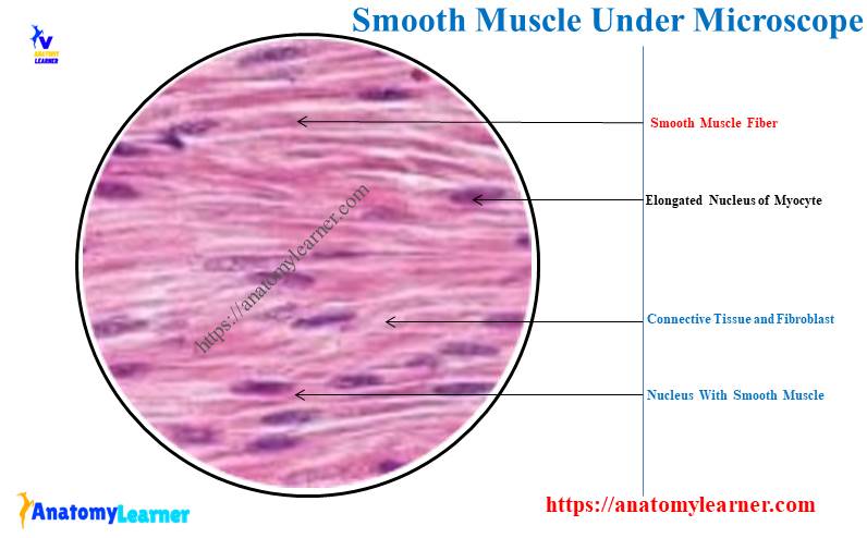
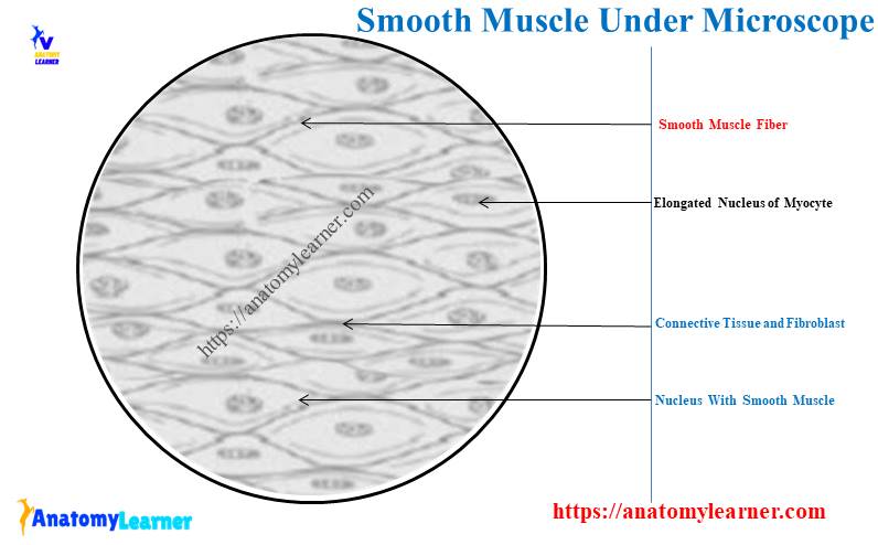
Typically, the longitudinal section of the smooth muscle shows small spindle-shaped cells with two tapered ends. You will find a dark stain in the cytoplasm of each smooth muscle fiber.
Again, an elongated or ovoid nucleus is in the center of each smooth muscle fiber. The connective tissue surrounds the muscle fiber and smooth muscle bundles.
Like skeletal muscle, you will not see the regular cross-striated pattern of actin and myosin filaments in smooth muscle structure. These actin and myosin filaments are oblique throughout the myocyte in the form of a lattice network.
So, the irregular arrangement of the contractile elements of the smooth muscle fiber appears smooth or nonstriated. Sometimes, the electron microscopic view of the smooth muscle shows numerous vesicles on its cell membrane.
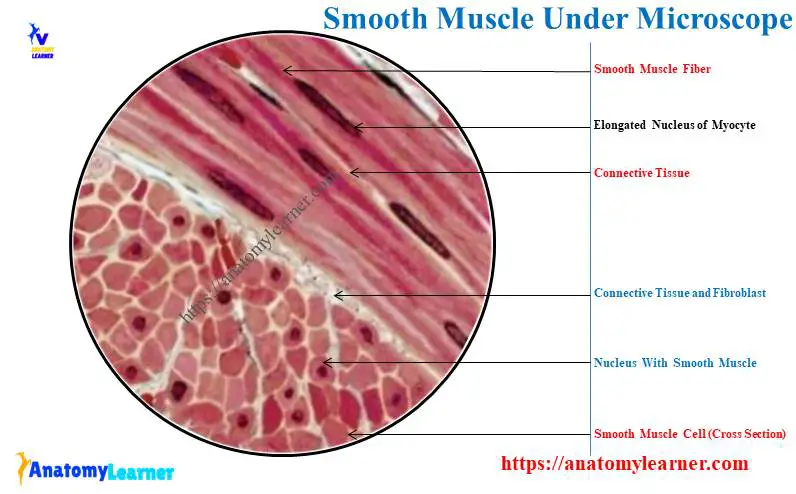
But, in the cross-section of these elongated spindle-shaped cells of the smooth muscle cut at various places along the length. Thus, you may find various shapes and sizes of the smooth muscle fiber under the light microscope.
Again, in the cross-section of the smooth muscle, you will not find a nucleus in each cell. You will find oval nuclei in these smooth muscle cells that are cut through the center.
On the other hand, if you cut the smooth muscle at the ends, the microscopic view will not show any nucleus.
Summary of smooth muscle microscope slide
While identifying the smooth muscle microscope slide, you might be considered the appearance of cells, nucleus, and other features from both longitudinal and cross-sectional views. Here, in Table 1, you will find the summary of the smooth muscle microscope slide –
| Smooth muscles under a microscope | Longitudinal section | Transverse section |
| Smooth muscle fibers | Spindle shape | Various shape |
| Nucleus shape | Elongated | Oval |
| Location of nucleus | Center, single | Center, single |
| Branching in muscle fibers | No branch | No branch |
| Cross striation | Absent | Absent |
The arrangement of the smooth muscles in various visceral hollow organs is different. You may get an idea about the general arrangement of the smooth muscle fiber in the hollow or tubular organs from the below-mentioned article –
In the article, as mentioned earlier here in the Anatomy Learner, you will find various arrangements (circular and longitudinal) of the smooth muscle fibers. That might help you to understand how these smooth muscle fibers look like a light microscope.
Smooth muscle microscope slide identification points
Now, I will enlist the smooth muscle microscope slide identification points for both longitudinal and transverse (cross) sectional samples. Okay, let’s identify the smooth muscle microscope slides with the below-mentioned identifying points.
First, I will enlist the identifying points for a longitudinal section of the smooth muscle histology slide –
- The provided sample shows the elongated spindle-shaped muscle fibers with two tapered extremities,
- Here, the muscle fibers are comparatively small and possess an elongated or ovoid single centrally placed nucleus,
- The sample tissue section (muscle fibers) doesn’t show any cross-striation,
- Again, the sample tissue shows connective tissue that surrounds the individual muscle fiber and also the bundles,
So, this is the longitudinal section of the smooth muscle microscope slide. Here, the spindle-shaped cell with an elongated central single nucleus is the key feature of the longitudinal section of smooth muscle.
Now, let’s see the identifying points for a transverse (cross) section of the smooth muscle fiber –
- The sample tissue section shows various shapes and sizes of the individual muscle cell (this is due to cut the spindle-shaped muscle fibers at different places along its length),
- The single and ovoid nucleus is seen in these muscle fibers that are cut through their center,
- Most of the muscle fibers or cells do not show any nucleus,
- Again, the provided muscle fibers show some other features like the presence of connective tissue that surrounds the individual myocyte and numerous blood vessels,
So, this is the transverse section of smooth muscle fibers at a light microscope. Here, the oval centrally placed single nucleus with various shaped cells is the key feature for the transverse section of smooth muscle.
Smooth muscle tissue location in an animal body
You will find smooth muscle in the various parts of the animal. The best example of the location of smooth muscle is in the blood vessels and different parts of the intestine.
The blood vessels and smooth muscle fibers are arranged in a circular pattern. This circular pattern of the smooth muscle control the blood pressure by altering luminal diameters.
Again, the smooth muscle fibers are arranged in various concentric layers around the specific part of the intestine. Okay, let’s enlist the location of the smooth muscle tissue from the animal’s body –
- #1. Smooth muscle is ideally seen in the wall of hollow visceral organs, including different parts of the compound stomach, simple stomach, various parts of the intestine, urinary bladder, and uterus,
- #2. You will also find the smooth muscle layers in these organs or structures that form various narrow tubes; for example – arteries, veins, bronchi, uterine tubes, and various ducts of various organs or glands,
- #3. The smooth muscle also present in the eye help to constrict and dilate the pupil,
- #4. Again, you will also see the smooth muscle of other various parts of the body like – the orbit of the eye, upper eyelid, and near the hair follicle (arrector pili),
But, the wall of the visceral organs will show both the longitudinal and transverse arrangement of the smooth muscle.
The smooth muscle under the microscope description
The other name for smooth muscle is non-striated, involuntary, or plain muscle. This muscle comprises long spindle-shaped (fusiform) cells or myocytes with a broad central part and two tapered ends.
The nucleus of a smooth muscle cell is elongated or oval that lies in the center of the cell. Again, the length of the smooth muscle cell or fibers is highly variable and measures 14 – 250 micrometers.
Under the light microscope, the sarcoplasm of smooth muscle fiber appears to have indistinct longitudinal striations. But, you will not see any type of transverse striations in the smooth muscle at a light microscope.
The smooth muscle cells are typically aggregated from the muscle fasciculi. Again, these fasciculi aggregated from the layers of variable thickness.
In this variable thickness layer, cells are arranged so that the thick central part of one cell is opposite the thin tapered ends of adjoining cells. Each smooth muscle myocyte is surrounded by a network of fine fibers (made of collagen, reticular, and elastic fibers).
Again, these thin networks of the individual smooth muscle cell continue with more abundant connective tissue. Now, the connective tissue separates the smooth muscle fasciculi or various layers.
The cytoplasm (acidophilic) of the smooth muscle is stained evenly with eosin in a routine H&E preparation. Again, the nucleus of the smooth muscle fiber takes a basophilic stain and often has a corkscrew appearance in the longitudinal section.
This characteristic of a smooth muscle cell’s nucleus results from the cell’s contraction during fixation. With the help of this feature, you may distinguish smooth muscle cells from fibroblasts in routine histologic sections.
Fine or ultrastructure of smooth muscle cells
The ultrastructure of smooth muscle fibers possesses a contractile apparatus of thin and thick filaments and a cytoskeleton of desmin and intermediate filaments. Here, the thin myofilaments of the smooth muscle contain actin and tropomyosin but lack troponin.
But, in the structure of the thin myofilaments of the skeletal and cardiac muscles, you will find troponin. Again, the thick myofilaments of the smooth muscle fiber comprise myosin II.
This thick myofilament of the smooth muscle is scattered throughout the sarcoplasm of the cells. They are extremely labile and tend to be damaged during smooth muscle tissue preparation.
Some special techniques may also apply to view the details finer structure of the thick and thin myofilaments from the smooth muscle. But, the structural integrity of the thick myofilaments from smooth muscle can be demonstrated with the help of an electron microscope.
The thin filaments of the smooth muscle fiber attach to the cytoplasmic dendrite (dense bodies). In an electron microscopic view, these dense bodies are visible among the thin myofilaments of the smooth muscle.
A network of intermediate filaments is also present in the structure of the smooth muscle fibers. These intermediate filaments are part of the cytoskeleton of the cells and contain desmin protein.
Components of contractile apparatus
You know the component of the contractile apparatus of smooth muscle contains thin and thick filaments and dense bodies. Let’s discuss these components of the smooth muscle’s contractile apparatus.
- #1. Thin myofilaments of the smooth muscle,
- #2. Thick myofilaments of the smooth muscles, and
- #3. Dense bodies of smooth muscle,
Thin myofilaments of the smooth muscle
In the structure of thin myofilaments of the smooth muscle, you will find the followings –
- Actin,
- The smooth muscle isoform of tropomyosin, and
- Two smooth muscle-specific proteins – caldesmon and calponin,
But, there is no troponin associated with smooth muscle tropomyosin. Here, actin plays an important role in force-generating interaction with the smooth muscle myosin molecule.
Again, the tropomyosin position on the actin filaments is regulated by the phosphorylation of myosin heads. Caldesmon and calponin are the actin-binding proteins that also block the myosin-binding sites.
Finally, the action of these proteins is calcium depended. Again, this action within the smooth muscle fiber is controlled by the phosphorylation of the myosin head.
Thick myofilaments of the smooth muscle fibers
In the structure of the thick myofilaments of smooth muscle fibers, you will find the followings structures –
- #1. Smooth muscle myosin – slightly differ from skeletal muscle myosin,
- #2. Two polypeptide heavy chains and four light chains,
Overall, the structure of the thick myofilaments of the smooth muscle fibers is different than these of the skeletal muscles. Here, the smooth muscle myosin molecules are oriented in one direction (shown) on one side of the filament and in the opposite direction on the other side of the myofilament.
Thus, the myosin molecules are staggered in parallel between two immediate neighbors. Here, the side polar myosin is seen, and it has no central bare zone.
You will find several more proteins that are associated with the contractile apparatus of the smooth muscle fibers. These numerous proteins are essential to initiating or regulating smooth muscle contraction.
Okay, let’s see these several proteins in the smooth muscle’s contractile apparatus –
- #1. Myosin light chain kinase – it initiates the contraction cycle after the activation by the calcium and calmodulin complex,
- #2. Calmodulin – it is the calcium-binding protein and regulates the intercellular concentration of calcium ions,
- #3. Alfa actinin – this is another protein of the smooth muscle fibers that provide structural components too dense bodies,
Now, let’s learn a little about the dense bodies of smooth muscle fibers.
Dense bodies of smooth muscle fibers
The dense bodies of the smooth muscle fibers serve as an anchor site for the myofilaments. These dense bodies remain in the cytoplasm and cell membrane of the individual myocytes of smooth muscle.
These dense bodies contain a variety of attachment plaque proteins like alfa actinin. They help anchor both thin myofilaments and intermediate filaments directly or indirectly to the sarcolemma.
These dense bodies of smooth muscle fibers are important in transmitting contractile force inside the cell to the cell surface. In the ultrastructural view, dense bodies may find a small, isolated, irregular, and electron-dense appearance in smooth muscle fibers.
Contraction of smooth muscle
The smooth muscle’s contraction mechanism is different from those of the skeletal or cardiac muscle. Let’s see the mechanism of smooth muscle contraction with labeled diagrams.
As you know, the myosin of the smooth muscle is chemically different and binds to the actin. The myosin binds to actin only if its light chain is phosphorylated.
And this phosphorylation of the light chain is essential for the contraction of smooth muscle fibers. Again, the smooth muscle has very little energy for contraction compared to the skeletal muscle fibers.
The calcium ion flow mechanism in the smooth muscle fibers also differs from those of the skeletal muscle. Here, the small vesicles of the smooth muscle (known as caveolae) present on the cell surface play an important role in this process.
Here, actin and myosin form bundles and attach at both ends to the points on the cell’s membrane. Thus, they form the anchoring points or focal dendrites in the smooth muscle fibers.
You may also learn the details muscle contraction process from another article of anatomy learners with labeled diagrams and videos.
Smooth muscle under microscope labeled diagram
Now, you will find more labeled diagrams on smooth muscle under the light microscope. I will identify all of the features from both longitudinal and transverse sections of smooth muscle fibers with labeled diagrams.
Before that, let’s know the variation in the arrangement of the smooth muscle in different organs. Depending on the functional requirements, the smooth muscle fiber may arrange in various ways –
In some organs of animals (for example – the intestine), the smooth muscle arrange in the form of two distinct layers – inner circular and outer longitudinal layers. Within each layer, the smooth muscle bundles lie parallel to each other.
And you know, these particular types of arrangement of smooth muscle allow peristaltic movement to take place for propulsion of content from a tube.
Again, in some of the organs (like the uterus), the longitudinal layer of smooth muscle remains internal to the circular layer. That means the arrangement of the smooth muscle layers is reversed in the wall of some internal organs.
In some organs, like the urinary bladder, you will find three layers of smooth muscle – inner and outer longitudinal and middle circular layer. You may also find other different types of arrangements of the smooth muscle in various tubes or ducts.
In a tube or duct, you may see a thick circular smooth muscle layer surrounding the tube or duct and forming a sphincter. Again, the smooth muscle remains a narrow band in the skin and other places of the animal’s body.
Longitudinal and transverse section of smooth muscle 400x
In the single labeled diagram (with 400X magnification), I tried to show you both longitudinal and transverse sections of smooth muscle fibers. I have taken this diagram from the wall of the small intestine of an animal.
Here, the upper part of the labeled diagram represents the inner circular layer of smooth muscle fiber that cuts into a longitudinal section. The diagram shows the spindle-shaped or fusiform smooth muscle fibers (cells) with tapered extremities.
The cytoplasm of each muscle fiber shows a dark stain. An elongated or ovoid single nucleus is identified from the upper part of the labeled diagram.
Now, let’s see the lower part of the smooth muscle’s adjacent longitudinal section that represents the transverse section. As the spindle-shaped smooth muscle fibers are sectioned at different places along their length, their nuclei and cells exhibit various shapes and sizes.
The smooth muscle fibers that are sectioned through their center show the larger oval nuclei. Again, the smooth muscle fibers sectioned at different places do not show any nucleus and appear as a deeply stained area of clear cytoplasm.
Some focus of the labeled diagram shows a small cytoplasm with a small oval nucleus. The labeled diagram also shows the minimum amount of connective tissue fibers and fibrocytes between two layers of smooth muscle fibers.
Again, the smooth muscle labeled diagram also shows numerous capillaries, neurons, and myenteric nerve plexus. You may find more labeled diagrams on the smooth muscle microscope slide here on social media of anatomy learners.
As you know, every single hollow organ of the animal represents the smooth muscle layers on its wall. So, you should see and understand the arrangement of the smooth muscle fibers from these organs.
How to differentiate smooth muscle from cardiac and skeletal muscles?
Under the light microscope, you may easily differentiate smooth muscle fibers from cardiac and skeletal muscles. In Table 2, I will differentiate smooth muscle fibers from cardiac and skeletal muscle fibers based on their microscopic findings.
Let’s see the main characteristic features and differentiation among smooth, cardiac, and skeletal muscles –
| Features | Smooth muscle | Skeletal muscle | Cardiac muscle |
| Muscle fibers | Fusiform | Long cylindrical | Short narrow |
| Branching fibers | Unbranched | Unbranched | Branched |
| Nucleus | Single oval | Multiple flats | Single oval |
| Location of nucleus | Center | Periphery | Center of cells |
| Location | Visceral organs, Blood vessels, Skin, | Tongue, Esophagus, Diaphargham, | Heart, Pulmonary veins, Vena cava |
| Control | Involuntary | Voluntary | Involuntary |
| Striations | Absent | Well-defined | Poorly defined |
| Sarcoplasmic reticulum | Absent | Present | Present |
| Intercalated discs | Absent | Absent | Present |
| Regeneration | Occurs | Limited | Not Occur |
So, you will find a great variation in the appearance of the cells and nucleus of the smooth muscle fibers compared to other muscles. Again, the location of the smooth muscle fibers varies from the cardiac and skeletal muscle fibers.
There are no sarcoplasmic reticulum and intercalated discs in the structure of the smooth muscle fibers. Regeneration of the smooth muscle fibers frequently occurs, whereas it is limited for the skeletal muscle.
Again, regeneration is not commonly found in the cardiac muscle fibers. You may learn more about the other two different muscles from the below-mentioned articles of anatomy learners –
- The cardiac muscle under a microscope with a labeled diagram – common features, structure, and comparison with skeletal muscle, and
- The skeletal muscle under a microscope with labeled diagram – common identifying features, and structure,
Frequently asked questions on smooth muscle under a microscope
Now, you will find the most common questions on smooth muscle under a microscope that the learners ask. I will enlist the frequently asked questions on the smooth muscle fibers microscope slide with their concise answer.
But you might go through the whole article as I tried to show the basic structural features of the smooth muscle above. Okay, let’s find the commonly asked questions on smooth muscle fibers with their answers –
What does a smooth muscle look like under a microscope?
A smooth muscle looks like an elongated spindle-shaped or fusiform cell under a light microscope. Each cell of the smooth muscle fibers contains a single elongated center nucleus.
The diameter of the smooth muscle cells ranges from 5 to 20 micrometers. In contrast, the length of these smooth muscle cells ranges from 20 micrometers to one millimeter.
The longitudinal section of the smooth muscle cells is highly variable and shows typical features. I have already described all the typical smooth muscle tissue features in the light microscope above.
How do you identify smooth muscle tissue?
You may easily identify the smooth muscle tissue with the help of the appearance of its cells and nucleus. The cytoplasm of the smooth muscle cells is acidophilic and contains a basophilic single elongated nucleus in the center.
At the light microscopic view, you may also easily differentiate the longitudinal section of the smooth muscle from the transverse or cross-section. You should identify the longitudinal and transverse sectional view of smooth muscle cells or fibers based on the appearance of smooth muscle cells and nuclei.
I have already shown the typical features of the longitudinal and transverse sectional view of smooth muscle fibers in this article. The microscopic view of the smooth muscle transverse section is more variable than those of the longitudinal section.
What are the characteristics of smooth muscle?
The longitudinal section of the smooth muscle possesses the typical characteristic features. That means you will find the fusiform or spindle-shaped cells with the two tapered extremities at the microscopic view.
Again, these smooth muscle fibers are small and possess a single central oval (elongated) nucleus. But, there are no cross striations and intercalated discs in the smooth muscle fibers like the cardiac muscle fibers.
Few cross-sectional views of the smooth muscle fibers lack nuclear features because of the extent of muscle cells beyond the central nuclear region.
Again, the individual smooth muscle cell (myocyte) is surrounded by a fine or delicate network of reticular fibers, blood vessels, and nerves.
Conclusion
I hope you got the basic idea of the structure of smooth muscle under a light microscope. The characteristic features of smooth muscle fibers provided in this article might help you quickly identify them.
The provided labeled diagrams on the smooth muscle under the light microscope also help you understand its features. You may identify and differentiate the smooth muscle microscope slide from the other two muscles at your histology learning laboratory.