The blood vessels of the macrovasculature include arteries and veins. Arteries of the macrovasculature carry the blood from the heart to the microvasculature. These arteries are commonly defined by their position and further characterized histologically by their structure. In this short article, I will discuss artery histology with a labeled diagram.
First, I will describe the general histological structure of a blood vessel or artery. Then I will describe and identify the elastic and muscular arteries histology slides with their identifying features.
I will also show you a few differences between a vein and artery histology at the end of this article. You will also get the answers to some common inquiries on arteries histology in this article.
Artery histology
Arteries are the thick-walled blood vessels that carry blood from the heart to the capillaries. You may ask to identify the three different types of arteries slides (large, medium, and small) at the histology laboratory.
No worries; I will make it simple to identify any artery histology slide with proper identifying points. Here, I will show you the histological features of the following three types of arteries with the labeled diagrams.
Large artery or elastic artery
Medium-sized artery or muscular artery and
Small arteries or arteriolar arteries.
First, let’s know the structures you might identify from the elastic, muscular or small artery histology slides.
Three main layers of arteries (tunica intima, tunica media, and tunica adventitia)
Endothelium of tunica intima
Subendothelial connective tissue
Internal elastic lamina or membrane
Elastic fibers (in the elastic artery)
Circular smooth muscle fibers
External elastic lamina (in a muscular artery)
Connective tissue (collagen and elastic) in the tunica adventitia
Vasa vasorum in tunica adventitia
All these structures will find in the three different layers of arteries. So, it is very important to know the general structural organization of a blood vessel or artery. Then you will differentiate the elastic and muscular arteries easily.
You will find an internal elastic lamina both in the structure of the elastic and muscular artery. But, the external elastic lamina only present in the structure of muscular artery histology.
The general structure of blood vessel or artery
The wall of all blood vessels consists of three different concentric layers – inner tunica intima, middle tunica media, and external tunica adventitia. Arteries and veins differ based on the various tunics’ composition, particularly in the tunica media.
Now, I will discuss these three layers of a blood vessel with their special histological features. I request you to read this part with great care to understand the basic structure of an artery.
Tunica intima histology
The tunica intima is the innermost tunic or layer of a blood vessel or artery. You will find a simple squamous epithelium that lines the tunica intima layer. This simple squamous of the tunica intima is known as the endothelium.
A subendothelial layer containing collagen, elastic fibers, fibrocytes, and smooth muscle cells is also present at the tunica intima layer. In addition, you will find an internal elastic lamina in the outermost part of the tunica intima of a blood vessel or artery.
But the internal elastic lamina is often indistinct under the light microscope. On the other hand, you may identify the internal elastic lamina of tunica intima readily by electron microscope.
The tunica intima is avascular and nourished through the transendothelial transport of substances from the circulating blood.
Tunica media histology
Tunica media is a middle layer of the blood vessel structure. It consists of several layers of smooth muscle in a spiral arrangement, interspersed with varying numbers of the elastic lamina, elastic fibers, and collagen fibers.
You will find a thick elastic lamina in the outermost part of the tunica media of the artery. This thickens elastic lamina is known as the external elastic lamina. But, this external elastic lamina is distinguishable in only the largest muscular arteries histology.
So, this layer is very important to differentiate the elastic artery from a muscular artery (describe below). The tunica media receives nutrients from the tunica intima and also from the vasa vasorum.
Tunica adventitia histology
Tunica adventitia is the outermost tunic of the blood vessel. You will mainly find the collagen fibers and elastic fibers in this tunica adventitia layer of an artery. But, sometimes, you may find some smooth muscle cells in the tunica adventitia.
You will also find the vasa vasorum in the tunica adventitia that extends into the outer layer of the tunica media. Sometimes, you may find some nerve plexuses in the outer part of the tunica adventitia of most larger arteries.
A few axons penetrate the tunica media and terminate near smooth muscle cells of tunica adventitia of an artery.
Elastic artery slide identification
The elastic arteries are the largest blood vessels in the animal’s body, including the pulmonary trunk, aorta, and major branches. Now, I will enlist some important identifying points to identify the elastic artery histology slide easily.
The sample tissue section shows the distinguished three different layers – tunica intima, tunica media, and tunica adventitia.
The tunica intima is lined by the simple squamous epithelium (endothelium).
There is a well-developed subendothelial layer in the tunica intima of the provided sample.
There is a comparatively thin tunica media with many elastic laminae. These elastic lamina form the bulk of the tunica media with smooth muscle fibers.
Vasa vasorum is present in the tunica adventitia of the tissue sample.
So, this is a slide of a large or elastic artery.
Elastic artery histology
Do you want to learn the details histology of an elastic artery? Well, please continue this article to know the histological features of an elastic artery.
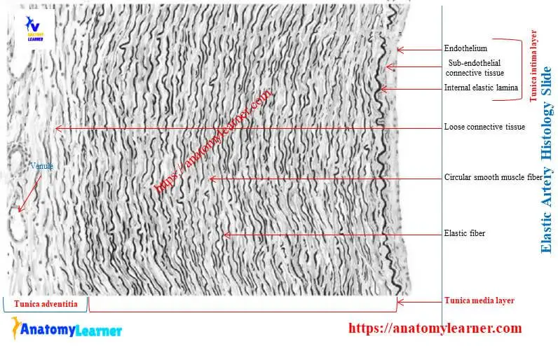
The tunica media of an elastic artery is thicker than that of the other arteries of the animal body. The endothelial cells often appear to have a brick-like shape.
You will find smooth muscle cells, fibroblasts, primarily longitudinally oriented collagen fibers, and numerous fine elastic fibers in the subendothelial layer of the elastic artery. In addition, the internal elastic lamina has often split that merge with the elastic laminae of tunica media.
The tunica media is the thickest layer that contains primarily concentrically arranged, fenestrated elastic lamina. You will also find some smooth muscle cells that lie between the adjacent laminae.
All intercellular fibers and ground substances are synthesized by the smooth muscle cells. The external elastic lamina of tunica media is indistinct or absent in the elastic artery.
You will find longitudinally arranged bundles of collagen fibers, a few elastic fibers, and fibroblasts in the tunica adventitia of the elastic artery. The interlacing of the collagen fibers limits the elastic expansion of the artery.
In some domestic mammals, you may find a comparatively thicker subendothelial layer in the tunica intima. The transition from the elastic artery to the muscular artery may either gradual or abrupt. Some arteries like the carotid, femoral, vertebral, brachial commonly begin with an elastic artery and gradually transform into the muscular type artery in the peripheral region of the body.
Muscular artery slide identification
Sometimes, you may also be asked to identify the muscular artery histology slide at a laboratory. Here, I will enlist some identifying points for the muscular artery to identify it easily.
You know, the muscular arteries are the most numerous vessels in the animal’s body. They distribute blood to various parts of the animal’s body. Branches of the carotid artery, radial artery, and ulnar artery are examples of muscular arteries.
Okay, let’s identify the muscular artery histology slide under a light microscope with the following identifying characteristics.
The sample tissue section shows the three different layers – tunica intima, tunica media, and tunica externa or adventitia.
The tunica intima is lined by the simple squamous epithelium (endothelium).
There presence a thick tunica media with many smooth muscle fibers.
Presence of well-developed internal elastic lamina and also external elastic lamina in the tunica media layer of the sample tissue section
There are elastic fibers, collagen fibers, vasa vasorum in the tunica externa or adventitia of the sample tissue section.
So, this is a slide of a medium-sized muscular artery.
Muscular artery histology
In this part of the article, you will know the details of muscular artery histology. But, first, I will describe the different histological features from the three layers of a muscular artery.
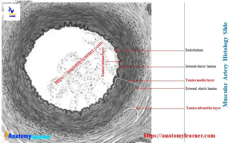
The tunica intima is comparatively thin than elastic arteries and consists of endothelium with a thin subendothelial layer. The subendothelial layer of the muscular artery contains collagen and elastic fibers. You may also find few fibroblasts and smooth muscle cells in this layer of the muscular artery.
The prominent, thick internal elastic membrane has fenestration through which cytoplasmic processes of endothelial cells contact the smooth muscle of tunica media.
You will find a thick tunica media in the structure of a muscular artery. It consists of mainly smooth muscle cells in the form of circular or spiral arrangements. You may find three to forty layers of smooth muscle cells in the tunica media of the muscular artery.
You will also find the elastic fibers or lamellae and collagen fibers in between the smooth muscle cells. The external elastic membrane is often discontinuous and not always clearly visible in a muscular artery. Instead, it consists of a dense network of elastic fibers adjacent to the tunica adventitia of the muscular artery.
You will find a moderate amount of collagen fibers, elastic fibers, and fibroblasts in the outermost tunica adventitia layer. But the amount of these component decrease with the decreasing the size of the muscular artery.
Arteriole histology
The arteriole is a small artery having a diameter of fewer than 0.5 millimeters. Therefore, you will find a relatively thick wall in arteriole histology.
The tunica intima of an arteriole consists of endothelium, a thin subendothelial layer of elastic and collagen fibers, and an internal elastic membrane. The internal elastic membrane of the arteriole is fenestrated and eventually disappears in the smallest arterioles.
But, some authors stated that there are no subendothelial layers and no internal elastic lamina in the tunica intima of an arteriole.
You will find one or three layers of smooth muscle cells and collagen fibers in the tunica media of the small artery or arteriole. There is no external elastic lamina in the tunica media of the arteriole.
The tunica externa or adventitia is very thin and poorly developed. You will find loose connective tissue and few nerve fibers or plexuses in the tunica externa of an arteriole.
Vein histology labeled and identification.
Veins are the thin-walled blood vessels that carry blood from capillaries to the hear of the animals. Compare with the arteries, veins are typically numerous and have a thinner wall, larger diameter, and greater structural variations.
Let’s identify the large or small veins under a light microscope with the proper identifying characteristics –
The presence of three different layers (tunica intima, tunica media, and tunica adventitia) provides a tissue sample.
The wall of the sample tissue section is thin, and the lumen is larger.
Tunica intima of the sample tissue is lined by the simple squamous epithelium (endothelium).
There is a thin tunica media with few smooth muscle fibers and less elastic fibers (in medium-sized veins).
There is a poorly developed tunica media (in large vein).
There is a thick tunica adventitia with longitudinally oriented bundles of smooth muscle fibers (in large vein).
So, this is a large or medium-sized vein.
General features of a vein
You may find small veins, large veins, and medium-sized veins in the animal’s body. The histological features of these veins are almost similar.
The tunica intima of the medium-sized vein consists of an endothelium lining and a thin subendothelium layer of collagen and elastin fibers. In addition, you may find the internal elastic lamina in the larger veins of the body.
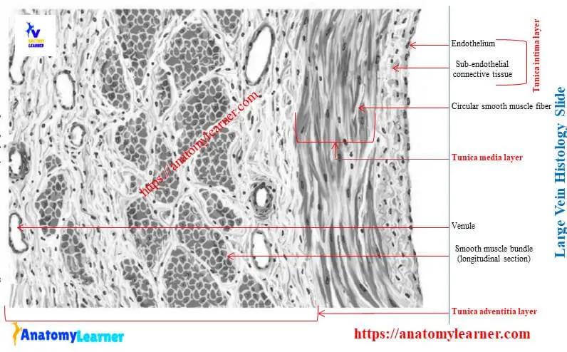
The tunica media of the medium-sized vein consists of several layers of smooth muscle associated with collagen and elastic fibers. They arrange in a circular or spiral fashion in the tunica media of vein.
You will find a thick tunica externa in a medium-sized vein that consists of longitudinally oriented smooth muscle bundles. You may also find collagen fibers and longitudinally oriented elastic fibers in the tunica externa layer of the medium-sized vein.
Elastic and muscular artery diagrams
Now, I will also show you more labeled diagrams on elastic and muscular artery histology. If you want to get notification on any changes on information or new diagrams, you may follow anatomy learner on social media.
If you find any mistakes on the artery labeled diagrams, please let me know.
Frequently asked questions on artery histology.
Now, I will try to answer all the common inquiries on artery histology. I hope you will find your desire questions and answers on an artery in this part of the article.
What are the 3 layers of the artery?
All arteries have the same basic structure and consist of three tunics or layers – innermost tunica intima, middle tunica media, and outermost tunica externa or adventitia. The tunica intima consists of endothelium and subendothelial connective tissue.
The tunica media is made of smooth muscle and connective tissue (collagen and elastic fibers). You will find more variation in the composition of tunica media in between muscular and elastic arteries. Again, the tunica externa or adventitia consists of fibroelastic connective tissue with vasa vasorum.
What are the histological differences between arteries and veins?
Arteries are the thick-walled blood vessels that carry blood from the heart to the capillaries. On the other hand, veins are the thin-walled blood vessels that carry blood from capillaries to the heart.
In arteries, you will find three well-developed tunics – tunica intima, media, and adventitia. You will also find the same three tunics in the structure of the vein, but the composition may vary. Again, you will find a larger lumen and thin tunica media in vein histology.
The internal elastic lamina is usually absent in the histology of the vein. Thus, again, the tunica adventitia of vein is more developed and thickest than the tunica adventitia of the artery.
What are the histology of the blood vessels?
As I mentioned earlier, you will find three different tunics in the histology of a blood vessel – tunica intima, tunica media, and tunica externa. Therefore, please read the histological features of different layers of the blood vessel again.
What are layers of artery?
There are three layers of an artery (histologically). They are tunica interna, tunica media, and tunica externa. I hope you already learn about these three layers of an artery in detail.
What is the largest artery of the body?
You know the aorta is the largest artery in the animal’s body. You will find the ideal histological features in the histology of an aorta.
Conclusion
This short guide might help you to get a basic idea of artery histology. In addition, I hope this guide also helps you identify the elastic and muscular artery histology slides under a light microscope.
You might use the arteries as mentioned above-labeled diagrams and compare them with the slide pictures of your histology laboratory.