The external and internal anatomy of a chicken is somewhat different compared to mammals. Therefore, you might get interested to know the unique features of chicken anatomy. In this article, I will discuss the peculiar anatomical facts from different organ systems of a chicken.
First, I will show you the external anatomical features of a chicken with a labeled diagram. Then I will go with the detailed anatomy of different organ systems from a chicken.
So, if you want to know the particular anatomical features from a chicken, then continue this article till the end.
Chicken anatomy
You will find a lot of variation in the chicken bones anatomy, muscles, digestive organs, respiratory organs, and other different organ systems compared to mammals. Most of the bones from a chicken skeleton are fused and possess some unique features. You will also find two different parts (proventriculus and gizzards) in the stomach of a chicken.
Again, you will find different air sacs in the respiratory system of a chicken. There are also some unique features in the organ of a male and female chicken.
Here, I will enlist some of the critical anatomical features from a chicken. These might help you to get the basic idea of the chicken anatomy in short. But, first, you might read the detailed article to know more about a chicken.
Vital anatomical features of chicken
So, what are the unique anatomical features that make the chicken different from other animals? Fine, let’s read the below-mentioned points.
- Most of the bones of the chicken skull are fused and possess a sizeable orbital cavity. You will also find a single occipital condyle in the chicken skull.
- Last few caudal vertebrae of chicken fuse to form a pointed bony projection, known as pygostyle.
- Each rib of the chicken has dorsal and ventral segments. The dorsal segment posses a caudal segment (known as the uncinate process) that supports the thoracic cage.
- There are no teeth in chicken. You will also find two distinct parts in the chicken stomach – glandular (proventriculus) and muscular (gizzards).
- There presence some unique structures like ceca, cecal tonsil, colo-recteum, and cloaca in the intestine of a chicken.
- There are eight air sacs in the chicken that serves to keep the body light.
- You will not find any bladder in the chicken and the ureter directly open into the cloaca (urodeum).
- The testi-(cles) are located on either side of the midline of the sublumbar region of the abdomen. You will find the copulatory apparatus of male chicken at the ventral aspect of the caudal end of the cloaca.
- There is only one ovary and one oviduct (left) found in a chicken.
- You will find a cone-shaped heart at the midline in the cranial part of a chicken’s thoracic abdominal cavity. The wing vein and shank vein are most important for collecting blood from a chicken.
- There are no gyri and sulci in the cerebral hemispheres of the chicken brain. You will find the ischiatic nerve (sciatic) medial to thigh region, between iliotibialis and iliofibularis muscles.
To know more about other different special anatomical features from a chicken, please continue this article.
External anatomy of a chicken
It is also important to known the external anatomy of a chicken. First, you should become familiar with the different regions of the chicken body. Now, I will show you the skull, face, back, rump, tail, belly, arm, thigh, leg, and foot region from a chicken.
But you will find some other special external features in a chicken body.
- A comb of a chicken
- Wattle of chicken
- Ear lobe of chicken
- Beak of chicken
- Cere of chicken
- Metatarsal spur of chicken leg
- Frontal process (actually found in turkey)
- Beard of chicken and
- Uropygial or oil gland of chicken
You will find a single comb in a chicken that consists of base, body, points, and a blade. The color of the comb may vary, but generally, it is bright red in pullet.
The wattle is a pendant, a double fold of skin suspended from the mandibular molar region. You will find an ear lobe at the caudal end of the malar region and below the ear opening. This is an elevated portion of the dermal connective tissue.
The beak is the part of the chicken skeleton of the upper and lower jaws and the hard keratinized sheet covering them. The cere lies at the base of the beak as a soft tissue covered by a membranous corneum.
You will find some scales on the feet of the chicken. But these scales differ from those of reptiles or fishes.
The claws are the terminal part of the pedal digits in chicken and adapt for scratching.
Sometimes you may find the frontal process and beard in some chicken species. But these features are not usually standard in chicken.
There presence a uropygial gland at the tip of the pygostyle. You could quickly identify the opening of the uropygial gland externally.
Internal anatomy of chicken
Before going to the detailed anatomical features of different organ systems, I would like to summarize the internal anatomy of the chicken. This might help you to get an idea of different organs from different systems of a chicken.
Here, I will show you all the organs from a male and female chicken with a diagram. But I will discuss each organ system of chicken individually.
Not enough for the identification of different organs from a female chicken? Well, now I will show the different organs from a male chicken with a labeled diagram.
Great, let’s discuss on different organ systems of a chicken. You will get information on different organ systems, but I will mainly focus on the following organ systems of chicken.
Chicken skeletal system
Muscles of chicken
Digestive organs of the chicken
Respiratory system organs of the chicken
Urinary organs of the chicken
Organs from male and female chicken
The cardiovascular and nervous system of chicken
But if you want to know more about other different organs system, please go to the avian anatomy section and find your inquiries.
Chicken skeleton
You will find almost one hundred and seventy bones in the chicken skeleton. As I mentioned earlier that most of the chicken bones are fused and possess some unique features. I have also published a complete guide on chicken skeleton anatomy. If you are interested to learn details on chicken bones, please read that article.
Here, I will enlist the most important anatomical features from the chicken skeleton. Please read these points carefully and try to find out these features from the chicken skeleton.
The chicken skeleton is compact and lightweight but robust. You will find two types of bone in a chicken skeleton – pneumatic bones and medullary bones.
The pneumatic bones are hollow that connects with the chicken respiratory system, and adaption for respiration and flight.
Most of the skull and vertebrae bones fused and forms some unique structures.
You will find a S-shaped cervical region of the vertebral column of a chicken.
At four-month of age, the last cervical vertebrae and first three thoracic vertebrae fuse and form the single bone notarium.
You will also find another unique bony structure in the chicken skeleton – synsacrum. The synsacrum of chicken consists of fused thoracic, lumbar, and sacral vertebrae.
You will find some free caudal vertebrae that help to the movement of the tail of a chicken. But the last three or six caudal vertebrae fuse to form a pygostyle that provides attachment for the innermost tail feathers.
There presence a well-developed keel, lateral notch, and medial notch in the sternum of a chicken. In addition, you will find five or six pairs of ribs that consist of sternal and vertebral segments. The vertebral segment posses a caudal extension that provides attachment for muscles, ligament, and strength of the thoracic wall.
Now, I will discuss other features of chicken bones.
Other features of chicken bones
The scapula of a chicken is blade-like and relatively immovable. It is parallel to the vertebral column of the chicken. Cranially, this scapula articulates with the clavicles, coracoid bone, and humerus. The right and left clavicle fuse ventrally and forms the furcula.
The body of the chicken humerus bone is less twisted compare to mammals. You will find a vast interosseous space in between the radius and ulna bone of a chicken. You know, the ulna bone is massive than the radius bone in a chicken. However, the length of these two bones is equal in chicken.
There presence two carpal bones in the proximal row in the chicken. But in the distal row, there are no carpal bones. This is because the first-row carpals bone fuses with the metacarpal bone in chicken.
Three metacarpal bones fuse with the carpals and form an irregular D-shaped structure (carpometacarpus).
You will find three-digit in the wing of a chicken. The first and second digits consist of two phalanges, and the third digit consists of only one phalange.
The pelvic girdle of chicken consists of ilium, ischium, pubis that fuses with sacrum bones. In most of the chicken species, the pelvic girdle is incomplete ventrally. You will find an extra-large aperture – ilioischiatic foramen, that transmits the ischiatic nerve of chicken.
The distal end of the chicken femur bone slopes craniolaterally. The knee joint of the chicken forms between the distal end of the femur, patella, and proximal end of the tibiotarsus and fibula.
The tibiotarsus is the longest bone in the chicken skeleton anatomy. This bone refers to as the drumsticks and consists of the splint-like fibula bone. In addition, you will find a well-developed cranial cnemial crest in the tibiotarsus bone that provides an attachment for the main extensor muscles of the knee joint.
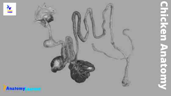
Chicken anatomy digestive system
The chicken digestive system begins at the mouth and ends at the cloaca, and has several intervening organs in between. This is an essential organ system of chicken anatomy. Therefore, you might also learn the anatomical features of all organs from chicken’s digestive system with great care.
I have published a detailed guide on the chicken digestive system anatomy. If you want to learn the details of the different organs from a chicken digestive system, you may read that article.
But, if you want to know the basic and the topmost important anatomical features from the digestive system, you may continue this article. Here, I will enlist all the organs and some important anatomical facts from the chicken digestive system.
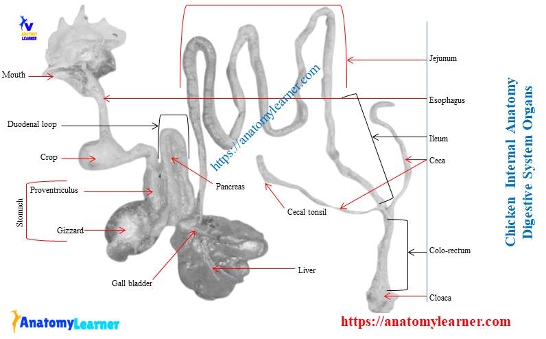
Organs of the chicken digestive system
Okay, first, start with the organs of the chicken digestive system. You will find the following different organs in the chicken digestive system. Please use the chicken digestive system labeled diagram and find out all the organs from the actual sample.
- Mouth cavity (tongue and beak)
- Pharynx of chicken
- Esophagus and crop
- Different parts of the stomach (proventriculus and gizzards)
- Parts of small intestine (duodenum, jejunum, and ilium)
- Parts of the large intestine (ceca, cecal tonsil, colorecteum, and cloaca)
- Other organs or structures (liver, pancreas, Meckel’s diverticulum) from the chicken digestive system
Well, I hope the chicken digestive system labeled diagram might help you get a better idea of the arrangement of organs.
Special features of chicken digestive organs
Now, I will enlist some of the important anatomical facts of different organs from the chicken digestive system.
You will find a rigid triangular tongue that consists of a fixed base and a considerable free apex.
Soft palate and teeth are absent in chicken.
The esophagus of chicken is the flexible tube that connects the mouth with the rest of the digestive tract.
You will find a ventral diverticulum of the esophagus, and that is a crop. But, again, the crop is highly developed in pigeons than chicken.
The stomach of the chicken consists of a cranial glandular component and a caudal muscular component. The glandular component of the chicken stomach is the proventriculus, and the muscular part is gizzards.
You will find some well-developed papillae that project into the lumen of the stomach.
Most of the gizzard wall is made of smooth muscles rich in myoglobin and arranged into four semi-autonomous masses.
The duodenum is arranged as a narrow U-shaped loop with an ascending and descending part. You will find a unique structure (Meckel’s diverticulum) about the jejunum’s middle portion. Another name of Meckel’s diverticulum is the vitelline diverticulum. It is the short remnant of the yolk duct and opens into the lumen of the small intestine.
You will find two ceca that have three different parts –proximal, middle, and distal.
The straight portion of the intestine that extends from the ilium to cloaca is the coloreceteum.
In cloaca, you will find three different compartments – coprodeum, urodeum, and proctodeum. But it is tough to distinguish these three chambers in chicken grossly.
A spherical, dark-red spleen lies on the right side of the junction between proventriculus and gizzard.
The U-shaped duodenal loop encloses the pancreas. The right liver lobe is larger than the left one in chicken.
Other features of chicken digestive organs
You will find different salivary glands in chicken. The duct of some salivary glands like maxillary, palatine, lingual, submandibular open into the mouth cavity. Other salivary glands like the pterygoid, laryngeal open into the cavity of the pharynx.
Between the two compartments of the chicken stomach, you will find an intermediate zone that marks the outside by a constriction. This constricted portion is known as the isthmus.
Do you know where the spleen is located in chicken? The spleen is located at the right side of the junction between the proventriculus and the gizzard. It is a small rounded and reddish-brown organ in chicken. The shape of the spleen may vary in duck or pigeon compare to the chicken spleen. You will find a triangular spleen in duck and an elongated spleen in pigeon.
At hatching, the liver lobes have a bright yellow color. However, the color gradually changes to the mahogany-brown between eight and fourteen days chick.
The pancreatic and the bile ducts of the chicken open into the distal part of the ascending limb of the duodenum. In the chicken, the common hepatopancreatic ducts drain the right and left lobes of the liver. In addition, the right hepatic duct gives off a branch that enters into the gall bladder.
You will find this gall bladder in the right lobe of the chicken liver, but the left lobe has no gall bladder. Pancrease is the elongated, lobulated, yellowish-red gland in chicken.
Respiratory organs of the chicken
You will find some peculiarities in the respiratory system of chicken anatomy. Here, I will also enlist the unique features of the respiratory organs of a chicken, along with a short description.
You might identify the following organs from the respiratory system of a chicken. Make sure you use the below-mentioned diagram and also the actual sample.
- Slit like nostril of chicken
- Larynx and its cartilages (four cartilages)
- Complete tracheal ring on the trachea
- Syrinx (a cartilaginous compartment of the trachea)
- The rectangular lung of chicken
- Nine air sacs of chicken
I hope you will identify these organs from the chicken respiratory system with the help of the labeled diagrams.
Special features of chicken respiratory organs
Do you want to know the unique anatomical features of chicken respiratory organs? Well, now I will enlist the particular anatomical features of the organs from the chicken anatomy respiratory system.
You will find a slit-like nostril at the upper beak of a chicken. The dorsal border of the chicken’s nostril is enclosed by a keratinized flap (known as operculum).
The nasal cavities are narrow, and each has three compartments – vestibule, respiratory, and olfactory parts. You will also find three cartilaginous nasal conchae in the nasal cavity of a chicken.
The cartilage and muscle of the chicken larynx form a prominent laryngeal mound that protrudes into the caudal part of the floor of the oropharynx.
You will find four different laryngeal cartilages in the structure of the chicken pharynx – cricoid, pre-cricoid, and one pair of arytenoid cartilages.
The tracheal cartilages of chicken are complete rings and consist of broad and narrow parts. The broad part of one cartilage ring overlaps the narrow part of the two adjacent rings.
You will find a special cartilaginous compartment (syrinx) at the junction of the trachea and the right and left primary bronchi of a chicken. The syrinx of chicken is responsible for the source of the sound. But in duck, this syrinx is known as the syringeal bulla.
The lung of the chicken is small, bright red, and triangular or quadrilateral in shape. You will not find any distinguish lobes in the chicken lung.
Unlike mammals, the chicken lacks a diaphragm to inflate and deflate the lung. Instead, the chicken has nine air sacs that distribute from the neck region to the body cavities. But no gessoes exchanges take place through the wall of the air sacs.
These are the essential features from the chicken respiratory system anatomy.
Other features from chicken respiratory organs
Now, I will discuss some other anatomical features from the respiratory organs of a chicken. You will find the laryngeal muscle below the mucosa on the dorsal surface of the laryngeal mound. The tracheolateral muscles in the chicken extend from the caudal part of the trachea to the larynx and combine with the sternotracheal muscle.
In most chicken, the syrinx is partly tracheal and bronchial origin and highly variable in structure. It is composed of variably ossified cartilages, vibrating membranes, and muscles.
The upper border of the chicken lung has a row of grooves in which the vertebral ribs are embedded. Unlike the mammals, the chicken lung undergoes minor changes in volume during breathing. If you remove the lung from the chicken, you will find some intercostal nerves. These intercostal nerves lie below the parietal pleura and caudal to the ribs.
In the ventral surface of the chicken lung, you will find the hilus where the primary bronchus, the pulmonary artery, and pulmonary vein enter into the lung. The bronchi extend outside of the lung in thin-walled transparent chambers called the air sacs.
The air sacs of the chicken consist of the unpaired cervical sac, unpaired clavicular, paired cranial thoracic, paired caudal thoracic, and paired abdominal sacs.
The cervical air sac of the chicken has a tubular diverticulum that extends up to the neck of each side of the vertebral column. In addition, the abdominal air sacs have a perirenal diverticulum that extends between the kidney and pelvis.
Male chicken anatomy
The male chicken anatomy is very different from that of the mammals. The chicken male organ system is all inside their body, unlike mammals with testis outside of the body.
You will also find those kinds of peculiarities in the male chicken. Here in this part, I will show you the organs from the male chicken and their peculiar anatomical features.
Great, let’s identify the following organs or structures from a male chicken.
- Bean shaped testes of chicken
- Ductus deference of male chicken
- The rudimentary copulatory organ of a male chicken
I hope you will identify these organs with the help of a male chicken organ labeled diagram. Let’s discuss the peculiar anatomical features of male chicken.
A peculiar feature of a male chicken
Now, I will enlist some of the peculiar anatomical features from the male chicken. Later, I will provide a bit of extra information on different organs from male chicken.
The testi-(cles) of male chicken are oval or bean-shaped yellowish structures and overlie the cranial end of the kidney.
These testi-(cles) are located symmetrically on either side of the midline at the sublumbar region in the chicken’s abdomen.
You will find a long epididymis in the chicken that locates at the dorsomedial aspect of the testis.
The ductus deference of male chicken is a zig-zag tube and locates medial to the corresponding ureter. It opens at the dorsal wall of the cloaca together with the ureter.
In some chicken, you will find a zig-zag or convoluted ductus deference at breeding season, but in the non-breeding season, it is a straight tube-like structure.
You will find a rudimentary copulatory organ at the ventral aspect of the caudal end of the cloaca of male chicken. The copulatory apparatus of male chicken consists of two papillae of ductus deferences, two vascular bodies, a phallus, and two lymphatic folds.
You may be familiar with the term canonization; it is the process of ultimately removing the male chicken testes. It is done to make the chicken docile, less active, and for better meat production. If you are a veterinary student or poultry practitioner, you might know the exact location of the testi-(cles) of male chicken to perform the canonization.
“If you don’t know this process –canonization; please don’t try to perform it.
Other features of a male chicken
During the peak period, the testes of male chicken become triple in size and become white. The increase in the size of the testes is more marked in the wild chicken.
The ductus deference extends caudally on the ventral surface of the kidney and terminates in the cloaca of the chicken. You will find the epididymis that lies dorsal to the testis of male chicken. In wild chicken, the epididymis also enlarges considerably during the breeding season.
There are no accessory glands like seminal vesicles or prostate in the male chicken organ system.
Female chicken anatomy
The female chicken organ system is divided into two main parts – the ovary and the oviduct. In chicken, the left ovary and oviduct are functional. Although, the right ovary and oviduct present embryologically and regress during the development.
You will find a lot of special anatomical features in female chicken. First, I will show you the different parts of female chicken with a labeled diagram.
I will also enlist the particular anatomical features of the organs from female chicken.
Okay, let’s identify the following structures or parts from the female chicken anatomy labeled diagram.
- Ovary of a chicken
- Infundibulum of chicken oviduct
- Magnum of chicken oviduct
- Isthmus of chicken oviduct
- Shell gland of the chicken uterus
- Vent of a chicken
Ovary of chicken
The ovary of chicken is a cluster of developing yolks or ova. You will find many grapes with four or five large, mature follicles and thousand of immature follicles in the chicken ovary.
The mature follicles are yellow due to the presence of yolk, protein, and lipid. Ovulation is the release of mature ovum from the ovary into the second part of the female chicken reproductive system, the oviduct.
Immediately after ovulation, the follicle becomes a thin-walled sac. Unlike mammals, there is no corpus luteum in the ovary of a chicken.
During the molt in chicken in the non-breeding season, the ovulation ceases, and the follicle is absorbed. So the ovary comes to resemble the shrunken structure.
Oviduct of a chicken
The second major part of the female chicken anatomy is the oviduct. The oviduct of a female chicken is a long convoluted tube that divides into five major parts.
So, the five significant parts of the oviduct are the infundibulum or funnel, magnum, isthmus, shell gland or uterus, and vent. So, now I will discuss these five parts of the chicken oviduct.
The first part of the oviduct is the infundibulum. It is the membranous expanded funnel-shaped cranial end of the chicken oviduct. This part of the oviduct receives the ovum release from the ovary of the chicken. It also serves as a reservoir of spermatozoa so that fertilization can take place.
The next part of the chicken oviduct is the magnum, and it is the most significant part of the tube. The wall of the magnum is thicker than the wall of the infundibulum. It produces thick white or albumen.
The third part of the chciekn oviduct is isthmus. It is short in length and has less diameter compare to other parts of the oviduct. The isthmus produces the inner and outer shell membrane to the egg.
The shell gland or uterus is a little thicker and expanded in the laying condition of the chicken. Uterus pumps the egg, forms the shell and cuticles, and determines the shell pigment.
The last part of the chicken oviduct is the veg. It is a short narrow, and curved tube in chicken. This part of the oviduct does not play any role in forming an egg but helps to push the egg out of the chicken’s body.
The cloaca is the common chamber through which egg passes and also responsible for the expulsion of feces and urine. A vent is the exterior opening through which passes occurs from the digestive system, urinary tract, and reproductive tract.
Chicken cardiovascular system
The chicken heart is comparatively larger than that of the mammals. It lies in the ventral part of the thorax surrounded by the lobes of the liver. Compared to the chicken heart’s left ventricle, the right ventricle has a much thinner wall, and the endocardial surface is smooth.
Unlike in mammals, the aorta in chicken develops from the right fourth arterial arch and bends to the right. The subclavian artery supplies the wing of a chicken.
The right jugular vein of the chicken is much larger than the left vein. This right vein can be seen through the skin and is used for collecting blood. Sometimes the left jugular vein may be absent in chicken.
But the wing vein and caudal tibial vein are most convenient for collecting blood from a chicken. The caudal tibial vein crosses the medial aspect of the tarsal joint, where it is easily accessible.
Chicken anatomy diagrams
Here I provide different chicken anatomy diagrams that might help you to get the basic knowledge. I will update the information and diagram if necessary. You will get a notification on social media if I update any topics or diagrams.
Frequently asked questions on chicken.
In this part of the article, you will find answers to the frequently asked question on chicken anatomy. I will update this part with more information.
Do chickens excrete feces and lay eggs from the same passage?
Yeah, the chickens excrete feces and lay eggs from the same passage. As you know, the cloaca is the terminal part of the intestine of the chicken. It has three different parts – coprodeum, urodeum, and proctodeum.
Urogenital ducts open on the dorsolateral wall of the urodeum. The ureter opens dorsally, and the genital ducts open laterally.
What organ systems do chickens have?
Chicken has different organ systems like the mammals – skeletal system, muscular system, digestive system, respiratory system, urinary system, male organ system, female organ system, immune system, cardiovascular system, and more.
What is the function of a chicken gizzard?
The gizzard is the unique part of the chicken digestive system and is often referred to as the mechanical stomach. Consumed feed and the released juice pass from the proventriculus to the gizzard for grinding, mixing, and mashing.
Conclusion
This guide might help you to get a basic idea of chicken anatomy. I hope you got the most important anatomical features of different organ systems from a chicken. Now, it’s time to learn the details anatomy of different organ systems from a chicken.
I always suggest using the chicken internal and external organs labeled diagram and the actual sample for better understanding.