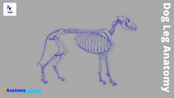
Most first-year veterinary students have a misconception of the term “leg.” Anatomically, the term leg means the part of the hind limb that extends from the stiffle joint to the hock joint (knee to ankle or tibia and fibula bones region). This short post will try to cover the dog leg anatomy in detail with labeled diagrams.
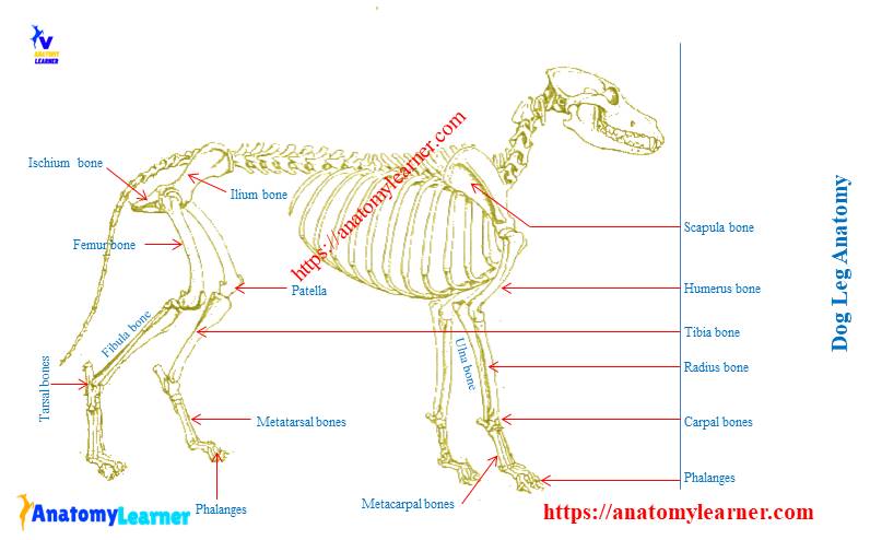
The leg of a dog consists mainly of the two long bones – tibia and fibula. So, here you will get the detailed anatomy of the leg region of a dog (bones, muscles, and vessels). But, I will also discuss the anatomy of other parts of the dog’s hind limb.
You will get short information on the dog front leg anatomy, dog leg joints anatomy, and dog leg ligaments. I will also enlist some essential dog leg muscles anatomy here in this post.
Dog leg anatomy
First, you might have a basic idea of the different bones of the forelimb and hindlimb of a dog. Now I will provide you the few information on the other bones of dog leg anatomy with their unique features.
The front leg of a dog consists of the clavicle, scapula (arm), radius and ulna (forearm), carpals, metacarpals, and phalanges (forepaw). You will also find some palmar sesamoid bones in the front leg of a dog.
Again, each pelvic limb or hindlimb of a dog consists of half of the pelvic girdle composed of ilium, ischium, and pubis bones. There are also thigh bones (femur), leg bones (tibia and fibula), and hind paw or pes (tarsals, metatarsals, and phalanges) found in the hind leg of a dog. You will also find some planter sesamoid bones in the hind leg of dog anatomy.
Following are the unique anatomical features found in the leg bones of a dog. But, these are not enough to learn the limb bones anatomy of a dog.
Special features of front leg bones of a dog
There are many special anatomical features in the front leg bones of a dog. But, I will enlist the most important anatomical features of different bones from the dog’s front leg.
The lateral surface of the scapula divides into two halves by the spina scapulae. The coracoid process is absent in dog scapula.
You will find a comparatively long, less twisted humerus bone in the front leg of a dog. The musculospiral groove is not so prominent in dog humerus bone. Again, you will find a particular feature (supratrochlear foramen) at the distal part of the dog humerus.
The radius and ulna of dog are two separate bones that contact each other by their ends. They possess narrow interosseous space that extends throughout the length of the bones.
Great, there are seven bones in the carpus of a dog, three at the proximal row and four at the distal row. The radial carpal fused with the intermediate carpal and located at the proximal row.
There are five metacarpal bones in the front leg, where the first one is the shortest. Again the third and fourth metacarpal bones are largest, whereas the second and fifth are almost equal in length.
You will find five digits in the front leg of a dog. The first digits of a dog consist of two phalanges and four digits consisting of three phalanges.
The distal part of the third phalanx is laterally compressed and forms a cone (ungula process). Again, you will find an ungula crest at the lateral and dorsal part of the base of the ungula process.
There are two sesamoid bones for each metacarpal bone of a dog. Again, you will find nine planters and five palmar sesamoid bones in the digits of a dog.
Special features of hind leg bones of a dog
Now, let’s discuss the particular anatomical features of the hind leg bones of a dog. To know more about the osteological features of back leg bones, please read the detailed anatomy of these bones.
The dog’s hip bone consists of four distinct bones – ilium, ischium, pubis, and acetabular bone. The left and right ilium bones of a dog are almost parallel to each other. You will find a concave area in the gluteal surface of dog ilium bones.
You will also find some other special anatomical features in the hip bones of a dog. The presence of twisted ischium is another most important feature of a dog’s hip bones.
The greater trochanter of dog’s femur is located at a lower level to that of the head. You will not find supracondyloid fossa in the dog femur bone. The patella of the dog is comparatively more prolonged, and the anterior surface is more convex.
You will find some unique features in the leg bone anatomy of a dog. In addition, the leg bone of a dog consists of the tibia and fibula bones. The upper part of the body of the dog tibia is prismatic, and the lower part is cylindrical. You will find a very prominent tibial crest in the proximal portion of the dog tibia.
The fibula of a dog is a thin and long bone that extends the whole length of the tibia bone. You will find a flat proximal end in the dog fibula that articulates with the lateral condyle of the tibia. The distal end of the dog fibula bone is thick.
There are seven tarsal bones in a dog that arrange in three rows. You will find five metatarsal bones in the dog where the first one is ill-developed.
Dog front leg anatomy
In this part of the article, I will discuss the dog front leg anatomy in detail. If you are interested in learning details anatomical features of dog front leg bones, you may continue.
Here, you will find the anatomical features of the following bones from the dog front leg –
Scapula and clavicle of a dog
The humerus of a dog
Radius and ulna bones of a dog
Bones of forepaw (carpals, metacarpals, and phalanges)
Okay, let’s start to learn the anatomical features of dog front leg bones.
Scapula of a dog
The scapula of a dog is a flat, large bone in the shoulder joint. The lateral surface of the dog scapula is divided into nearly two equal fossae by the spine of the scapula.
You will find a broad supraspinous fossa and triangular infraspinous fossa at the lateral surface of the dog scapula. Okay, now you will find two rectangular areas (facies serrate) and shallow subscapular fossa at the medial or costal surface.
You will find a thin cranial border in the dog scapula except in the extremities. The caudal border is thick, and you will find an infraglenoid tubercle at the dorsal to the ventral angle.
You will also find three angles (caudal, cranial, and ventral) found in the ox scapula. At the ventral angle of the dog scapula, a shallow glenoid cavity receives the head of the humerus and forms the shoulder joint.
The supraglenoid tuberosity is the largest tuberosity of the dog scapula. You will also find a beaklike process (coracoid process) that leaves the medial side of the scapular tuberosity.
The clavicle of the dog is located at the tendentious intersection of the brachiocephalic muscle. The medial end of the clavicle attaches to the sternal fascia by a distinct ligamentous band.
The humerus of a dog
Thanks for continuing, the humerus is the bone of the arm or brachium of a dog’s front leg. You will find head, neck, body, and condyles in dog humerus bone.
The head of the dog humerus is oval and located at the proximal extremity. You will also find a cranio lateral projection (greater tubercle) at the proximal extremity of the dog humerus bone. The lesser tubercle is the flattened medial enlargement of the proximal medial part of the dog’s humerus.
The neck of the humerus is usually indistinct in a dog. You will find four different surfaces in the body of the dog humerus – lateral, medial, cranial, and caudal surfaces.
The lateral surface of the dog humerus presents a tricipital line and elongated deltoid tuberosity. There is a less twisted brachialis or musculospiral groove in a dog humerus.
The deltoid tuberosity is the most prominent feature of the lateral surface of humerus anatomy in a dog. It serves for the insertion of the deltoideus muscle of the dog.
Radius and ulna bones of a dog
Radius is the main weight supporting bone in the front leg anatomy of a dog. The radius of the dog divides into a proximal head, neck, body, and trochlea distally.
You will find an irregular oval-shaped head at the proximal part of the dog radius bone that posses a concave articular fovea. A constricted segment of the radius (known as the neck) joins the head to the body.
The body of the radius bone is compressed and posses two surfaces and two borders. You will find a convex cranial surface in the dog radius bone. The caudal surface divides into two flat, concave areas by a verticle interosseous border.
You will not find any special anatomical features in the medial and lateral borders of the dog radius bone. The trochlea is the distal extremity of the radius and more massive parts of the bone in a dog.
You will find an ulnar notch at the lateral surface of the trochlea and a wedge-shaped projection (styloid process) medially.
The proximal extremity of the dog ulna bone consists of the olecranon. You will find the olecranon tuber, anconeal processes, and the proximal part of the trochlear notch there. At the distal extremity of the ulna bone, there is a pointed head (styloid process).
Forepaw of a front dog leg
Forepaw is the distal part of a dog’s front leg that consists of the carpus, metacarpus, phalanges, and certain sesamoid bones associated with them. Another name of the dog forepaw is maneus.
You will find seven bones in the carpus of a dog’s front leg that arranges in two rows. There is also a small medial sesamoid bone in the carpus of a dog.
The bones of dog carpus are –
The intermediate carpal bone of a dog
The ulnar carpal bone of a dog
An accessory carpal bone of a dog
A first, second, third, and fourth carpal bones of a dog
The intermediate carpal bone locates at the medial aspect proximal row and the largest of the carpal elements.
You will find five cylindrical metacarpal bones in the dog front leg anatomy. The metacarpal bones I and V are the prominent bones in the forepaw.
They are irregular rod-shaped bones with a uniform diameter. You will find sesamoid fossae, sesamoid impression, and sagittal crest at the palmar aspect of metacarpals.
There are five digits in the forepaw of a dog’s front leg which four are fully developed, and one is rudimentary. The rudimentary first digit is also known as dewclaw in a dog. Sometimes, you may find more than one dewclaw in some dog breeds.
Except for the first digit (which contains two phalanges), the rest have three phalanges – proximal, middle, and distal. At the palmar surface of each metacarpophalangeal joint, you will find slightly curved sesamoid bones.
Dog hind leg anatomy
Each hind leg or pelvic limb of a dog consists of half of the pelvic girdle, femur, tibia, fibula, and hind paw. The pelvic girdle of a dog consists of ilium, ischium, pubis, and acetabular bone. Again, the hind paw of a dog consists of tarsal, metatarsals, digits containing phalanges, and the sesamoid bones.
- Os coxae of dog (ilium, ischium, and pubis bones)
- The thigh bone of the dog (femur)
- A leg bone (tibia and fibula bones) and
- The hind paw or pes of a dog (tarsal, metatarsal, phalanges, and associated sesamoid bones)
Now, I will describe the anatomical features of the bones as mentioned above in detail. If you are interested to learn the dog hind leg anatomy, you may continue this part.
Os coxae of a dog
Os coxae of a dog is also known as hip bone that consists of ilium, ischium, and pubis bones. There are many unique anatomical features in the hip bone of a dog compared to ox hip bones.
Make sure you know the following terms and parts from a hip bone of any animal –
- Pelvic cavity, pelvic inlet, and pelvic outlet of animals
- Transverse diameter and conjugated diameter of the hip bone
- Terminal line of the hip bone
- Pelvis symphysis of animals
The ilium is the most prominent bone of the dog hip that consists of a concave area, a wing, and a more irregular caudal body. The iliac crest consists of tuber sacralae and tuber coxae and forms the cranial border of the ilium.
You might identify the following structures or parts from the dog ilium, ischium, and pubis bone of a dog –
- The cranial and caudal dorsal iliac spines
- A cranial ventral iliac spine
- Alar spine of ilium bone of dog
- The gluteal surface, sacropelvic surface of ilium bone
- Greater ischiatic and lesser ischiatic notch
- Arcuate line of ilium bone
- Iliopubic eminence of the hip bone
- Acetabular fossa and lunate surface
- Ischial tuberosity and ischiatic arch
- Obturator foramen of dog hip bone
- Ischiatice table of a dog
- Ventral pubic tubercle and pecten
The ischium of a dog consists of body, ramus, table, and tuberosity. It forms the caudal third of the os coxae. Then it enters into the formation of the acetabulum, obturator foramen, and pelvic symphysis.
The pubis of a dog is a dorsoventrally compressed bone that extends from the ilium and ischium laterally to the symphysis pubis medially.
Dog femur bone
The dog femur is the heaviest bone in the hindlimb. You will find a nearly hemispherical head, neck, two processes, or trochanters at the proximal part of the dog femur. The distal portion of the dog femur is quadrangular and posses condyles and trochlea.
You will find a small, indistinct circular pit on the medial part of a hemispherical head of dog’s femur. The greater trochanter, the largest tubercle of the proximal extremity of the femur bone, locates directly lateral to the head and neck.
There is a deep trochanteric fossa in the dog femur. The lesser trochanter is a distinct, pyramidal-shaped eminence that projects from the caudomedial surface of the proximal extremity of the dog femur. You will find a low but wide arciform intertrochanteric crest in the femur of a dog.
The body of the femur bone is nearly cylindrical and is straight proximally and cranially arched distally. There are medial and lateral supracondylar tuberosities at the distal end of the dog femur bone.
The lateral condyle of the distal end of dog’s femur is convex, and the medial condyle is smaller and less convex. The condyles of dog femur are separated by the intercondylar fossa, which is slightly oblique in direction.
You will find a smooth, wide articular groove (femoral trochlea) on the cranial surface of the distal extremity of the femur.
Dog leg bone anatomy
You know, the dog leg bone consists of tibia and fibula bones. In this part of the article, I will describe the dog leg bone anatomy in detail.
The tibia is a long, thick bone that lies in the medial part of the leg. The proximal half of the tibia is triangular and more massive than the distal part. Again, the distal portion of the dog tibia is nearly cylindrical.
You will find a relatively flat and triangular proximal end in the tibia that consists of two condyles. There also present intercondylar eminence, medial and lateral intercondylar tubercles, and popliteal notch. The tibial tuberosity is the extensive, quadrangular, proximocranial process in the tibia of a dog.
There are three surfaces in the body of a dog tibia bone – caudal, medial, and lateral surfaces. The caudal surface of the dog tibia presents an oblique popliteal line.
The fibula is a thin, long, laterally compressed bone that articulates with the caudolateral part of the lateral condyle of the dog tibia proximally and with the tibia and talas distally. You will find a transversely flattened head in the dog fibula bone.
A short neck blends with the body of the fibula with no specific distinction. The body of the dog fibula is slender and irregular. You will find a twisted appearance at the proximal half of the dog fibula bone. Again, this bone becomes wider, thinner, and more regular at the distal half.
There are two surfaces in the dog fibula bone – the medial and the lateral surface. The medial surface of the fibula is rough and lies close to the tibia of the dog. Again, the lateral surface of the dog fibula is smooth and lies embedded in the muscle of the leg region.
Hindpaw or pes of a dog
The hind paw or pes of a dog’s back leg consists of tarsal, metatarsal bones, phalanges, and sesamoid bones associated with the phalanges.
There are seven tarsus bones in dog hind leg anatomy that are arranged in two transverse rows. The tarsus is more than three times as long as the carpus in a dog. You will find a prolonged and most considerable calcaneus (os calcis) tarsal bone in the hock joint of a dog.
The second-largest tarsal bone of a dog is the talus that posses a head, neck, and body. The body forms the proximal half of the talus bone of a dog. You will find a transversely elongated head in the dog talus bone.
The central tarsal bone lies in the medial part of the tarsus between the proximal and distal rows. Do you know which one is the smallest tarsal bone in a dog? Fine, the second tarsal bone is the smallest tarsal bone in a dog’s hind leg.
The metatarsal bones resemble the corresponding metacarpal bones in general forms. Dog metatarsus bones are compressed transversely.
You will find four typical metatarsal bones (except the first metatarsal) in the hind leg of a dog. The first metatarsal is atypical and considered the first digit of a dog. Again, the first digit of a dog is also known as a dewclaw.
A typical metatarsal bone consists of a proximal base, triangular shaft or body, and ball-shaped distal extremity. You will find a deep, transverse sesamoid fossa that separated the head from the body of the metacarpal bones. The phalanges and the sesamoid bones of the hind paw of a dog are similar to these of the forepaw.
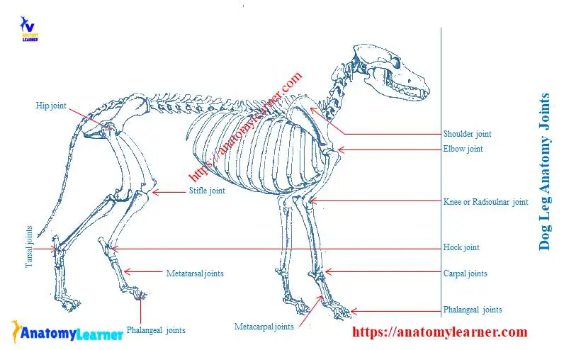
Dog leg anatomy joints
You know, joints are formed when two or more bones are joined by fibrous, elastic, or cartilaginous tissues or by a combination of these. I have a detailed article on the joint anatomy of an animal here.
Ensure you know the anatomy of different types of joints like fibrous, cartilaginous, and synovial joints of an animal body. I hope you also have a deep knowledge of the structure of a synovial joint of animals.
Here, I will not describe all the joints of a dog’s body in detail. Instead, I prefer to enlist few important joints of a dog’s body with a bit of information.
Dog front leg joints
It is essential to know the joint’s name with their bone involvements from both the dog’s front and hind legs anatomy. Let’s find the following joints from the dog front leg anatomy.
- The shoulder joint of a dog
- Elbow joint of a dog
- Knee or radioulnar joint of a dog
- Carpal joint of a dog
- A metacarpal joint of a dog and
- The phalangeal joint of a dog
The shoulder joint of a dog is a ball and socket joint between the glenoid cavity of the dog scapula and head of the humerus. You will find two main ligaments – medial and lateral glenohumeral ligaments in this shoulder joint of the dog.
The humeral condyle forms the dog’s elbow joint with the head of the radius bone. In this dog’s joint, you will find lateral collateral ligaments, medial collateral ligament, annular ligament of the radius bone, olecranon, and oblique ligament.
The knee joint of dog forms by the radius, ulna, and distal part of the humerus bone. You will find a thick but short collagenous interosseous ligament in the knee or radioulnar joint of the dog.
The carpal joints of dog are composite joints that include proximal, distal, and middle, and intercarpal joint surfaces. You will find different essential ligaments in the carpal joints of a dog.
- Lateral and medial collateral ligaments of carpal joint
- Dorsal radiocarpal ligament
- Palmar ulnocarpal ligament
- Palmar radiocarpal ligament and
- Accessory metacarpal ligament
The intermetacarpal joints are the close-fitting joints between the proximal ends, bases of the adjacent metacarpal bones. In a metacarpophalangeal joint, you will find short sesamoids ligament and cruciate ligaments of the sesamoid bones in the front dog leg.
Dog hind leg joints
Now, let’s discuss the dog hind leg joint anatomy. But I will not describe all the anatomical features of these joints from the back leg. I will only show you an essential joint with little information.
In the hind leg of a dog, you will find the following vital joints –
- Joints of pelvic girdle of a dog
- The hip joint of a dog
- Stifle joint of a dog
- Tibiofibular joint or hock joint of a dog
- Tarsal, metatarsal, and phalangeal joints of a dog
The pelvic symphysis of a dog consists of the pubic symphysis and the ischiatic symphysis. You might know the detailed anatomy of the hip joint of a dog. The hip joint of the dog formed by the head of the femur joins with the acetabulum of os coxae.
You will find essential ligaments in the hip joint of a dog like – transverse acetabular ligament, iliofemoral ligament, ischiofemoral ligament, and ligament of the head of the femur.
The stifle joint is another important joint of a dog that is a complex condylar synovial joint. You will find more than ten crucial ligaments in the stifle joint of a dog.
Dog hind leg anatomy ligaments
You will find the following important ligament in the stifle joint (dog hind leg joint anatomy). Please, learn the detailed anatomy of the hip, stifle, and hock joints of a dog. These joints are essential for field practices.
- Meniscal ligament of stifle joint of a dog
- Cranial tibial ligament of the medial meniscus
- Caudal tibial ligament of the medial meniscus
- Cranial and caudal tibial liagaments of the lateral meniscus
- Femoral ligament of the lateral meniscus of stifle joint
- Femorotibial ligament of the stifle joint of a dog
- Transverse ligament of stifle joint of a dog
- Cruciate ligament of the stifle joint of a dog
- Medial and lateral collateral ligaments of the stifle joint of a dog
- Cranial and caudal cruciate ligaments of the stifle joint
- A patellar ligament in the stifle joint of a dog
This information is not enough to get a complete idea of the stifle joint or the ligaments of a stifle joint of a dog. You might learn the detailed anatomy of the stifle joint of a dog to get a complete idea of the ligaments.
Dog front leg muscles anatomy
You will find the extrinsic and intrinsic muscles in the front leg of a dog. I will show you the essential muscles from the dog front leg anatomy with a labeled diagram. Fine, if you want to know more about the dog muscle anatomy, you may read the article here.
Muscle of the shoulder and arm region of a dog (lateral aspect and medial aspect)
- Supraspinatus muscle of the dog
- Infraspinatus muscle of the dog
- Deltoideus muscle of a dog
- Triceps brachii (long and lateral head)
- Subscapularis muscle of the dog
- Teres major muscle of the dog
- Biceps brachii muscle of the dog
- Brachialis muscle of the dog
- Tensor fascia antebrachial muscle of a dog
- Triceps brachii medial head
That’s fine; let’s identify the superficial muscles of the dog’s forearm (craniolateral aspect).
- Extensor carpi radialis muscle of a dog
- A extensor digitorum communis muscle of the dog
- Extensor digitorum lateralis muscle of the dog
- Ulnaris lateralis muscle of dog front leg
- Abductor digit I longus muscle of dog front leg
- Extensor digit I and II muscles of a dog
Now, you should learn the detailed anatomy of the dog’s front leg muscles with labeled diagrams and videos.
Frequently asked questions on a dog leg
Again, in this part of the article, you will get the answers to the most asked question on dog leg bones, joints, and muscles.
What are the parts of the dog’s leg?
Anatomically, the dog leg means the area between the stifle joint to the hock joint. The dog leg bone consists of the tibia and fibula bone. These are the long, massive bones in the hindlimb of a dog.
You will find different critical anatomical features in the dog leg region. I have already described these anatomical facts of the dog leg region.
Again, sometimes the dog leg means the front leg and also the hind leg. I hope you know the name of all the bones from the front and back legs with their special anatomical features.
How do I know if my dog’s legs injury is serious?
Sometimes, you may find sudden injuries in the dog’s leg. You may find abnormalities in the posture and gait of a dog. There may occur sudden swelling, dislocation, and hot area, pain on palpation in the specific part of any leg.
ISo, if you find these signs in your dog, the injuries of your dog’s leg may be serious. It is recommended to get your dog to the local veterinarian.
Where is the ACL on a dog’s back leg?
ACL on a dog’s back leg means the anterior cruciate ligament. There are 2 cruciate ligaments in the stifle joint of a dog – anterior and posterior cruciate ligaments. These cruciate ligaments locate within the joint cavity of the stifle joint.
This is a thin white connective tissue of the stifle joint of a dog that articulates the leg bone (tibia and fibula) with the distal part of the femur bone.
What is the back leg of a dog called?
Generally, the back leg of a dog is called the hindlimb or the pelvic limb. You will find different parts in the pelvic limb of a dog. The thigh, leg, and hind paw are three other parts of the back leg of a dog.
The thigh region of a dog’s back leg consists of the femur bone. Again, the leg region of a dog consists of the tibia and fibula bones. You will find the tarsus, metatarsal, and phalanges in the hind paw of a dog.
Conclusion
So, if you want to learn the details anatomy of the dog leg, you might know the anatomical features of bones and the muscles, joints, and vessels. This article presents the summary of the dog leg anatomy, but you may learn the specific anatomy of each part from other articles.
It is always recommended to practice with the actual anatomical samples you learn from both the front and hind leg of a dog.