The cat mouth anatomy includes the opening between the lip into the vestibule of the oral cavity. Here, the oral cavity of a cat divides into a vestibule and oral cavity proper.
The vestibule of a cat’s oral cavity is a space between the internal surface of the lip and external to the teeth. Again, the mouth cavity proper of a cat extends from the inner surface of the teeth to the oropharynx.
So, in the cat mouth cavity proper, you will find the gums, teeth, tongue, palates (hard and soft), and salivary glands. This article might help you to learn the anatomy of these structures from a cat mouth cavity with the labeled diagram.
I will enlist and describe all the features of these structures of the cat mouth shortly. That might play a significant role in identifying them practically from the actual samples.
So, let’s learn the facts of feline mouth cavity anatomy with the labeled diagram.
Cat mouth anatomy
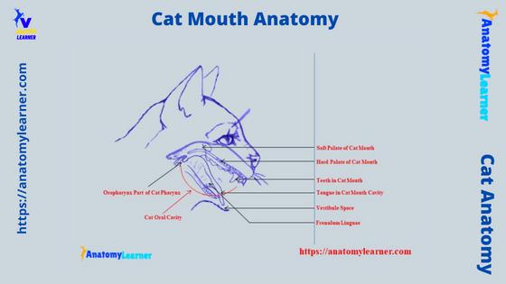
The cat mouth anatomy is defined externally by the lips and extends up to the entrance of the oropharynx. So, first, let’s try to identify the boundary of a cat’s mouth (both vestibule and mouth cavity proper) with the help of a labeled diagram.
Here, in the labeled diagram, you will find the following essential organs or structures in the cat’s mouth cavity –
- Lips (externally) with mucous membrane (internally) and hairs (externally),
- Cheek – lateral wall of the vestibular cavity,
- The floor of the cat mouth – consists of the sublingual caruncle, sublingual fold, incisive papilla, incisive duct, and frenulum linguae,
- The roof of the cat mouth – consists of palates (soft palate, hard palate), palatoglossal arch, palatoglossal muscles, palatine glands, vessels, and nerves,
- Teeth and gums of the cat,
- Cat tongue – consists of papillae, muscles, glands, and
- Salivary glands of a cat – consists of mandibular, lingual, and parotid,
Above these are the main organs or structures of the cat mouth. But, you will find some other minor structures in the cat mouth cavity. I will try to discuss these minor features of the cat mouth structure in the specific part of this article.
Parts of cat mouth cavity
But, for description purposes, you may divide the parts of the feline mouth anatomy are follows –
| Cat Mouth Anatomy | Parts |
| Vestibule | Lips Cheek Gums |
| Mouth Cavity Proper | Teeth Tongue Palates Floor of Mouth Salivary Glands |
You may compare the anatomical features of the cat mouth with the dogs. The below-mentioned article might help you to differentiate the basic anatomical features of the cat and dog mouth –
- Dog mouth anatomy with the labeled diagrams – lips, tongue, teeth, and salivary glands
Now, I would like to provide the unique features of the cat mouth cavity structure. Let’s see the special features of the cat mouth that make them different from other animals.
Unique features of cat mouth and oral cavity
The unique features of the cat mouth and oral cavity are listed below –
- The lips of a cat mouth are the paired folds whose inner surface is covered by the mucous membrane and outer surface is hairy,
- You will find a small vestibular cavity in cat that extends from the inner surface of the lips to the outer surface of the teeth,
- There is a labial frenulum in the cat vestibule that connect the upper and lower lips at the midline to the respective gums,
- The number of teeth in the cat mouth cavity is about 30 (please see the dental formula),
- The cat tongue is a very mobile muscular organ compared to the other animals that attach to the floor with the frenulum linguae,
- You will see four major types of papillae on the dorsal surface of the cat tongue – filiform, fungiform, vallate, and foliate,
- The filiform papillae of the cat are numerous and pointed structures on the apex of the tongue,
- Again, the fungiform are the mushroom-shaped structure that is less numerous and scattered among the filiform papillae,
- Vallate are more prominent and rounded papillae in the cat tongue that locates at the root,
- The foliate papillae are leaf-shaped and locates on the posterolateral aspect of the cat tongue,
- You will find the anterior soft palate and hard posterior palate on the roof of the cat’s mouth cavity,
In the hard palate of a cat, there are a series of connective tissue folds known as the rugae or transverse folds. You will find the incisive ducts at the anterior end of the cat’s hard palate. You will find some other special features in the oropharynx of a cat.
Cat lip anatomy
Lip is the first structure of a cat mouth cavity anatomy. These are the paired mucosal folds that form the rostral and lateral external boundaries of the vestibule of the cat mouth.
The anatomy of the cat lip is almost similar to that of dogs. You will find the superior and inferior lips in a cat that meet at the angle of the mouth. The structure that binds the superior and inferior lips with the angle of the mouth (or vestibule) is the labial frenulum.
So, you can understand that the lips bound the cat’s oral fissure or the external opening of the mouth into the vestibular part. The superior (upper) lip of a cat is narrow and sometimes devoided of tactile hair except for the rostral two third.
So, in the rostral two, a third of the cat’s upper lips contain hairs whose few are tactile. These tactile hairs of the cat’s lips are arranged into four rows. The direction of the hairs of a cat’s upper and lower lips are caudoventral.
You will find the orbicularis muscle (main) in the cat’s lip. But, some other facial muscles insert into the cat’s lips.
External to the upper lip of a cat, there is a deep, straight, and narrow cleft known as the philtrum. This narrow cleft makes the union of the two halves of the upper lip rostrally.
As you know, a cat’s upper and lower lips are connected to the gums by the frenulum or mucosal fold. However, this mucosal fold is not so developed in the cat’s mouth.
Thus, the inner mucosal surface of the lip forms a space with the outer surface of the teeth. And this space is known as the cat vestibular space or cavity.
Cat cheeks anatomy
The caudal part of the lateral wall of the cat’s vestibular cavity is formed by the cheeks. You will find the smaller cheeks in the cat’s mouth as the opening is larger. The cat’s cheeks run medial to the masseter muscle and extend caudally to the buccinator’s muscle.
Anatomically, the cat cheeks are composed of skin, muscles, papillae, and glands. Let’s see the essential and unique features of these structures of the cat cheeks.
The external layer of the cheek is the skin and contains hairs. You will find the coarse tactile hairs at the caudal part of the skin of a cat’s cheeks.
The main muscles of the cat cheeks are buccinators and masseter. Again, you will find the zygomaticus muscle and some fasciculi of the cutaneous fascia at the lateral aspect of the buccinator muscle.
You will find three types of buccal glands in the cat cheek region –
- Dorsal buccal glands,
- Ventral buccal glands, and
- Middle buccal gland,
The dorsal buccal glands of the cat are covered by the buccinators and masseter muscles. This is a yellowish structure that extends from the angle of the mouth to the maxillary tubers.
The ventral buccal glands are a few small and solitary glands in the cats mouth cavity. These glands are located rostral to the masseter muscle and medial to the fibers of the ventral part of the buccinator.
Again, the middle buccal glands of a cat have arranged that lie deep to the buccinator loosely. The buccal nerves innervate all these buccal glands.
You will also find some pointed conical papillae directed to the pharynx. The angle of the mouth possesses the more prominent conical papillae. Again, you will also find the shorter conical papillae in the caudal part of the cheeks.
Cat mouth oral cavity proper anatomy
The cat oral cavity proper is the area of mouth extending from the lingual side of the teeth (inner) to the cranial part of the oropharynx. From the cat’s oral cavity, you might learn the anatomy of the below-mentioned structures or organs –
- The floor of the cat’s oral cavity,
- Structure and features of the cat tongue,
- Structure of the hard and soft palates,
- Features of a cat tooth and dentition,
- Three types of salivary glands, and
- Parts of the cat oropharynx,
If you notice, the cat mouth cavity proper is dorsally bounded by the hard palate and a small part of the soft palate. You will find the dental arches and teeth at the lateral and rostral surface of the cat’s oral or mouth cavity proper.
Now, with the labeled diagrams, let’s see the details of these structures or organs from the cat’s oral cavity.
The floor of the cat’s oral cavity
First, let’s try to identify (shown in the diagram) the following features from the floor of a cat’s oral cavity –
- On each side of the cat’s tongue,
- Frenulum linguae – wide fold of mucous membrane that connects the floor of the mouth and ventral surface of the tongue,
- Sublingual folds – line of large papillae and microscope orifices of minor sublingual duct, and
- Sublingual caruncle – where the mandibular and major sublingual ducts open,
The main organs or structures of the floor of a cat’s oral cavity are the tongue and mucous membrane. It nearly fills the mouth cavity proper when it is closed. I will discuss the anatomical facts of the cat tongue later.
Ventral to the body of a cat tongue, you will find two important features – frenulum linguae and sublingual caruncle. The ventral surface of the cat tongue is attached to the floor of the oral cavity by a wide fold of mucous membrane. This wide fold of the mucous membrane is called the frenulum linguae.
You will find the large papillae which possess the serrated edge. These large papillae have located on either side behind the third incisor teeth of the cat. Anatomically, it is a wide, rigid structure known as the sublingual caruncle.
You will find the orifices lateral to the sublingual caruncle. The major sublingual and mandibular ducts open lateral to this sublingual caruncle.
Again, there is a sublingual fold just caudal to the sublingual caruncle and lateral to the frenulum linguae. This structure lies close to the body of the mandible and consists of underlying mandibular and major sublingual ducts.
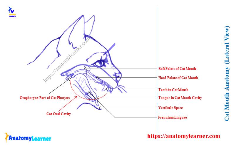
Cat mouth hard and soft palates
The floor of a cat mouth anatomy consists primarily of the hard and soft palates. You will find the rostral bony hard palate, whereas the soft membranous palate is in the caudal position.
You should identify the following anatomical (important) features from the cat’s hard palate –
- Palatine ridge or ruge – covers two third of the hard palate,
- Median groove on the hard palate – separate right and left ridges,
- Palatine glands – at the caudal third of the hard palate, and
- Incisive papillae – a diamond-shaped structure where the incisive ducts open,
The cat’s hard palate is nearly flat and formed by the process of palatine, maxillary, and incisive bones on each side. Laterally and rostrally, the cat’s hard palate inclines slightly ventrally.
You will find the pseudostratified ciliated columnar epithelium lining on the mucosa membrane of the nasal side of the hard palate. Again, the mucosa on the oral side consists of stratified squamous epithelium.
The hard palate is covered by a series of folds known as the ruge or palatine ridges. You will see a median groove on the hard palate that separates the right and left palatine ridges.
A small diamond-shaped structure is at the anterior end of the cat’s hard palate. This is the papillae incisive of the cat.
There is a deep fissure on both sides of the incisive papillae. You will see the pairs of incisive ducts that open directly on the deep fissure of the papillae. These ducts branch out and lead to the nasal cavity and the vomeronasal organ.
Cat soft palate anatomy
The cat soft palate anatomy consists of the connective tissue, muscles, glands, and vessels. It extends from the caudal end (proximal) of the cats hard palate to the last superior molar teeth.
The cat soft palate is comparatively shorter than the dog. It becomes gradually thickens at the junction of its middle and caudal thirds.
There are essential features (palatoglossal arch) that connect the body of a cat tongue to the initial part of the soft palate. This structure is the boundary between the oral cavity and the oral pharynx.
The caudal border of the cats soft palate continues to the dorsolateral wall with the help of the palatopharyngeal arch. Another name for the palatopharyngeal arch of a cat is a caudal pillar of the soft palate.
This caudal pillar of the soft palate serves as a boundary between the nasal pharynx and the laryngopharynx. You will find the palatopharyngeal muscle that covers the caudal pillar of the soft palate.
Cat mouth soft palate structure
The cat mouth soft palate structure possesses the following features –
- Epithelium – stratified squamous to pseudostratified ciliated columnar,
- Palatine glands – mixed glands,
- Palatine muscles of the cats soft palate,
- Numerous palatine vessels and nerves, and
- Cat’s palatine aponeurosis,
The characteristics of these structures of cat’s soft palate are almost similar to those of dogs. Let’s see the unique anatomical features of the different structures of the soft palate.
Cat soft palate epithelium
As you know, most of the part of cats is a hard palate lined by the stratified squamous epithelium. The continuation of this lining epithelium covers the ventral surface of the soft palate. You will see numerous longitudinal and few transverse folds in the cat’s soft palate.
The caudodorsal end of the soft palate doesn’t contain the stratified squamous epithelium. Here, you will see the pseudostratified ciliated columnar epithelium.
Numerous mucosal folds are present on the surface of the soft palate. That features indicate the mobility and slight elasticity of the soft palate.
Palatine glands in the cat’s soft palate
There are numerous palatine glands present on the oral surface of the cat’s soft palate. These mixed glands form the thickest layer in the soft palate.
Again, the caudal margin of the soft palate possesses fewer palatine glands. The number of the palatine gland decrease at the caudal surface of the soft palate.
But, the thickness of the palatine gland on the rostral and oral surface is less thick. They become gradually wider in the middle; again, they become thinner at the end of the caudal margin of the soft palate.
The cat’s palatine glands are bounded by the large medial pterygoid and styloglossus muscle on each side.
Muscles of the feline soft palate
Let’s see the muscles of the feline soft palate –
- Paired palatine muscles (palatine muscle),
- Tensor veli palatine muscles,
- Levator veli palatine muscles,
- Pterygopharyngeal muscle of the feline soft palate, and
- Palatopharyngeal muscles,
You will see the paired palatine muscles in the soft palate of a cat. The end of the muscle divides into a paired tensor and levator veli palatine muscles.
The right and left palatine muscles of the cats soft palate lie on each side of the median plane and ventral to the palatine aponeurosis. Again, the tensor and levator veli palatine muscles (extrinsic) blend with the palatine aponeurosis.
You will also see the right and left pterygopharyngeal muscles in the soft palate of a cat. These muscles are originated from the pterygoid bone and pass lateral to the caudal part of the cat’s soft palate.
Again, right and left palatopharyngeal muscles are present in the cat’s soft palate. These two soft palate muscles arise near the median plane from the palatine aponeurosis.
The right and left palatopharyngeal muscles pass laterally and dorsocaudally in the pharynx. These muscles help to form the base of the palatoglossal arches.
Cat soft palate vessels and nerves
You will see the following major vessels in the soft palate of a cat –
- The right and left major palatine vessels (arteries),
- A right and left minor palatine arteries, and
- The palatine veins,
Again, the major nerves of the feline soft palate are –
- Branches (major and minor palatine) of the maxillary nerve (a major division of trigeminal nerve), and
- Branches of glossopharyngeal and vagus nerve,
The main arteries of the soft palate of a cat are right and left minor palatine. Again, the hard palate of a cat possesses two main arteries – right and left major palatine.
The minor palatine of the cat’s soft palate has a direct connection with the major palatine and ascending pharyngeal arteries. You will see the major vein (palatine) in the soft palate of a cat that lies mainly lateral to the two slender palatine muscles.
But, this palatine vein or plexus arises from the venous plexus of the hard palate. They are located in the mucosa covering the bony plate of the hard palate.
The major palatine nerve will from the maxillary nerve courses through the major palatine canal. This major palatine nerve innervates the sensory fibers to the oral side of the hard palate.
Again, the minor palatine nerve arises from the rostral end of the soft palate. You will also find the branches of glossopharyngeal and vagus nerves that also innervate to the soft palate of a cat. Here, these glossopharyngeal and vagus nerves innervate the pterygopharyngeal, palatopharyngeal, and palatinus muscles of the cat.
Again, the branches of the glossopharyngeal nerve innervate to the lateral wall of the oral pharynx and the lesser part of the soft palate. They also innervate to the caudal part of the cat tongue.
What is the palatine aponeurosis in cats’ soft palate
The cat palatine aponeurosis comprises a thin, fanned-out terminal tendon of the right and left tensor veli palatine muscle. You will see this structure between the ventral margin of the right and left perpendicular parts of the palatine bone and the pterygoid bone.
The palatine glands directly lie dorsal to the palatine aponeurosis. A pseudostratified ciliated columnar epithelium covers the palatine aponeurosis and continues rostrally.
Cat mouth soft tissue anatomy
Sometimes you may be asked to write the cat mouth soft tissue feature. Do you know what the soft tissue is under the cat’s mouth? Well, let’s see –
The cat mouth soft tissue anatomy consists of lips, cheeks, gum, and the tongue. Each soft tissue comprises the connective tissue, muscles, and numerous vessels and nerves.
I have already described the anatomical facts of the lips and cheek in this article. Now, it’s time to explain the anatomical features of the cat tongue in detail.
The tongue is the more vital soft tissue or muscular organ in the cat’s mouth cavity. It plays an essential, versatile role in the life of a cat. This structure is used as the organ for food manipulation, swallowing, and drinking.
Again, the gum is another soft tissue structure in the cat’s mouth that surround the teeth. Both the tongue and gum of the cat’s mouth cavity possess numerous blood vessels and nerves.
Let’s see the unique features of feline tongue anatomy.
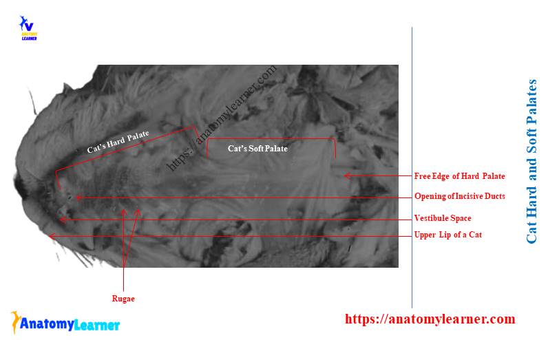
Cat or feline tongue anatomy
The cat tongue is primarily comprised of the skeletal muscle. It occupies the more significant part of the cats oral cavity proper that extends up to the oropharynx.
Before going to the details description of the different features of the cat tongue, you might identify the followings –
- An apex, a body, and root of the cat tongue,
- Frenulum linguae of the cat tongue (ventrally),
- Mechanical papillae – consists of filiform,
- Gustatory papillae – consist of fungiform, vallate, and foliate,
- The extrinsic muscle – includes styloglossus, genioglossus, hyoglossus, and
- The intrinsic muscle – has superficial and deep longitudinal muscle, transverse muscle, and perpendicular muscle,
The cat tongue is an elongated muscular organ covered by stratified squamous epithelium. It extends from basihyoid bone to its free ends. The free end of the cat tongue is the apex.
Again, the lingual body and root of a cat’s tongue are separated by a row of vallate papillae. The cat tongue also possesses two surfaces (dorsal and ventral) and two lateral borders.
The dorsal surface of the cat tongue is very rough due to the presence of different types of papillae. Mainly four types of papillae (filiform, fungiform, foliate, and valet) are present on the dorsum of a cat tongue.
The two lateral margins of a cat tongue separate the dorsal and ventral surfaces. These two margins meet at the apex and form the thinnest and narrowest ends.
The filiform and fungiform papillae are located on the mucous membrane of the oral part or body. In contrast, the foliate and vallate papillae locate on the caudal third, or root of the tongue’s body.
The ventral surface of the cat tongue shows another unpaired median mucosal fold known as the frenulum linguae. It attaches the cat tongue to the dorsal surface of the floor of the mouth cavity.
Papillae of the cat tongue
First, I would like to summarize the anatomical features of the four different types of papillae from the cat tongue anatomy –
Most of the dorsal surface of a cat tongue (apex to body) possesses numerous filiform papillae. These papillae are spiky and pointed. But they become less pointed at the caudal third of the body.
Some mushroom-shaped and less numerous papillae are scattered in the filiform; they are the fungiform papillae of the cat tongue.
Vallate is the larger and rounded papillae isolated by a shallow groove. They are arranged in a V configuration near the root of the cat’s tongue.
There are some leaf-shaped foliate papillae present in the cat tongue. These foliate papillae are located on the posterolateral aspect of the tongue root.
The spiky and pointed filiform papillae of the cat tongue are used in grooming or as a rasping device. They help to remove the tissue from the bone.
You will find the taste buds (microscopic features) in the cat’s fungiform, vallate, and foliate papillae. In contrast, the filiform papillae of the cat’s tongue don’t possess any taste buds.
Cat filiform and fungiform papillae
If you observe the dorsal surface of the cat tongue, you will see numerous pointed papillae. These are the smallest lingual papillae in the cat’s tongue and remain throughout the apex and body.
Sometimes the filiform papillae of the cat may extend caudally to the level of vallate papillae. The filiform papillae are inclined, so their tips become pointed caudally.
The cornification of the filiform papillae on the cranial part of the body is more than on the caudal part. The lingual nerve (branch of the mandibular nerve) innervates the filiform papillae of the cat tongue.
Normally the fungiform papillae of a cat spread on the rostral two third of the tongue. Occasionally, some fungiform papillae may be found caudal to the vallate papillae.
The fungiform papillae of the cat tongue are the second most numerous papillae. You will not find any fungiform papillae on the median or longitudinal groove of the tongue (at the body).
Fungiform papillae are shorter and broader than these filiform. You will also find the cornification on the cats fungiform papillae. But, the cornification of these fungiform papillae is less than these of the filiform.
The cat’s fungiform papillae are well vascularized and possess microscopic taste buds. You will see the centrally located primary dermal core at the sagittal section of the fungiform papillae (microscope).
Cat vallate and foliate papillae
The cat vallate papillae are located at the tongue’s caudal third of the dorsum. These papillae are marked between the body and root of the cat tongue.
Generally, you will find four to six vallate papillae in the cat tongue. Then these vallate papillae of the cat tongue are arranged in the form of V evenly located on both sides of the median sulcus.
But, sometimes, you may find three or five vallate papillae in the cat tongue. They arrange asymmetrically on the caudal third of the dorsum of cat tongue.
There are two types of vallate papillae in the cat tongue – simple and complex. You will find the same arrangement of the simple and complex vllate papillae in the cat tongue as the dogs.
Here, the simple vallate papillae are more prevalent in the cat tongue, which may be four or six. Again, the complex vallate papillae occur occasionally in the cat tongue. You will find some serous gustatory glands at the base of the cat’s vallate papillae.
Like the dog, the cat tongue also possesses two groups of foliate papillae. They are also cornified papillae and remain 8 -12 in each group.
Each group of the foliate papillae locates on the dorsolateral aspect of the caudal third of the cat tongue. The long axes of these foliate papillae run obliquely from the side of the tongue towards the dorsum.
The microscopic view of the cat’s foliate papillae shows a central primary dermal core. Again, you will find the taste buds in both vallate and foliate papillae of the cat tongue.
Cat mouth and tongue muscle anatomy
You know the tongue is the muscular organ that fillup most spaces of cat mouth anatomy. So, you might know the details of the anatomical features of the cat tongue’s intrinsic and extrinsic muscles.
I have already mentioned the extrinsic and intrinsic muscles from the cat tongue. Again, you may learn more about these muscles from another article by an anatomy learner (cat muscle anatomy).
Here, I will summarize the anatomical features of the cat tongue’s intrinsic and extrinsic muscles –
Extrinsic muscles of the cat tongue
The styloglossus is the most lateral extrinsic muscle of the cat tongue. This extrinsic muscle of the cat locates at the caudal third of the tongue.
You will see a narrow proximal extremity and wide, thin distal end in the cat’s styloglossus muscle. There are three divisions of the styloglossus muscle in a cat tongue. All the fiber direction of these parts goes ventrally and rostrally.
The main action of this styloglossus muscle is to draw the cat’s tongue caudally. You will find the hypoglossal nerve innervation on the cat’s styloglossus muscle.
The hyoglossus is another main extrinsic muscle of the cat tongue that locates at the root. This muscle (hyoglossus) arises from the ventrolateral surface of the basihyoid and the adjacent extremity of the thyrohyoid bone.
Ventrally, you will find the myolohyoideus muscle and geniohyoideus laterally. It crosses the medial side of the styloglossus muscle at the base of the cat’s tongue.
This muscle of the cat retracts and depresses the tongue. Again, you will find the hypoglossal nerve innervation in the myolohyoideus muscle.
Genioglossus is a fan-shaped muscle that originates from the medial surface of the mandible. You will see the three bundles of fibers that form the cat genioglossus muscles –
- Vertical bundle – locates at the rostral part,
- Oblique bundle – lies caudal to the verticle bundle, and
- Straight bundle – lies lateral to both verticle and oblique bundles,
The cat genioglossus muscle depresses and protrudes the tongue. You may also find some other intrinsic muscles in the structure of a cat tongue.
Cat tongue intrinsic muscles
You will see the proper lingual muscle in the cat’s tongue. This is the one type of intrinsic muscle that form the core of the cat tongue.
This proper lingual muscle of the cat tongue is arranged bilaterally and divided into four groups –
- Superficial longitudinal muscle,
- Deep longitudinal muscle,
- Transverse muscle of the cat tongue, and
- Perpendicular muscle fiber of the cat tongue,
Deep into the dorsal lingual mucosa, you will see the superficial longitudinal muscle fibers. They are well developed at the caudal segment of the cat tongue. The superficial longitudinal muscle fibers are well viewed at the rostral end in a small group.
Again, at the ventral half of the cat tongue, you will find the deep longitudinal muscle fibers. These are less numerous and less organized than the superficial longitudinal fibers.
These muscle fibers connect with the extrinsic lingual muscle and the transverse and perpendicular fibers. Now, the transverse and perpendicular muscle fibers occupy a wide area in the center of the cat tongue.
The intrinsic muscle of the cat tongue plays a wide variety of functions. They protrude the tongue, bring complicated local movement, and prevent the tongue from being bitten.
If you see the microscopic figure of the cat tongue, you will find different salivary glands. Again, the glands associated with the base of the vallate and foliate papillae of the cat tongue are serous (gustatory gland).
Cat tongue vessels and nerves
The paired lingual arteries and lingual veins are the main vessels of the cat tongue. Here, the lingual artery enters the cat tongue deep into the hypoglossal nerve.
Cat lingual artery crosses the medial surface of the hypoglossal muscle at the root of the tongue. You will find different muscular branches of the lingual artery for the intrinsic and extrinsic muscles of the cat tongue.
The right and left lingual arteries of the cat tongue anastomoses in the apex, body, and root areas. There is also a sublingual artery that supplies to the genioglossus and geniohyoideus muscle of the cat tongue.
The lingual veins are the primary vein in the cat tongue that are innervated by the sympathetic nerve fibers. Anastomoses of the lingual vein may find at the apex of the cat tongue.
Cat lingual nerve, a branch of the mandibular nerve, possesses the general somatic fibers. It innervates to the rostral two third of the cat’s lingual mucosa.
You will also find the lingual branch of the glossopharyngeal nerve in the cat tongue that carries special visceral afferent fibers. These branches of the glossopharyngeal nerve innervate to the caudal third of the cat’s tongue.
Finally, the branches of the cats hypoglossal nerve innervate the extrinsic muscles of the tongue.
Cat mouth gums
The cat mouth gums are another important soft tissue that surrounds the teeth. In the structure of the cat mouth gums, you will find dense fibrous tissue that covers with smooth and highly vascularized mucosa.
There is thick gum around the neck of the teeth. Then it extends down into the alveoli to continue with the alveolar periosteum.
Due to the presence of numerous blood vessels in the cat’s mouth gums, it may bleed readily and heal quickly. There are two surfaces in the cat’s mouth gums –
- Labial surface or external part of the cat gums, and
- Internal surface of the cat gums,
The external surface of the cat mouth gums continues with the mucosa of the vestibule. Again, the internal surface of the cat’s mouth gums blends with the floor of the oral cavity proper. It also blends with the hard palate of the cat’s mouth cavity proper.
Now, I will discuss the anatomical features of the cat teeth. Let’s continue to read the cat teeth and know the unique features that help you to differentiate them from the other species.
Cat mouth and teeth anatomy
The teeth are the highly specialized hard tissue in the cat mouth anatomy. In the structure of a cat tooth, you will find the three distinguished parts – crown, neck, and roots.
The anatomical features of the cat tooth are similar to those of dogs. So, you may read the below-mentioned article to know all the anatomical facts of a general tooth with the diagram –
- Dog teeth anatomy with the labeled diagram
Let’s see (identify) some of the important features of the cat tooth structure –
- Crown, neck, and root of the cat’s tooth,
- Dentine and enamel of cats tooth,
- Cementum and cementoenamel junction,
- Pulp cavity and root canal of the feline tooth,
- Enamel buldge,
- Periodontal ligament of the feline tooth, and
- Gums and gingiva,
All of these features from the cat tooth structure are identified in the labeled diagram. Let’s learn a little about these features from the cat tooth structure.
Cat tooth structure
First, let’s see the enamel of the cat tooth structure. This is the pearly white outer layer of the cat tooth’s crow part. This is the hardest tissue in the cat’s body, consisting of hydroxyapatite crystals.
You know this hardest structure of the cat mouth cavity can not regenerate if they damage. The enamel of the cat tooth extends part at the lower and form the enamel buldge.
The dentin of the cat tooth is similar to the bone that encloses the pulp cavity. It forms the bulk of the cat tooth.
You will find a flow of fluid through the dentinal tubules from the pulp cavity to the surface of the tooth. The dentin of the cat tooth remains disorganized and stains faster.
Let’s know what the cementum of a cat tooth is. This is a thin covering found at the root of the cat tooth.
It is very hard to differentiate the cementum of the cat tooth from the dentin. You will see the periodontal ligament on the root that attaches to the cementum.
The middle of the cat tooth structure shows soft connective tissue, which is known as the pulp of the cat tooth. Here, you will find the sensory nerves, arteries, veins, lymphatic capillaries, and connective tissue. All these structures in the middle pulp cavity hold them together.
You will find the pulp within the cavity throughout the length of a cats tooth. Again, there is a root canal within the pulp cavity of the cat tooth.
The distal extremity of the root canal shows different small channels and forms the apical delta. These small channels allow free passages of vessels and nerves.
Cat mouth dental chart and formula
The dental formula of any mammals often indicates specialization for dietary habits. You will find the following dental formula in the cat’s mouth cavity –
| Cat Dental Formula | Upper Jaw | Lower Jaw |
| Incisor | 3 | 3 |
| Canine | 1 | 1 |
| Premolar | 3 | 2 |
| Molar | 1 | 1 |
| Total = 30 | 8 x 2 = 16 | 7 x 2 = 14 |
Table 2 shows the type of teeth with their number (both in the upper and lower jaws) of a cat. The upper number of the teeth (of this formula) indicates the teeth rooted in the half of the maxilla.
Again, the lower part of the cat dental formula represents the teeth rooted in one-half of the mandible. Now, let’s calculate the total number of teeth from the cat’s dental formula.
To calculate the total number of teeth, let’s multiply the teeth in each half of the jaw by 2. So, you will find 30 teeth in the cat’s mouth cavity.
These teeth of the cat are highly adapted for a carnivorous diet. The cat’s dental formula shows incisor, canine, premolar, and molar types of teeth present in the upper and lower jaws of a cat.
The small and wedge-shaped incisor teeth of the cat mouth cavity are responsible for the sharp bite. Again, the elongated conical canine teeth of the cat are for holding prey. Finally, the blade-like molariform teeth are responsible for cutting and shearing.
Tooth surface and groups
If you study the structure of a cat tooth, you may find the following surfaces –
- The vestibular surface of a cat tooth,
- Lingual surface of the tooth,
- The contact surface of the tooth,
- Mesial surface of the feline tooth,
- The distal surface of the feline tooth, and
- The occlusal surface of the tooth,
The vestibular surface is that portion of teeth that faces the lip or cheek. You may also be called this vestibular surface the buccal surface. Again, the part of the teeth that faces the tongue is the lingual surface.
The portion adjacent to the next tooth in the dental arch is the contact surface. You will see the mesial and distal surface in the contact surface of all teeth.
The mesial surface is the contact surface adjacent to the next rostral or medial tooth. Again, the distal surface is the contact surface adjacent to the next caudal or lateral tooth.
Finally, the surface that faces the opposite superior or inferior dental arch is the occlusal surface.
You already got the idea of the teeth grouping of a cat. A cat’s teeth are grouped into four main groups – incisor, canine, premolar, and molar.
You will find a great variation in the number of teeth in the dog dental formula compared to the cats. The dental formula of the dog is shown in Table 3 –
| Dog Dental Formula | Upper Jaw | Lower Jaw |
| Incisor | 3 | 3 |
| Canine | 1 | 1 |
| Premolar | 4 | 4 |
| Molar | 2 | 3 |
| Total = 42 | 20 | 22 |
Cat mouth anatomy and salivary gland
While studying the cat mouth anatomy, you might also have a good piece of knowledge on the salivary gland. There are three main salivary glands present in the cat – parotid, mandibular, and sublingual. But, you will find another two extra salivary glands in the cat anatomy.
Another two different salivary glands of a cat are – molar and zygomatic. But, you will not find these molar and zygomatic salivary glands in ruminants or horses.
The cat salivary glands are paired organs located along the lateral surface of the head beneath the connective tissue and skin. But, the duct of these salivary glands opens into the mouth cavity. So, I will discuss the unique features of these cats’ salivary glands here.
You may get more information on the structure of the salivary glands from the below-mentioned article –
- Animal salivary glands anatomy with the labeled diagram
Here, I will focus only on the location of the five different salivary glands in a cat with their ducts opening to the mouth cavity. Okay, let’s see where the cat’s salivary glands are located and where they open.
Cat parotid salivary gland
The parotid (first) is the largest salivary gland in the cat that locates ventral to the base of the ear. You will see the larger, diffused, and lobulated structure when you will explore the anatomical facts of this cat’s salivary gland.
The main ducts of the cat’s parotid salivary gland emerge from the midpoint of the cranial surface. This parotid duct of a cat crosses the masseter muscle and enters the vestibule of the mouth cavity.
The cat’s parotid duct opens opposite the third upper premolar tooth. Here, you will find the cranial and caudal branches of the facial nerve near the parotid duct.
Anatomically, the cat’s parotid glands divide into deep and superficial portions. You will find three angles (dorsorostral, dorsocaudal, and third ventral) and three borders (dorsal, rostral, and caudal) in the cats parotid salivary glands.
The accessory parotid glands may occasionally occur in the cat. But, it is commonly seen in the dog parotid gland structure. The parotid arteries supply the cat parotid glands.
Cat mandibular gland anatomy
Another name for the cat mandibular salivary gland is the submaxillary glands. This submaxillary or mandibular salivary gland is located just ventral to the parotid gland and posterior to the angular process of the cat’s mandible.
The mandibular salivary glands of a cat are also lobulated, but the lobes are less diffused. Thus, you may see the mandibular gland’s surface as smooth and more well-defined.
The cat’s mandibular duct exists from beneath the anterior edge of the gland. It continues laterally and beneath the digastricus and mylohyoid muscles.
Finally, this mandibular cat duct enters the floor of the oral cavity and opens at the base of the small papillae just cranial to the frenulum linguae. This cat mandibular gland sometimes may be confused with the lymph nodes.
The facial and caudal auricular arteries supply to the cat’s mandibular gland. Again, you will find the facial and lingual veins that supply to the mandibular gland.
Cat sublingual salivary glands
The cat’s sublingual salivary glands are the smallest of these three main glands. It adheres to the cranial surface of the cat’s mandibular glands.
The shape of cat sublingual gland is a conical shape and possesses a smooth surface. This sublingual gland of the cat wraps around the proximal extremity of the mandibular duct.
The cats sublingual duct is inconspicuous and run parallel to the mandibular gland. This duct also opens on the floor of cat oral cavity along with the mandibular duct. The specific location for the sublingual duct opening is the sublingual caruncle.
Cat zygomatic and molar salivary glands
The zygomatic and molar glands are also part of the cat salivary system. You will see the brownish-gray and granular molar glands in the cat.
These molar glands of a cat are located at the angle of the jaw immediately beneath the skin. Different molar salivary gland ducts open on the inner surface of the cheek.
On the other hand, zygomatic salivary glands are present in the cat salivary system. They are also known as the infraorbital glands located on the floor of the orbital cavity.
But, the opening of these zygomatic ducts finds in the posterolateral part of the roof of the cat mouth cavity.
Oropharynx of a cat
You know the oropharynx is also part of the cat’s mouth cavity. So, let’s know the unique anatomical features of the pharynx of a cat.
The cat pharynx is the space shared by both the digestive and respiratory systems. This space extends from the oral cavity of the cat to the larynx.
Like other animals, the cat pharynx is also divided into three segments –
- Nasopharynx of the cat,
- Oropharynx of the cat, and
- Laryngopharynx of a cat,
The nasopharynx is dorsal and extends from the lateral nares to the free edge of the cat soft palate. You will see the paired opening of the auditory tube on the lateral wall of the nasopharynx.
These paired auditory tubes connect the air-filled middle ear with the nasopharynx.
Again, the cat oropharynx is the space bounded laterally by the palatoglossal arches. This structure of the cat pharynx extends from approximately the base of the tongue to the free edge of the soft palate.
Between the oral cavity and pharynx of the cat, you will find the fauces. You know, air, food, and liquid pass through the oropharynx on their way to the trachea and esophagus. There are also palatine tonsils present on the laterodorsal wall of the cat’s oropharynx.
Finally, the cat laryngopharynx is the part of the pharynx that connects with the epiglottis and glottis of the larynx.
Cat mouth vs. human mouth anatomy
Sometimes you may get interested to know the anatomical features of the cat mouth vs. human mouth. However, I will not describe all the anatomical features of the human mouth. Here, I will point out some features that might help you know the basic difference between a cat mouth and a human mouth.
There is a great difference in the dental formula of the cat and the human oral cavity. The respective dental formula of humans are shown in Table 4 –
| Human Dental Formula | Upper Jaw | Lower Jaw |
| Incisor | 2 | 2 |
| Canine | 1 | 1 |
| Premolar | 2 | 2 |
| Molar | 3 | 3 |
| Total = 32 | 16 | 16 |
Again, humans are described as omnivorous, whereas cats are carnivorous. You will see the shorter and blunter canine teeth in humans compared to cats. Again, the molariform teeth of humans are flattened to adapt to grinding.
You will see the diastema in the cat’s oral cavity (this is the space between the lower canine and premolar). But, there is no diastema in the human oral cavity.
The structure of the human tongue is very close to that of cats. But, the filiform papillae are less numerous compared to the cat’s tongue. You will find numerous vallate papillae, but no foliate papillae are present.
You will also find similar anatomical features in the human palate like the cat except of the incisive duct and ruge. Again, you will find the same salivary glands (parotid, mandibular, and sublingual) in the human-like cat. But, there are no molar and zygomatic salivary glands present in the human oral cavity.
Cat mouth anatomy labeled diagram
Now, let’s see the cat mouth anatomy labeled diagram again to remember all the important features. Here, I will provide diagrams of different parts or segments of the cat mouth cavity.
That means you will find the separate labeled diagram on the cat’s lip, cheek, teeth, tongue, pharynx, and different salivary glands. First, let’s see the first cat mouth labeled diagram where I tried to show the upper and lower lip with the cheeks.
This cat mouth labeled diagram also shows proper vestibular space and mouth cavity. Now, let’s see the different features of the cat tongue anatomy. Here, I tried to show you the borders, surfaces, and papillae in the cat tongue labeled diagram.
Again, the cat mouth floor labeled diagram shows the frenulum linguae and sublingual caruncle. The cat mouth roof labeled diagram shows the different ruge on the hard palate and smooth, soft palate.
Here, on the floor of cats mouth cavity, you will find the opening site for the incisive ducts. Now, let’s see the cat teeth labeled diagram; all the important features of the cat tooth are identified.
Finally, the cat oropharynx is identified from the pharynx. And the three major salivary glands of cat are identified in the labeled diagram.
You may find more cat mouth labeled diagrams on the social media of anatomy learners.
More inquiries on feline mouth anatomy
Here, I will enlist the common questions on the feline mouth anatomy with the possible answers. If you are interested in knowing the anatomical features of the specific organs of cat mouth anatomy, you may read the article here.
What is the inside of a cat’s mouth supposed to look like?
The healthy cat’s mouth shows the pink (may be pale or bright) inside it. Again, you will see the reddish gums.
But, if you find any changes in the color of the cat’s mouth cavity or the gums, that might be a concern. You should immediately contact your pet veterinarian to get a valuable suggestion.
What is wrong with my cat’s mouth?
There are different problems found in the cats mouth cavity proper. You may find gingivitis, periodontics, and teeth resorption in your cat mouth.
These dental problems may cause serious discomfort to your cat. In this case, you should check the dental status regularly with a professional veterinarian.
What is the tip of a cat’s mouth called?
The top of the cat’s mouth cavity is known as the palate. You may also be called this area the floor of the cat mouth cavity.
The cat palate divides into hard and soft portions. In the hard palate (upper cranial) of the cat, you will see some of the transverse mucosal folds, known as the ruge (palatine ridges in other animals).
But, this palatine ridge on the cats hard palate is less numerous than the dogs or ruminants. Again, a longitudinal sulcus is present in a cat’s hard palate.
At the front aspect of the cat’s hard palate, you will find the opening of the incisive ducts.
What is the mouth part of a cat called?
The cat mouth cavity is divided into two major parts – the vestibule (vestibular space) and the mouth cavity proper. Again, the cat mouth cavity proper consists of different organs, structures, or parts like the teeth, tongue, gums, and oropharynx.
I have already described the vestibular space and the cat mouth cavity properly in this article. Please find the specific information from that segment of the article (cat mouth cavity proper).
What are the special features of a cat mouth cavity proper?
The cat mouth cavity proper contains 30 teeth, a well-structured tongue, floor, and roof. In the tongue, you will find four different types of papillae that extend from the apex to the root.
The hard and soft palate locates on the roof of the cat mouth cavity anatomy, where you will find some unique features (ruge) that differ from dogs or ruminants. The number of palatine ridges on the cat’s hard palate is less than in dogs.
Again, the other most important anatomical features of the cat mouth cavity are – frenulum linguae, sublingual caruncle, and another site of salivary ducts.
Conclusion
So in the cat mouth anatomy, you will find two primary divisions – vestibule and mouth cavity proper. I hope you can identify the different structures from the cat’s mouth cavity with the help of a labeled diagram.
From the cat mouth cavity anatomy, you might learn the basic features of the teeth, tongue, gums, and salivary glands. Now let’s practice and identify all these structures with the peculiar features from the actual sample of the cat mouth cavity.