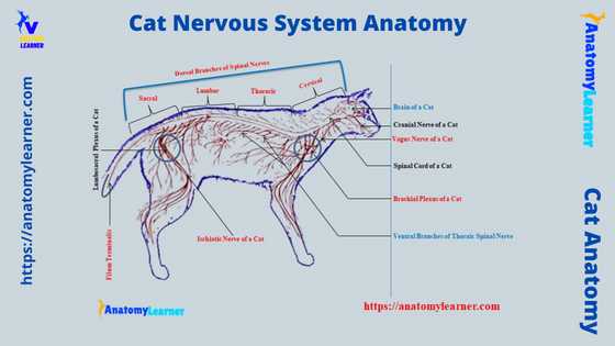
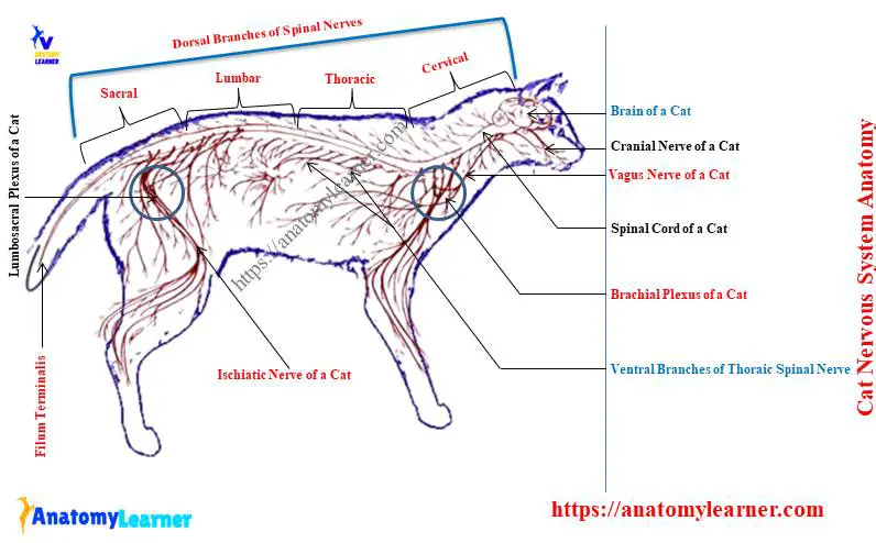
The cat nervous system consists of two main components – the central nervous system and the peripheral nervous system. In the central nervous system of cats, you might learn the anatomy of their brain and spinal cord. Again, you might know the features of the cranial, spinal, and visceral peripheral nerves from the cat peripheral nervous system anatomy.
You know, the ventral branches of the cervical and thoracic spinal nerves of the cat form the brachial plexus. In contrast, the ventral branches of the lumbar and sacral nerves form the lumbosacral plexus in a cat.
In this article, I will describe the anatomy of the brain, spinal cord, and different nerves from these two plexuses of the cat nervous system. Again, I will discuss some common neurological problems in the cats at the end.
So, if you want to know the anatomical features of the cats’ nervous system, let’s continue this article till the end. Don’t miss any labeled diagrams and videos I provided throughout the article to learn about the feline nervous system.
Cat nervous system
The nervous system is the most complex and least understood in the cat’s body. This system initiates, moderates, and coordinates the other body systems of the cat.
First, let’s see some of the unique features of the cat nervous system anatomy –
- The structural and functional unit of the cat’s nervous system is the neuron that connects by synapse to create a complex network.
- These complex networks convey electrical impulses all over the cat’s body.
- You will see the dense accumulation of the nervous tissue in the brain and spinal cord of the cat’s nervous system. These two structures (brain and spinal cord) form the control center of the nervous system.
- The cat brain divides into mainly the fore, mid, and hindbrain, where they perform different vital functions for survival.
- There are 12 pairs (twelve) of cranial nerves originating from the cat brain and generally innervate the glands, muscles, and different organs.
- These 12 cranial nerve pairs typically pass through the specific foramina of the cat skull.
- A dorsal hollow spinal cord extends from the brain, passes through the foramen magnum, and continues through the spinal column.
- In between individual vertebrae, paired spinal nerves emerge and associate with the innervation of the skin, surrounding structure, and muscle of the individual body segments.
- The brain and spinal cord are protected externally by the three layers of meninges and bony covering. Again, the ventricular system internally consists of a canal filled with cerebrospinal fluid.
- A more complex arrangement of the ventral branches of the segmental spinal nerves pattern is found in the forelimb (brachial plexus) and hindlimb (lumbosacral plexus).
- The cat’s autonomic nervous system divides into sympathetic and parasympathetic parts.
Parts of the nervous system in the cat
The brain and spinal cord are grouped as the central nervous system. In contrast, the cranial nerve, spinal nerve, and peripheral nerves are grouped under the peripheral nervous system of cats.
So, mainly you will find the following parts of the nervous system in the cat –
| Parts of Nervous System | Divisions |
| Central Nervous System | |
| Brain (4 divisions) | Telencephalon Diencephalon Mesencephalon Metencephalon |
| Spinal cord (5 segments) | Cervical part Thoracic part Lumbar part Sacral part and Caudal part |
| Peripheral Nervous System | |
| Cranial nerve (12 pairs) | Olfactory Optic Oculomotor Trochlear Trigeminal Abducens Facial Vestibulocochlear Glossopharyngeal Vagus Accessory and Hypoglossal |
| Spinal nerve (39 pairs) | 8 cervical spinal nerve 13 thoracic spinal nerve 7 lumbar spinal nerve 3 sacral spinal nerve and 8 caudal spinal nerve |
| Brachial plexus | Subscapular nerve (3) Suprascapular nerve Axillary nerve Musculocutaneous nerve Anterior ventral thoracic Posterior ventral thoracic Medial cutaneous Radial nerve Median nerve Ulnar nerve Long thoracic Lateral thoracic nerve |
| Lumbosacral plexus | Femoral nerve Saphenous nerve Obturator nerve Cranial and caudal gluteal Posterior femoral cutaneous Pudendal nerve Caudal hemorrhoidal nerve Ischiatic nerve Tibia and fibular nerves |
| Autonomic Nervous System | |
| Sympathetic | Thoracolumbar (T1 to L5) |
| Parasympathetic | Craniosacral division Cranial nerve – III, IV, IX, X Sacral – S1and S2 |
(table 1) shows different parts of the cat’s nervous system. I hope you will get an overview of all the features and structures of the cat’s nervous system from this table.
In the next section, you will learn the anatomy of the cat’s central nervous system (brain and spinal cord) and peripheral nervous system (cranial, spinal, and peripheral nerves) with labeled diagrams.
Cat central nervous system anatomy
The main component of the cat central nervous system anatomy is the brain and spinal cord. You will find different parts or segments in the cat’s brain and spinal cord (shown already). Now, in this section, I will describe the anatomy of these segments from the brain and spinal cord –
- Cat brain – includes telencephalon, diencephalon, mesencephalon, myelencephalon, and metencephalon,
- Cat spinal cord – divides into cervical, thoracic, lumbar, sacral, and caudal segments.
But, for the description purpose, you may divide the cat brain into the cerebrum, cerebellum, and brain stem (pons and medulla oblongata). You know, telencephalon and diencephalon of the cat brain are under the forebrain.
At the same time, the mesencephalon is included under the midbrain of a cat. The metencephalon and myelencephalon form the hindbrain in a cat.
Again, the different segments of the cat spinal cord vary in features in other particular areas. Both the spinal cord and brain possess three layered protecting covers that will discuss here in this article.
Cat brain anatomy and identification
First, I would like to show (identification) you the most essential features of the cat brain anatomy. This might help you to get an overview of the cat’s brain structure. Here, I will cover the following topics from the cat brain anatomy –
- Identification of the anatomical features from the dorsal view of the cat brain,
- Ventral view of cat brain and identification of anatomical features,
- Features from the right and left sagittal section,
- Different structures from the cats’ brainstems, and
- Parts of the brain covering (meninges),
Okay, let’s see and try to identify the following structures from the cat brain (dorsal view) –
- Cerebrum and cerebellum hemispheres,
- Longitudinal cerebral fissure,
- The transverse fissure between the cerebrum and cerebellum, and
- Cerebral gyrus and sulcus of the cat brain,
Again, from the ventral surface of the cat brain, you might identify the following important anatomical features –
- The olfactory bulb, lateral and medial olfactory bands,
- Optic tract, chiasma with the optic nerve,
- Area of the hypophysis (pituitary gland),
- Mammillary body,
- Cerebral peduncle of the cat brain,
- Pyriform lobes of the cat brain,
- Pons, and medulla oblongata of the cat brain, and
- Origins of different nerves like a trigeminal, spinal accessory, and others (described and identified in the specific part of the article),
The labeled diagram identifies all these structures from the dorsal and ventral view of a cat brain.
Sagittal section of cat brain and identification
Now, let’s see the sagittal section of the cat’s brain and try to identify the following important features –
- Genu, trunk, fornix, and splenium,
- Septum pellucidum of the cat brain,
- Mammillary body,
- Infundibulum and hypophysis of the cat,
- Superior and inferior colliculus,
- Cerebral peduncle,
- The pineal gland of a cat,
- Pons, medulla oblongata, and spinal cord (showed before), and
- Cerebellum with the arbor vita from the cat brain,
You might also identify the different ventricles and foramina from the cat brain. Let’s see what the ventricles and foramina are found in the sagittal section of a cat’s brain –
- Two lateral ventricles of the cat brain,
- Third ventricle from the cat brain,
- The fourth ventricle of the brain, and
- Foramen Monro and cerebral aqueduct in the cat brain structure,
If you remove the cerebrum and cerebellum from the cat brain, you will find the brainstem that consists of mesencephalon parts, pons, and medulla oblongata. From this cat brainstem, you might identify the below-mentioned anatomical features –
- Medial and lateral geniculate bodies,
- Thalamus and pineal bodies,
- Superior and inferior colliculus (corpora quadrigemina),
- Medial and posterior cerebellar peduncles,
- Fourth ventricular space,
- Dorsal median sulcus on the brainstem, and
- Roots of different cranial nerves in the cat,
I hope you got the basic idea (different parts) of the various features from the other parts of the cat brain anatomy. Now, I will discuss details on the five different parts of the cat brain. Let’s see the unique features of these five divisions of the cat brain.
Telencephalon of cat brain
The telencephalon of the cat brain from the forebrain consists of cerebral hemispheres. It covers a large part of the cats brain and can be seen on the dorsal, ventral, and lateral views.
The longitudinal fissure separates the cerebral hemispheres of the cat brain anatomy. You will see the numerous elevation (known as the gyrus) and shallow depression (sulcus) on the dorsal surface of the cerebral hemispheres.
You will see the olfactory bulb at the ventromedial aspect of the cerebrum of the cats brain. This olfactory bulb of the cat brain represents the olfactory nerves that carry sensory information to the cerebrum.
You will see the two distinct bands (lateral and medial olfactory bands) on the ventral surface of the telencephalon. The pyriform lobes of the cat brain are separated from the telencephalon part of the cat brain.
You will find a triangular (identified) area between the two bands. This triangular area on the ventral surface of the telencephalon is known as the olfactory trigone.
The telencephalon of the cat brain also divides into different functional lobes –
- A frontal lobe – involves an involuntary movement,
- The parietal lobe – works as pain, touch, and temperature receptors,
- Occipital lobe – works as the visual interpretation, and
- The temporal lobe – is associated with the auditory function, behavior, and memory,
The corpus callosum connects the left and right cerebral hemispheres. Ventral to the corpus callosum, you will see the septum pellucidum. The hippocampus and the mammillary bodies are related to the white matter (fornix).
Diencephalon of the cat brain
The diencephalon is another part of the forebrain and consists of the thalamus. It also consists of the smaller area of the epithalamus, subthalamus, metathalamus, and hypothalamus.
If you remove the cerebral hemispheres, you will quickly see the thalamus of the cat brain. Interthalamic adhesion may be seen as a solid oval tissue in the center of the third ventricle (in the median section).
Functions of the thalamus: it works as the sensory relay station, receiving general sensory impulses and transmitting them to the telencephalon of the brain.
Let’s see the anatomical features of the other components of the diencephalon of the cats brain –
- Epithelamus – locates on the dorsal midline of the caudal diencephalon. It includes the striae habenularis, a habenular nucleus, and the pineal body.
- Metathalamus – includes the lateral and medial geniculate bodies. These geniculate bodies grow caudally over the dorsorostral mesencephalon during development.
- Subthalamus – locates between the thalamus and hypothalamus of the cat brain,
- Hypothalamus – locates on the ventral surface of the cat brain that extends caudally to the optic chiasma. It associates with the autonomic nervous system in the cat.
The ventral surface of the diencephalon of the cat’s brain shows an x-shaped optic chiasma. It demarks the cranial end of the hypothalamus on the ventral surface of the cat’s brain. You will see the optic nerve on the cranial end of the optic chiasma of the cat brain.
You will also find the optic tracts on the respective sides of the optic chiasma. From the hypothalamus of the cat brain, a delicate slender tube extends to the pituitary gland. This slender tube of the ventral aspect of the diencephalon is known as the infundibulum.
Mesencephalon of the cat brain anatomy
The mesencephalon of the cat brain, also known as the midbrain, consists of the superior and inferior colliculus and cerebral peduncle. You may easily see the superior and inferior colliculus of the cat brain if you remove the cerebral hemispheres.
The superior colliculus of the cat brain attaches to the lateral geniculate body. Again, the inferior colliculus of the brain connects with the medial geniculate body. The superior and inferior colliculus of the cat brain works as the visual and auditory structure.
You will see the mesencephalic aqueduct on the dorsal surface of the mesencephalon of the cat brain. The root of the oculomotor and trochlear nerves are associated with the mesencephalon part of the cats brain.
You will also see the crus cerebri on the ventral surface of the mesencephalon. There is a depression in between the crura cerebri known as the interpeduncular fossa.
Metencephalon of the feline brain anatomy
The metencephalon forms the hindbrain in the cat or feline. It consists of the cerebellum dorsally and pons ventrally.
You will find the vermis in the center part of the cerebellum. Again, the cat cerebellum consists of two cerebellum hemispheres.
The dorsal surface of the cat cerebellum possesses highly convoluted folia and sulcus. You will see the peculiar features in the cortex and medulla of the cats’ metencephalon or cerebellum segment.
The medulla of the cat’s cerebellum is the white matter that shows branching arbor vitae. Again, the cortex of the cerebellum shows three distinct layers –
- The external molecular layer,
- A pyriform neuron (purkenji cell layer), and
- The granular layer,
You may know more about these layers from the below-mentioned article –
- Different layers of the animal’s cerebellum with the histological slide picture,
You will find three cerebellar lobes on the metencephalon segment of the cat brain – rostral, caudal, and flocculonodulus. These lobes again divide into different sublobes.
If you remove the cerebellar hemisphere from the cat brain, you will quickly identify the space of the fourth ventricle and pons. Again, you will see a longitudinal median sulcus on the dorsal surface of the pons and medulla oblongata (after removing cerebellum hemispheres).
The pons locates just caudal to the cerebral peduncle and includes a central area with infinite borders. You will see the prominent transverse fibers on the ventral aspect of the pons. From the most caudal region of the cats pons, the trigeminal nerve originates.
Myelencephalon and cat’s brainstem
The myelencephalon is another part of the cat’s hindbrain and is known as the medulla oblongata. The most caudal part of the cat’s brain connects with the spinal cord.
The dorsal surface of the myelencephalon shows the continuation of the fourth ventricle. Lateral to the fourth ventricle, you will see the vestibular nuclei, which are essential structures in the vestibular system of the cats brain.
The spinal tract (cat) of the cranial nerve V (trigeminal) can be seen on the dorsolateral aspect of the myelencephalon. Again, you will see the trapezoid body on the ventral surface of the myelencephalon part of the cats brain.
A midline ventral median fissure is present on the ventral and caudal aspects of the trapezoid body. You will find the roots of different cranial nerves at the medulla oblongata of the cat’s brain structure.
Let’s see the nerves that have a connection with the myelencephalon of the cat’s brain –
| Nerves | Number |
| Abducens | VI |
| Facial | VII |
| Acoustic | VIII |
| Glossopharyngeal | IX |
| Vagus | X |
| Spinal accessory | XI |
| Hypoglossal | XII |
But the spinal accessory also receives the fibers from the cranial end of the spinal cord of a cat.
The parts of the mesencephalon, pons, and medulla oblongata form the brainstem in the cat. It is the site for different realy and vital reflex centers. This myelencephalon of the cat’s brain acts as the cardiac, swallowing, respiratory, and vomiting centers.
Spinal cord from cat nervous system
The spinal cord of the cat nervous system is the more or less cylindrical structure that is continuous with the caudal part of the medulla oblongata. In the feline or cat spinal cord, you will find 8 cervical segments, 13 thoracic segments, 7 lumbar segments, 3 sacral, and 8 caudal segments.
But, in most cats, the spinal cord ends at the level of the second sacral vertebra. A dorsal median sulcus is present on the dorsal midline of the cat’s spinal cord. You will also see the deep ventral median fissure on the ventral midline of the spinal cord.
The transverse section of the cat’s spinal cord shows the outer white matter that surrounds the inner gray matter. You will see a butterfly or H-shaped central gray matter in the cat’s spinal cord.
Here, the dorsal horn of the H-shaped structure possesses the sensory function, whereas the ventral horn possess motor functions. You will see the dorsal and ventral horns in every segment of the cat spinal cord except in the last sacral.
There is a central hollow canal present at the center of the cats spinal cord. This central canal of the spinal cord continues with the fourth ventricle of the cats brain.
Thus, the cerebrospinal fluid circulates within the brain ventricles and also in the central canal of the spinal cord.
You will not find uniformity in the diameter of the cats spinal cord. There are two conspicuous enlargements in the spinal cord of a cat –
- One is at the cranial cervical and thoracic region – supply nerve to the forelimb of the cat, and
- Another is at the posterior lumbosacral region – which supplies nerves to the cat’s hindlimb.
Finally, this cord ends as a filum terminalis.
Segments of the cats spinal cord
As I told you before, there are five segments in the cats spinal cord –
| Segments | Spinal Nerves |
| Cervical | 8 Cervical Spinal Nerves |
| Thoracic | 13 thoracic spinal nerve |
| Lumbar | 7 lumbar spinal nerve |
| Sacral | 3 sacral spinal nerve |
| Caudal | 8 caudal spinal nerve |
All these segments of the cats spinal nerve from the spinal nerve. The typical structure of the spinal nerve is well understood from the cervical part to the first segment of the sacral vertebra.
Here, the diagram shows the white and gray matter of the spinal cord (cross-sectional view) along with the dorsal and ventral roots of the spinal nerve. Again, it also shows the spinal nerve and the dorsal and ventral ramus of this nerve.
How is the spinal nerve formed in a cat?
I think you have already understood the structure of the spinal nerve from the previous diagram. The dorsal and ventral branches of the nerves exit from the spinal cord. Here, the dorsal root carries the sensory function, whereas the ventral root carries the motor function.
Within the vertebral foramen, these two roots (dorsal and ventral) exchange their fibers and form the spinal nerve. Then, they exist through the intervertebral foramen as the dorsal and ventral branches of the individual spinal nerve.
According to the structure, each of these spinal nerves of the cat carries both sensory and motor functions. You will find the complex relationship of the ventral branches of the spinal nerves at the cervical and lumbosacral regions of the cat’s body.
Cat or feline peripheral nervous system
The feline peripheral nervous system consists of these nervous tissues other than the brain and spinal cord. Thus, the different types of nerves and ganglia throughout the body are included under the peripheral nervous system.
You know the nerves are the bundle of neuron processes in the peripheral nervous system of any animal. Any nerve’s specific nerves or branches may be motor, sensory, or mixed-in functions.
Again, the ganglion is a functionally related cluster of nerve cell bodies in the peripheral nervous system.
The anatomy of the feline peripheral nervous system is almost similar to the peripheral nervous system of the dog. Under the feline peripheral nervous system, I will discuss the following topics –
- Cranial nerves of the cat (12 pairs),
- Spinal nerves of the cat,
- Cat brachial plexus formations and their branches, and
- Formation of the lumbosacral plexus and its nerves,
Okay, let’s get started with the anatomical features of the cats cranial nerves.
Cranial nerves of the cat
There are 12 pairs of cranial nerves in the cats’ nervous system. They are associated with the different parts of the brain but are the parts of the feline peripheral nervous system.
The name of the cat cranial nerves are according to their sensory fibers and functions. Let’s try to identify the roots of the following 12 pairs of the cranial nerves from the cat –
| Name and number | Distribution |
| Sensory | |
| Olfactory I | Nose |
| Optic II | Eye (retina) |
| Cochlea-vestibular VIII | Ear |
| Mixed | |
| Trigeminal V | Face, jaw, ear |
| Abducens VI | Eye |
| Facial VII | Face, tongue, salivary glands |
| Glossopharyngeal IX | Pharyngeal muscle, parotid gland |
| Vagus X | Pharynx, larynx, esophagus, lung, heart, abdominal viscera |
| Motor | |
| Oculomotor III | Ocular muscles |
| Trochlear IV | Dorsal oblique muscle of eye |
| Spinal accessory XI | Neck, shoulder, larynx |
| Hypoglossal XII | Muscles of tongue |
This table shows the main distribution of these cats’ cranial nerves. But, you might learn the specific attachments and distribution of all the cranial nerves from a cat.
Attachments of the cats cranial nerves
The olfactory nerve of a cat is associated with the ventral part of the telencephalon (olfactory bulb). Again, the optic nerve is associated with the ventral part of the diencephalon (optic chiasma).
The oculomotor and trochlear nerves of the cat are attached to the ventral surface of the mesencephalon. Here, the trochlear nerve is the only nerve that leaves the dorsal aspect of the cats brain.
The trigeminal nerve of the cat attaches to the cranio lateral aspect of the pons by a significant sensory and a small motor root. You will find three major branches of the cats trigeminal nerves like the other animals – ophthalmic, maxillary, and mandibular.
The ophthalmic and maxillary branches of the trigeminal nerve pass through the foramen orbitorotundum, whereas the mandibular branches pass through the foramen ovale.
The abducens nerve attaches close to the corpus trapezoidal and passes through the foramen orbitorotundum. The rest of the cranial nerves (VII-XII) of a cat are attached to the lateral surface of the medulla oblongata.
But, the vestibule cochlear nerve attaches close to the junction of the pons and medulla oblongata in a cat. Again, you will see peculiar features in the attachment of the accessory spinal nerve of the cat.
The cranial root of the spinal accessory nerve attaches to the dorsolateral aspect of the medulla oblongata. In comparison, the spinal root of the accessory spinal nerve connects to the first four to five cervical segments of the spinal cord.
Finally, the hypoglossal nerve of a cat attaches to the ventral lateral aspect of the medulla oblongata and passes through the hypoglossal foramen. The glossopharyngeal, vagus, and accessory spinal nerves pass through the jugular foramen.
Distribution of cats cranial nerves
The cats olfactory cranial nerve distributes to the neurosensory cells of the nasal mucosa. In contrast, the optic nerve of a cat possesses the sensory fibers that distribute to the retina.
The oculomotor nerve of a cat innervates the following muscles of the eye –
- Dorsal, ventral, and medial straight muscles,
- Ventral oblique muscle of the eye,
- Retractor bulbi muscle,
- Levator palpebrae superiors muscle, and
- Intrinsic ciliary muscle,
The trochlear nerve of a cat will supply to the dorsal oblique muscle of the eye.
You will find a wide distribution of the cat trigeminal nerves as they provide three major branches – ophthalmic, maxillary, and mandibular. Here, the ophthalmic branch innervates the skin of the eye, nose, upper eyelid, eyeball, and lacrimal gland.
Again, the maxillary branch of the trigeminal innervates the cat’s upper lip, jaw, teeth, and palates. The mandibular branch of the trigeminal innervates to the lower lip, jaw, teeth, and cheek. It also supplies to the cat’s masseter, temporal, and pterygoid muscles.
The abducens cranial nerve of the cat innervates to the lateral straight muscle of the eye. At the same time, the facial nerve innervates the facial and digastricus muscles. Again, it innervates to the tongue, mandibular, sublingual, and lacrimal gland.
The vestibular branch of the vestibulocochlear nerve innervates to the utricle, saccule, and semicircular canals of the internal ear. Again, the cochlear branch of the vestibulocochlear nerve innervates organs of the Corti.
The cat glossopharyngeal nerve will supply the pharynx muscles, the tongue, and the parotid gland. You will find a wide distribution of the cat vagus nerve, especially on the pharynx, esophagus, heart, lungs, and abdominal viscera.
The accessory nerve of a cat will supply the cleidomastoideus, sternomastoideus, and trapezius muscle. Finally, the cat’s hypoglossal nerves are supplied to the tongue muscles.
Spinal nerves from the cat nervous system
Each segment of the cats spinal cord gives off pairs of spinal nerves. You know these two spinal nerves are formed by the dorsal and ventral roots on each side of the vertebrae.
You have already got (seen) the idea of how the spinal cord forms in an animal. Again, five segments in the spinal cord (already described) possess specific spinal nerves.
The formation of the brachial plexus and lumbosacral plexus are the essential features of the spinal nerves of the cat nervous system. Again, some other special nerves like the costoabdoinal, genitofemoral, and ilioinguinal also come from the ventral branch of the spinal nerve (will discuss later).
The first cervical nerve of a cat exists through the lateral vertebral foramen of the first cervical vertebra. Again, a cat’s other cervical spinal nerves (second to the seventh cervical) exist through the intervertebral foramen just cranial to the vertebra of the corresponding number.
The eighth cervical spinal nerve leaves through the intervertebral foramen between the seventh cervical and first thoracic vertebra. Again, the remaining spinal nerves exit the intervertebral foramen just caudal to the vertebra of the corresponding number.
Let’s see the example; a cat’s first thoracic spinal nerve will find between the first thoracic vertebra and second thoracic vertebra (intervertebral foramina). I hope you can understand this process of the existence of spinal nerves from the vertebral foramina.
But, some cat species show different anatomical features in the sacral and caudal spinal nerves.
Special features of cat’s cervical spinal nerves
The first cervical spinal nerve of the cat is small and difficult to identify. This is because of a large amount of connective tissue in this area (at the location of the first cervical).
The first cervical spinal nerve passes over the ventral surface of the longus Colli muscle and innervates to the ventral muscle of the neck.
The cat’s second to fifth cervical spinal nerves is also challenging to identify due to more connective tissue. After removing the connective tissue, you may easily differentiate these spinal nerves from each other.
The second to fifth cervical spinal nerves supply the tissue of the shoulder and neck regions of a cat. Again, fibers from the fifth and sixth cervical spinal nerves contribute to forming the phrenic nerve. You will find a close relationship with the vagus nerve, which passes the thoracic region and finally innervate to the diaphragm.
The sixth cervical spinal nerve extends to the area of the shoulder joint. It passes between the supraspinatus and infraspinatus muscles of the shoulder region.
Brachial plexus in a cat
The ventral branches of the sixth, seventh, and eighth cervical spinal nerve and the first thoracic nerves are fused to form the brachial plexus. This brachial plexus of a cat locates medial to the scapula and cranial midventral part of the thoracic cage.
You will find the following nerves in the brachial plexus of a cat –
| Brachial Plexus of Cat | Innervation |
| Subscapular nerve (3) | Subscapularis muscle, teres major |
| Suprascapularis nerve | Supraspinatus and subscapularis |
| Axillary nerve | Lateral shoulder muscle |
| Musculocutaneous nerve | Brachialis, biceps brachii |
| Cranial ventral thoracic | Superficial pectoral |
| Caudal ventral thoracic | Deep pectoral |
| Medial cutaneous | Skin |
| Long thoracic nerve | Serratus ventralis muscle |
| Thoracodorsal nerve | Latissimus dorsi muscle |
| Radial nerve | Triceps and extensor muscles |
| Median nerve | Flexor muscle of forelimb |
| Ulnar nerve | Flexor muscle of forelimb |
The table shows the different nerves from the cat brachial plexus with the main innervation. But, you may learn more about the innervation of these nerves from the cat.
Again, if you want to know the basic structure of the animal brachial plexus formation with their nerves, you may read the below-mentioned article –
- Brachial plexus formation in ox with the labeled diagram,
Innervation of cat’s brachial plexus nerves
You will find three subscapular nerves in the brachial plexus of a cat – first, second, and third. The first subscapular nerve of the cat arises from the ventral branches of the sixth and seventh cervical spinal nerve.
They travel with the subscapular vessel and innervate to the subscapular muscle of the cat scapula.
The ventral branch of the seventh (7) cervical spinal nerve contributes to forming the second subscapular nerve. This second subscapular nerve innervates the teres major muscle of the cat.
The third subscapular nerve arises from the ventral branches of the seventh and eighth cervical spinal nerves. It travels with the thoracodorsal vessels and innervates to the latissimus dorsi muscle.
The ventral branches of the sixth (6th) and seventh (7th) cervical spinal nerve contribute to forming the axillary nerve in a cat. It travels with the caudal humeral circumflex vessel and innervates the lateral shoulder muscles. You know the lateral shoulder muscle of a cat includes teres major, teres minor, deltoid, and cleidobrachialis.
The musculocutaneous nerve arises from the ventral branches of the sixth and seventh cervical spinal nerves. This musculocutaneous nerve of the cats brachial plexus innervates biceps brachii, brachialis, and coracobrachialis muscles.
The ventral branches of the seventh cervical spinal nerve form the cranial ventral thoracic nerve in a cat. This nerve travels with the ventral thoracic vessel and innervates to the superficial pectoral muscle. You may be called this nerve the cranial pectoral nerve.
The caudal ventral or caudal, pectoral nerve in a cats brachial plexus originated from the eighth cervical and first thoracic spinal nerves. It travels with the long thoracic vessels and supplies to the deep pectoral muscles.
Other nerves of cat brachial plexus and innervation
The ventral branch of the seventh (7th) cervical spinal nerve contributes to forming the long thoracic nerve in a cat. This long thoracic nerve of the cats brachial plexus innervates to the serratus ventralis muscle.
The radial, median, and ulnar nerves from the cats brachial plexus will supply different areas of the forelimb to the digits. You know the radial nerve is the largest in the cat brachial plexus and originated from the ventral branches of the sixth, seventh, eighth cervical, and first thoracic spinal nerves.
This radial nerve travels with the deep brachial vessel and divides into superficial and deep branches. Overall the radial nerve of the cat supplies to the triceps brachii, anconeus, tensor fascia antebrachii, and brachioradialis muscle.
The superficial branch of the cat radial nerve innervates the skin over the lateral forearm and skin over the dorsum of digits II, III, and IV. In contrast, the deep branch of cat’s radial nerve innervates extensor carpi radialis, lateral digital extensor, extensor carpi ulnaris, common digital extensor, supinator, and abductor longus muscles.
The median nerve arises from the ventral branches of the seventh, eighth cervical, and first thoracic spinal nerves. This nerve travels with the brachial artery and passes through the supracondyloid foramen.
The median nerve of a cat innervates the flowing flexor muscle of the forelimb –
- Flexor carpi radialis muscle,
- Pronator teres muscle,
- Pronator quadratus muscle,
- Deep digital flexor muscle, and
- Superficial digital flexor muscle,
The median nerve of the cat is also supplied to the cubital and carpal joint, skin over the palmar digits II, III, and IV.
The ventral branch of the eight cervical and first thoracic spinal nerves contributes to the ulnar nerve in a cat. It travels parallel to the median nerve and innervates to flexor muscles.
Lumbosacral plexus of cat nervous system
The lumbosacral plexus is another unique structure in the cat nervous system. This plexus is formed by the ventral branches of the last three lumbar and first two sacral spinal nerves.
You should identify the nerves from both the lumbar and sacral plexus of the cat separately –
- Lumbar plexus of cat – consists of lateral femoral cutaneous, femoral, saphenous, and obturator nerves,
- The sacral plexus of a cat – consists of cranial, caudal gluteal, sural, and ischiatic nerves. Again, the cat’s ischiatic nerves divide into two major parts – the tibial and fibular nerves.
The basic structure and formation process of the cat’s lumbosacral plexus is almost similar to that of a dog or other animals. You may learn the details of the formation of the animal lumbosacral plexus here.
Nerves from cats’ lumbar plexus
The lateral cutaneous femoral nerve arises from the ventral branch of the fourth and fifth lumbar spinal nerve. This nerve emerges beneath the psoas minor muscle and passes over the iliopsoas muscle.
Again, the lateral cutaneous femoral nerve of the cat travels with the deep iliac circumflex vessel. Finally, this cat’s nerve innervates the lateral surface of the hip and thigh regions.
The ventral branches of the fifth and sixth lumbar spinal nerve contribute to forming the femoral nerve in a cat. This femoral nerve passes between the iliopsoas and psoas minor muscle.
You will find two branches of the femoral nerve in a cat –
- Saphenous nerve – runs parallel to the femoral vessel and saphenous vessel,
- Lateral femoral branch – innervates to the skin over the medial thigh, stifle joint, and metatarsus.
The ventral branches of the fifth to seventh lumbar spinal nerves contribute to the obturator nerve in a cat. It passes into the pelvic region of a cat through the obturator foramen.
Then, it gives off branches to the adductor, gracilis, pectineus, and obturator externus muscles. You will see the lumbosacral trunk just medial to the obturator nerve of a cat. This lumbosacral trunk of the cat is formed by the ventral branch of the sixth and seventh lumbar spinal nerves.
Scaral plexus of a cat
The ischiatic nerve is the largest and distinctly observed nerve in the hindlimb of a cat. This nerve extends from the lumbosacral cord and courses over the lateral muscles of the cat’s thigh.
Before going to the details of the ischiatic nerve, let’s try to identify the cranial and caudal gluteal nerves from the cat lumbosacral plexus. The cranial gluteal nerve of a cat originated from the lumbosacral cord.
This nerve passes over the dorsal surface of the ilium bone and beneath the gluteus minimus muscle. The cranial gluteal nerve of a cat innervates to the gluteal medius, gluteus profundus, gamellus, and tensor fasciae latae muscles.
Again, the caudal gluteal nerve of a cat arises from the lumbosacral cord. This nerve passes caudally and innervate the caudofemoralis and gluteus medius msucles of a cat.
You will also find two other essential nerves in the lumbosacral plexus of a cat –
- A caudal cutaneous femoral nerve of a cat, and
- The pudendal nerve of a cat,
The caudal cutaneous femoral nerve arises from the ventral branches of the second and third sacral spinal nerves. This nerve passes with the caudal gluteal blood vessels and arches over the biceps femoris muscle. Finally, a cat’s caudal cutaneous femoral nerve innervates to the biceps femoris muscle and the tail’s skin.
The ventral branches of the second and third sacral spinal nerve contribute to the pudendal nerve in a cat. Practically, this pudendal nerve of a cat possesses tremendous clinical importance.
Now, let’s return to the cat’s ischiatic nerve and branches.
The ischiatic nerve of a cat
After the origin of the cat’s ischiatic nerve, it gives off different muscular branches – thick muscular, thin muscular, and small muscular. These muscular branches of the cat’s ischiatic nerves innervate to the biceps femoris, semitendinosus, semimembranosus, and thigh flexor muscles.
Again, you will see a small sural nerve originating from the ischiatic nerve. It passes across the gastrocnemius muscle to the ankle joint.
The ischiatic nerve divides into two main branches in the middle of the cat’s thigh – the common peroneal (fibular) and tibia nerve. The common peroneal nerve passes over the lateral head of the gastrocnemius, whereas the tibia passes between the two heads of the gastrocnemius.
The common peroneal nerve of a cat divides into superficial and deep parts that supply the following structures and muscles –
- Stifle joint, skin over the dorsal tarsus, metatarsus, and digits,
- Cranial tibial muscle of the cat,
- Long and lateral digital extensor muscles,
- Peroneus longus and brevis muscles, and
- Short digital extensor muscle,
Again, the tibial branch of the cats ischiatic nerve supplies the following essential structures and muscles –
- Stifle joint, skin over the plantar tarsus, metatarsus, and digits,
- Gastrocenemius, soleus, and popliteus muscles,
- Superficial and deep digital flexor muscles,
- Caudal tibial muscle,
- Abductor and adductor muscles of the digits II and V,
You will find the details of these muscles from the cat leg anatomy (both front and hind legs).
Autonomic nervous system of a cat
The autonomic nervous system of a cat is usually considered only in terms of its motor component. But, this system also includes the sensory output.
The feline autonomic nervous system is responsible for the innervation of viscera, glands, blood vessels, and smooth muscle throughout the body. If any changes in the external and internal environment are needed, the autonomic nervous system immediately responds.
Like other animals, the feline autonomic nervous system divides into sympathetic and parasympathetic divisions. Both the cats sympathetic and parasympathetic divisions of the autonomic nervous system work together. That means the tissue receiving autonomic innervation will also receive fibers from sympathetic and parasympathetic divisions.
The sympathetic division of feline ANS
The sympathetic division of the feline ANS is responsible for body reaction in response to an emergency. You know the sympathetic nervous system is responsible for dilating pupils, increasing heart rate, decreasing gut motility, and vasodilation of the limb vessels.
The nerve bodies of feline sympathetic preganglionic neurons are located in the zona intermedia, a lateral horn of the spinal cord T1 to L4. Thus, this sympathetic division of the autonomic nervous system is known as the thoracolumbar outflow.
The axon that forms the sympathetic division leaves the ventral root and joins the spinal nerve. You will find the cervicothoracic ganglion at the cranial end of the sympathetic thoracic trunk. These are the larger ganglion formed by the fusion of caudal cervical ganglions and T1 and T2 sympathetic trunk ganglions.
The thoracolumbar sympathetic trunk extends cranially (neck) by the vagosympathetic trunk. Here, the vagosympathetic and thoracolumbar trunks are connected by the ansa subclavia.
You will find the cranial cervical and middle cervical ganglia along with the length of the vagosympathetic trunk. Again, there is a postganglionic sympathetic fiber that leaves the cervicothoracic ganglia.
The sympathetic trunk provides different fibers to the abdominal viscera. The significant splanchnic nerve, minor splanchnic nerve, and lumbar splanchnic nerve come from the sympathetic division.
The parasympathetic division of feline ANS
The parasympathetic division of the feline ANS works with the body’s response in relaxed conditions. This parasympathetic division of the cat’s ANS is responsible for pupil constriction, increased gut motility, decreased heart rate, and increased salivation.
The nerve cell bodies of the preganglionic neuron locate in the parasympathetic nuclei of III, VII, IX, and X cranial nerves. Again, the preganglionic neuron comes from the zona intermedia of the sacral spinal cord segments. Thus, the parasympathetic division of ANS is also known as the craniosacral outflow.
The parasympathetic pathway begins in the central nervous system (III, VII, and IX) and innervates the specific organs or structures of the head. These pathways innervate to the smooth muscle of the eye, lacrimal, nasal, and salivary glands.
The parasympathetic preganglionic fibers of the sacral spinal nerves travel with the pelvis nerve. Here, the pelvic nerve forms the pelvic plexus and the hypogastric nerve. There are different pelvic ganglions in this area.
Feline nervous system diagram
I know you already got some of the labeled diagrams on the feline nervous system. Now, I will also provide some other diagrams that might help you learn more about the nervous system.
So, what are the major labeled diagrams you need to study the feline nervous system? You should need a diagram of the central nervous system, peripheral nervous system, and finally, a diagram of the autonomic nervous system.
Here, in the cat’s brain and spinal cord, all the essential structures or features identified serially are identified. The dorsal, ventral, lateral, cross-sectional, and transverse sectional views of the cat brain are included here.
Again, the diagram identifies the different structures of the cat spinal cord.
All the roots of the cat cranial nerve are shown in the labeled diagram. It is essential to understand the structure of a spinal nerve in a cat. Here, the different features of the spinal nerve formation are identified in the labeled diagram.
It is tough to show the autonomic nervous system labeled diagram from any animal. Here, I provide a diagram that shows the different fibers from the sympathetic and parasympathetic divisions with their major suppliers.
You may find more labeled diagrams on the feline nervous system here on social media of anatomy learners.
Cat nervous system problems
You will find different problems in the central and peripheral nervous systems of a cat. Here, I will show some common neurological issues of a cat.
But, this article will not deal with a details description of any neurological problems of a cat. You may learn more about the neurological issues of the cats from another article by an anatomy learner.
Let’s discuss some of the common neurological problems of a cat –
- Cerebellar hypoplasia,
- Spinal cord lesions – ischemic neuromyopathy and spinal lymphosarcoma,
- Demyelination and degeneration in ischiatic nerve,
- Peripheral nerve disorder,
- Problems in the brainstem,
In utero panleukopenia virus infection, you may find cerebellar hypoplasia in your cat (queen). Again, this cerebellar hypoplasia also may occur due to the application of treatments to the queen.
Due to the trauma, the spinal cord problem more frequently occurs in a cat. You may often find the ischemic neruomyopathay and spinal lymphosarcoma in your cat.
Again, if your cat has a feline leukemia virus infection, there is a great chance to occur spinal lymphosarcoma. Young cats (below two years) are more susceptible to spinal lymphosarcoma problems.
The demyelination of degeneration of the ischiatic nerve may occur in the cat. You may also find the different neurological problems in a cat’s cranial and peripheral nerves like the dogs.
Common questions on the feline nervous system
Now, let’s see anatomy learners’ most common questions on the feline nervous system. Here, I will try to solve all these questions with little information. But, I would like to suggest you read the whole article to get a basic idea of the feline nervous system.
How does the nervous system work in a cat?
The presence of a nervous system allows a cat to respond in a coordinated manner. This system takes the information from the external and internal environment, analysis it, and then initiates an appropriate response.
So, the cat’s nervous system function as follow –
- It receives stimuli (pain or touch) from the external and internal environments,
- Analysis and integration the collected stimuli of the environments, and
- Bring out the necessary response,
How do I know (learn) if my cat has a neurological problem?
The cat may show different symptoms if it has a neurological problem. You may find the behavioral change in your affected cat. If the cat has a neurological problem, it will show muscle tremors, pain, lack of coordination, and weakness.
Sometimes the cat shows paralysis in the specific affected area. Again, if any accidental damage occurs in the spinal cord, the symptoms may vary with the degree of damage.
What causes damage to the nervous system in cats?
You may find the different causes of damage to the nervous system in cats. The most common causes of nervous system damage are congenital that transmit from the parents. Again, some of the nutritional, environmental, and even viral infections may cause numerous neurological disorders in your cat.
Do cats have a sympathetic nervous system?
Yeah, the cats have a sympathetic nervous system that is also known as the thoracolumbar outflow. This system work in an emergency in the cat’s body. I have already described a little about the feline sympathetic system with its distribution.
Conclusion
The basic structure of the cat nervous system is similar to the dog. But, you may find differences in the formation and distribution of cranial and spinal nerves between cats and dogs. Again, a significant difference is located in the brachial and lumbosacral plexus between the cat and dog.
I hope this article will help you learn the basics of the cat or feline nervous system basics. But, please know all these nerves and structures from a dissected cat sample. The labeled diagrams on the cat’s nervous system might help you learn from the actual samples.