The dog shoulder anatomy consists primarily of a ball and socket joint between the glenoid cavity of the scapula and the head of a humerus. You will also see the infraspinatus, supraspinatus, biceps brachii, and teres major muscles in the dog shoulder structure.
Again, you will find the capsular, medial, and lateral glenohumeral ligaments in the structure of the dog or canine shoulder joint. I will describe all the anatomical features from the dog shoulder joint with the labeled diagram.
Again, I will share some common shoulder injuries in dogs like biceps tendon luxation, shoulder dislocation, traumatic cartilage injury, and others. So that you may apply your anatomical knowledge to identify the shoulder injuries in the dogs.
Dog shoulder anatomy
In the dog shoulder anatomy, you might describe the details of joints, muscles, and ligaments. Here, the shoulder joint is formed between the distal end of the scapula and the proximal end of the humerus bone.
In the canine shoulder joint description, you might point out its types, movement, bones involvements, and ligaments. There are some of the vessels and nerves that are involved with the canine shoulder joint anatomy.
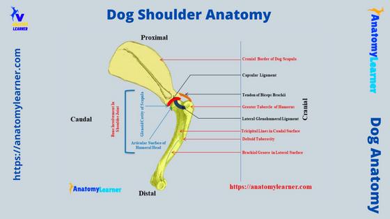
First, I would like to provide you with the details structures of the dog’s shoulder with the labeled diagram. Let’s try to identify the below-mentioned structures from the dog’s shoulder –
- The distal extremity of the dog’s scapula bone (especially the glenoid cavity and coracoid process),
- The proximal extremity of the dog’s humerus bone (head, neck, and tuberosities),
- Muscles of the shoulder joint – tendon of supraspinatus, infraspinatus, biceps brachii, teres major, and deltoideus,
- A capsular ligament of the dog’s shoulder joint,
- The lateral and medial glenohumeral ligaments, and
- Transverse humeral retinaculum,
I will show the involvement of the vessels and nerves in the canine shoulder lateral with the labeled diagram. Now, I will summarize the facts about the canine shoulder joint.
You know the glenohumeral (shoulder) joint of any animal is a poly axial, ball, and socket joint (synovial joint). It is a highly moveable joint (movement in any direction) in the dog’s body.
So, before going to summarize shoulder facts, you may go through the below-mentioned article to get a clear idea of the synovial joint structure –
- Typical structure of an animal synovial joint with the labeled diagram
Now, let’s move to the important facts about the glenohumeral joint of dogs.
Facts of dog shoulder structure
The anatomy of the dog shoulder joint consists –
Bone involvement: glenoid cavity of the dog scapula, and head of the humerus bone,
Joint: ball and socket type,
Movement of the shoulder joint: it shows the features of the spheroidal joint type. That means this shoulder joint may move in various directions. But, you will find extension and flexion as the chief movement in the shoulder joint.
There are also abduction and adduction movements found in the canine shoulder joint. But, they are very restricted in dogs.
The position of the shoulder joint is not fixed as both bones can move easily. Again, none of these bones articulate with the axial skeleton of the dog.
Ligaments of the shoulder (humeral or glenohumeral) joint: primarily two important ligaments find in this joint –
- Joint capsule or capsular ligament, and
- Two diverging (lateral and medial) elastic glenohumeral ligaments,
These two diverging elastic glenohumeral ligaments arise from the supraglenoid tubercle and end on the tuberosity of the dog’s humerus. Again, you will also see the transverse humeral retinaculum in the structure of a canine shoulder joint.
Now, let’s see what the muscles that involve in the structure of the canine shoulder joint are –
- Tendon of biceps brachii muscle,
- The supraspinatus and infraspinatus muscle,
- Subscapularis muscle (at medially),
- Omotransversarius and deltoideus (muscles and tendons), and
- Tendons of teres major and teres minor muscles,
These are the muscle that contribute to the shoulder anatomy and provide stability in the joint.
Now, let’s see the vessels and nerves that are also involved in the structure of a canine shoulder joint –
- Axillary, subscapularis, brachial and radial nerves and arteries (cranially),
- Subscapularis artery and nerve (caudally),
All these nerves and arteries from the shoulder will describe later with a diagram.
Dog shoulder bone anatomy
The dog shoulder bone anatomy only consists of the distal extremity of the scapula and proximal extremity of the humerus. So, let’s see what the osteological features that are involved in the structure of the canine shoulder joint are –
- Glenoid angle with its cavity,
- Supraglenoid tubercles of dog scapula,
- An elongated oval head of the dog’s humerus bone, and
- Lesser and greater tubercles with the intertuberal groove,
Again, other osteological features from the dog’s scapula and humerus bones are also responsible for the shoulder anatomy. Different muscles originated from the different areas of the scapula or humerus and inserted into them.
So, it will be better if you have a piece of basic knowledge of the overall osteological features of the dog scapula and humerus bone. I would like to suggest you read the below-mentioned articles to get the proper idea of dog scapula and humerus bones –
- Dog scapula anatomy with the labeled diagram, and
- Osteological features of dog humerus bone with the labeled diagram,
In the next section, you may also get the main osteological features of the canine scapula and humerus bone. But, I will only provide the basic and essential osteological features of the scapula and humerus here.
Canine scapula anatomy
The scapula is the large, flat, triangular bone in the canine shoulder joint. The dorsal part of the scapula lies ventral to the level of the free end of the spinous process of the first or second thoracic vertebrae.
Again, it extends from the manubrium of the sternum to the fourth or fifth thoracic vertebrae longitudinally. You will not find any articulation of this canine scapula bone with the axial skeleton.
To describe the anatomical facts of the dog or canine scapula, it may divide into two surfaces, three borders, and three angles. All these surfaces, borders, and angles are not important to form the shoulder joint.
But, you need a clear concept to know the confirmation and direction of the scapula bone from the canine skeleton. Let’s see some of the osteological features of the dog scapula –
Features of scapula surfaces
You know two defined surfaces (lateral and medial) in the canine scapula possess some important osteological features. Here, the diagrams show the different osteological features from the lateral surface of the canine scapula anatomy.
An essential osteological feature of the lateral surface of the canine scapula is the presence of the spine. This is a slender projection of the lateral surface of the scapula bone.
Again, the spine on the lateral surface of the canine scapula starts from the middle of the dorsal border and extends distally to the glenoid end. You will find both the cranial and caudal surfaces in the spine of the dogs scapula.
In other animals like the ruminant (cow, sheep, goat), the spine forms a pointed structure at the distal end, known as the acromion process. You will also find the acromion process in the canine scapula, but this structure is not as pointed as the ruminant.
The canine spine forms a thick, blunt end at the distal part of the scapula. You may easily palpate this structure from the surface approach on the canine shoulder.
Let’s see what the muscle that attaches (originate or insert) on the lateral surface and in the spine of the canine scapula are –
- Trapezius, omotransversarius muscles,
- Supraspinatus and infraspinatus muscles,
- Biceps brachii muscles of canine scapular region, and
- Teres major and minor muscles,
You will find the details of these muscles in the next part of this article (with their origin and insertion). Now, let’s see the osteological features from the medial surface of the canine scapula.
Supraspinatus and infraspinatus fossae
The supraspinatus fossa is located at the upper part of the lateral scapula. It is bounded by the cranial surface of the canine scapular spine and the adjacent lateral surface.
This supraspinatus fossa of the canine scapula is wider in the middle and becomes narrow at the distal extremity. You will find the scapular notch at the distal extremity in the canine scapula.
In this supraspinatus fossa, you will find the muscle that originated from all the parts. But, they are mainly derived from the distal portion of the supraspinatus fossa.
Let’s see the infraspinatus fossa of the canine scapula, which is almost triangular. You know the supraspinatus muscles arise from the proximal part of this fossa.
Medial surface of the canine scapula
From the medial surface of a canine scapula, you might learn two important osteological features – the subscapular fossa and facies Serrata. As this medial surface of the scapula lies adjacent to the first five ribs of the thoracic cavity, you may also be called this surface the costal surface.
There are two small rectangular areas located dorsocranial aspect of the medial surface. These small rectangular areas are the facies serrate. From this facies Serrata of the canine scapula, the serratus ventralis muscle arises.
Except for these facies Serrata on the medial surface, the remaining part forms the subscapular fossa. The subscapular fossa is nearly flat and presents a relatively straight muscular line. These muscular lines on the subscapular fossa attach the muscle tightly.
Again, the subscapularis muscle arises from the muscular lines and subscapular fossa. This is the only important muscles at the medial aspect of the canine scapula.
Borders of the canine scapula
The canine scapula shows the dorsal, cranial, and caudal borders. Here, the dorsal border is known as the base or vertebral border. This vertebral border of the canine scapula extends between the cranial and caudal angles.
You know the two borders of any objects or bones from the angle. Learn details about the angles in the scapular angles section.
You will see a narrow band of scapular cartilage on the dorsal border of the dog scapula. There are also some pitted areas on the dorsal border of the scapula.
You will see the rhomboideus muscle that attaches to the dorsal border of the canine scapula. Now, let’s find the osteological features from the other two borders of the scapula.
The cranial border (anterior) of the dog scapula is thin and from an arc. Distally, this cranial border forms the concavity, which is known as the scapular notch.
Again, the proximal border of the canine scapula becomes thicker and smoother. It joins with the dorsal edge of the scapula at the cranial angle.
Finally, the caudal border of the dog scapula is thicker and almost straight (except at the distal part). You will see a much broader tuberosity or bony prominence at the distal portion of this caudal border.
It faces the coastal surface and is known as the infraglenoid tubercle. You will also find two distinct muscular lines at the caudal border of the canine scapula.
At this caudal border (distal part), you will see the origin of the triceps brachii and teres minor muscles. Again, from the proximal segment of the caudal border, teres major muscle arises.
Angles of scapula and dog shoulder anatomy
The scapular angles are essential as the ventral angle is directly involved in dog shoulder anatomy formation. Again, the glenoid cavity is the unique osteological feature of the ventral angle of the canine scapula.
Thus, this ventral angle of the dog or canine scapula is also known as the glenoid angle. In addition, as this angle helps to form the canine shoulder joint structure, it is also known as the articular angle. Finally, some of the authors defined this as the lateral angle.
I hope you have a good idea of how these angles are formed. Let’s see the cranial and dorsal borders meet together at the cranial aspect and create the cranial angle. Again, they meet caudally from the caudal angle.
When the cranial and caudal border of the canine scapula meet at the distal portion of the bone, they form the ventral or glenoid angle. So, you can understand the three different angles from the dog scapula – ventral, cranial, and caudal.
In the cranial angle, the thin cranial border meets with the thick dorsal border and thus forms the thin convex angle. You will not find any direct muscle involvement (origin or insertion) on the cranial angle of the dog scapula.
Now, let’s see the thick caudal and dorsal border from the thick caudal angle. You will find the origin of the teres major muscle at the wide caudal angle and the adjacent areas of the scapular bone.
As the osteological features of the ventral angle are essential in forming the shoulder anatomy, let’s discuss them separately.
Ventral angle of dog scapula
The ventral or lateral angle of the canine scapula possesses a shallow cavity. This cavity is known as the glenoid cavity.
It receives the head of the dog’s humerus and forms the shoulder joint. The cranial border of the glenoid cavity attaches to the supraglenoid tuberosity.
Again, the lateral border of the glenoid cavity is flattened, whereas the medial border forms a larger arc. The supraglenoid tubercle is the largest tuberosity that projects cranially with a medial inclination.
The tendon of the cat biceps brachii arises from the supraglenoid tuberosity. Again, there is bony prominence at the medial aspect of the supraglenoid tuberosity. This medial bony prominence on the supraglenoid tuberosity is the coracoid process.
You know, the coracobrachialis muscles of a dog arise from the coracoid process of the scapula. Here, the coracoid process is the remnant of the coracoid bone.
Now, you will learn the anatomical features of another bone (humerus) of the canine shoulder joint. Let’s see what the unique features of the proximal and distal extremity of a canine humerus bone are.
Canine humerus anatomy
The canine or dog humerus is the most important bone in the forelimb as it is from two vital joints –
Proximally – it forms the shoulder joint, and
Distally – it includes the elbow joint.
The anatomy of the dog humerus bone is already described in the below-mentioned article –
Dog humerus bone anatomy with the labeled diagram
You will find everything about the canine humerus anatomy in that article. Again, I will share some of the essential features of the canine humerus bone (primarily focusing on the proximal extremity) with the labeled diagram.
For description purposes, the canine humerus bone may divide into two extremities (proximal and distal) and an elongated shaft. Again, the proximal extremity of the canine humerus bone contains an oval and long head, constricted neck, lateral and medial tubercles, and intertuberal groove.
There are different important osteological features found at the distal extremity of the canine humerus bone. Here, the unique features of the humeral distal extremity are the trochlear, capitulum, supracondylar foramen, and radial and olecranon fossa.
Again, this extremity of the dog humerus bone anatomy shows lateral, medial epicondyle, and supracondylar crest. The body of the canine humerus bone possesses four distinct surfaces where you will find some important osteological features.
Here, the lateral surface of the humerus bone shows the deltoid tuberosity and the brachial groove. Again, the other essential osteological features of the canine humerus bone include the teres major tuberosity, tricipital line, and crest of greater tubercles.
The labeled diagram shows all these osteological features from the canine humerus bone anatomy. Now, I will briefly describe the two extremities and elongated body of the canine humerus bone.
The humeral head and canine shoulder anatomy
You will see an oval head and constricted neck at the proximal extremity of the dog humerus bone. The head of the humerus provides a larger articular surface for the glenoid cavity of the scapula and forms the dog shoulder anatomy.
The cranial articular surface of the humeral head is more flattened than the caudal part. Again, the articular surface of the humeral head continues distally by the intertubercular groove.
Here, you will find the shoulder joint capsule and the bicipital tendon on the intertubercular groove at the proximal end. The other vital features from the proximal end of the canine humeral bone are the greater and lesser tubercles.
You will see the craniolateral more giant bony projection if you notice the dog humerus bone labeled diagram (proximal extremity). This craniolateral more giant bony projection of the humerus is known as the greater tubercle.
You will find another body elevation on the greater tubercle, which is smooth and extend proximal to the head. Two essential muscles (supraspinatus and pectoralis) are inserted into the greater tubercle of the dog humerus bone.
The upper smooth eminence of the greater tubercle serves as the insertion of the supraspinatus. But, you will see the partial insertion of the pectoralis muscle on the greater tubercle of the dog humerus bone.
Again, you will also see the bony prominence at the medial aspect of the proximal humerus bone. This is the medial or lesser tubercle of the dog humerus bone.
The lesser tubercle of the canine humerus possesses the convex border. This convex border of the lesser tubercle serves as the insertion of the subscapularis muscle.
You will see the neck (constricted part) laterally and caudally just below the head of the humerus bone.
Body of the dog humerus bone
The osteological features from the body and distal extremity are not essential to learning the canine shoulder joint anatomy. But, if you want to identify the different surfaces and confirm the dog humerus bone, then you may continue it.
For description, different authors divide the dog humerus bone into four distinct surfaces – lateral, medial, cranial, and caudal. Let’s find the important osteological features from these four different surfaces of the dog’s humerus bone.
The lateral surface of dog humerus
From the lateral surface of the dog humerus, you may easily identify two important and unique osteological features. Here, the lateral surface of the canine humerus bone possesses the deltoid tuberosity and the brachial groove.
The tricipital line proximally marks the lateral surface of the canine humerus bone. There is an elongated bony prominence at the lateral surface, known as deltoid tuberosity.
This deltoid tuberosity of the dog humerus bone divides the lateral surface into two areas –
- A narrow, slightly convex area that faces craniolateral, and
- A broader, smooth surface that is somewhat concave that faces caudolateral,
The brachialis groove (also known as the musculospiral groove) forms a little concave smooth area on the lateral surface of the humeral bone. But, this musculospiral groove is a distinct osteological feature in other animals’ humerus bones.
So, the only distinct and prominent osteological feature on the lateral surface of the dog humerus bone is deltoid tuberosity. And this deltoid tuberosity is the insertion of the deltoideus muscle in a dog.
Again, you will find the brachialis and parts of the triceps brachii muscle on the brachialis groove. Now, let’s discuss the osteological features from the other three surfaces of the dog humerus bone.
Medial surface of dog humerus
The medial surface of the dog’s humerus is almost rounded. But, you may find the triangular area at the proximal fourth of the medial surface.
There are less important osteological features present in the medial surface of the dog humerus bone. You will see the crest of lesser tubercles at the caudal aspect of the medial surface. Again, at the distal end of the medial surface of a dog humerus bone, there is a presence of tuberosity for the teres major.
The coracobrachialis muscle inserts on the crest of the lesser tubercle just adjacent to the teres major tuberosity. You will also find the insertion of the medial head of the triceps brachii muscle on the crest of the lesser tubercle.
Again, two important muscles (teres major and latissimus dorsi) are inserted on the teres major tuberosity of the dog humerus. Finally, you will find the biceps brachii muscle that loosely attaches to the medial surface of the dog humerus bone.
Cranial and caudal surfaces of dog humerus
The cranial and caudal surfaces of the dog humerus bone possess fewer osteological features. Here, the cranial surface begins at the crest of the greater tubercle.
Proximally, the cranial surface of the dog humerus is wider and attaches to the medial aspect of the deltoid tuberosity. But, distally, the cranial surface narrows and forms the humeral crest.
The humeral crest is prominent in other animals like the ruminant and horse. Again, the distal extremity expands and joins with the condyle of the humerus.
The pectoralis superficial muscle attaches to the entire surface of the crest of the greater tubercle. Again, a part of the pectoralis profundus muscle attaches to the proximal portion of the greater tubercle.
The caudal surface is rounded and begins from the neck of the humerus. Distally, the caudal surface continues with the lateral supracondylar crest.
The accessory head of the dog triceps brachii muscle arises from the caudal surface of the humerus bone. Another essential features of the caudal surface are the tricipital line. Here, the teres minor muscle is inserted in the tricipital line of the dog humerus.
You will also see the nutrient foramen at the caudal surface of the canine humerus bone. The brachioradialis, anconeus, and extensor carpi radialis muscles of the dog’s forelimb attach to the distal part of the caudal surface of the humerus bone.
The distal extremity of the canine humerus bone
The osteological features of the distal extremity of the canine humerus bone are essential as they form the elbow joint with the radius and ulna bones. You may find the details anatomical facts of the dog elbow joint from the below-mentioned article –
- Dog elbow joint anatomy with the labeled diagram,
You will find the lateral and medial (two) articular surfaces in the distal extremity of the canine humerus bone. The lateral articular surface of the distal humerus is known as the capitulum. In contrast, the medial later pully-shaped articular surface is known as the trochlear.
The lateral capitulum and medial trochlear will form the humeral condyle at the distal extremity. Again, you will also find the lateral and medial epicondyle in the distal extremity of the canine humerus bone.
Here, the lateral epicondyle is more prominent on the lateral capitulum. Different extensor muscle groups originated from the lateral epicondyle of the humerus bone. I have previously described these muscles (ulnaris lateralis, extensor digitorum communis, and lateralis).
As most of the extensor group of muscles of the antebrachium originated from the lateral epicondyle, it is also known as the extensor epicondyle. In comparison, the medial epicondyle of the dog humerus bone is known as the flexor epicondyle.
It is located on the medial aspect of the medial condyle or trochlea of the dog humerus bone. The flexor carpi radialis, flexor digitorum superficialis and profundus, and flexor carpi ulnaris muscle arises from the medial epicondyle of the dog humerus bone.
The cranial surface (anterior) of the distal extremity of dog humerus possesses a deep radial fossa. Opposite the radial fossa, there present another deep olecranon fossa caudally. The supratrochlear foramen joins the radial and olecranon fossae.
Dog shoulder muscle anatomy
The dog shoulder muscle anatomy consists of these muscles that directly or indirectly provide stability to this joint. You will find the supraspinatus, infraspinatus, biceps brachii, deltoideus, and teres major muscles in the structure of a dog shoulder joint.
Again, some other muscles like subscapularis, coracobrachialis, triceps brachii, brachialis, and teres minor also attach to the dog shoulder joint. First, I will share the information on some of the main muscles that directly provide stability and confirmation of the shoulder joint.
Let’s see the muscles that are arises from the distal end of the scapula or proximal end of the dog humerus –
| Dog Shoulder Muscle | Origin | Insertion |
| Supraspinatus | Supraspinous fossa | Greater tubercle |
| Infraspinatus | Infraspinous fossa | Greater tubercle |
| Deltoideus | Acromion process | Deltoid tuberosity |
| Biceps brachii | Supraglenoid tuberosity | Humeral cranial surface |
| Triceps brachii (long head) | Infraglenoid tuberosity | |
| Coracobrachialis | Coracoid process | Minor tubercle |
| Teres major | Scapular caudal angle | Teres major tuberosity |
| Teres minor | Infraglenoid tuberosity | Lesser tubercle |
| Subscapularis | Subscapular fossa | Lesser tubercle |
| Omotransversarium | Distal end of spine | |
| Trapezius | Acromion process |
Table 1 shows these muscles arise both from the distal end of the scapula and the proximal end of the humerus. Again, from table 1 you will easily understand the insertion site of some of the important muscles of the shoulder joint.
Now, let’s see the anatomical facts of these muscles related to the structure of canine shoulder anatomy.
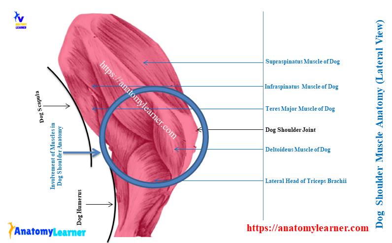
Supraspinatus muscle of dogs shoulder
The supraspinatus and infraspinatus are the lateral muscles of the scapula in the respective fossae. You will see the deltoideus and teres minor muscle along with these muscles that transverse the flexor angle of the shoulder joint.
Superficially, the supraspinatus muscle of the canine shoulder covers by the trapezius and omotransversarius muscles. This muscle fills the supraspinatus fossa and also arises from it.
Some part of the supraspinatus muscle also arises from the spine and edge of the scapular neck. You will see a strong muscular belly of the supraspinatus muscle that curves around the scapular neck. Thus, you will see this muscle at the medial surface of the canine shoulder joint.
The distal end of the supraspinatus muscle forms a thick tendon that inserts on the greater tubercle of a humerus bone. But, how does this muscle contribute to the canine shoulder joint structure?
Well, this supraspinatus muscle help in extending the shoulder joint; thus, the advancement of the limb movement occurs.
If you want, you may learn the origin, insertion, fiber direction, and innervation of all the forelimb muscles from the dog muscle anatomy (another article by anatomy learner).
Infraspinatus muscle of the canine shoulder
The infraspinatus muscle is also directly involved in the canine shoulder structure. The deltoideus muscle superficially covers this muscle.
The infraspinatus muscle of the dog arises from the infraspinatus fossa, scapular spine, and the caudal border or angle of the scapula. Again, you will find an aponeurotic part of the infraspinatus muscle that arises from the deltoideus.
Near the dog shoulder joint, the infraspinatus muscle forms a thick tendon and crosses the caudal part of the greater tubercle. This tendon of the infraspinatus muscle inserts on the lateral or greater tubercle of the humerus bone.
The infraspinatus muscle plays a significant role in the dog’s shoulder joint structure. It helps to flex or extend the canine shoulder joint. The tendon of the infraspinatus muscle acts as the lateral collateral ligament of the canine shoulder structure.
Teres minor muscle of the shoulder
The teres minor muscle lies on the flexor side of the shoulder joint. Here, the teres minor muscle is covered by infraspinatus and deltoideus muscles.
The primary origin of the teres minor muscle will be from the distal third of the caudal edge of the scapula. Again, you will see that this muscle also originates from the aponeurotic part of the long head of the triceps brachii muscle and infraglenoid tubercle.
Finally, this muscle forms a strong tendon that inserts on the crest of the greater tubercle proximal to the deltoid tuberosity. What is the function of the teres minor muscle in a dogs shoulder joint?
The main action of the teres minor muscle is flexion of the canine shoulder joint.
Deltoideus muscle of dog shoulder
The deltoid muscle of the canine shoulder joint consists of two major parts – scapular and acromion. Here, the scapular part of the deltoideus muscle is superficial, directly deep into the scapular fascia between the spine and proximal portion of the humerus.
It arises from the scapular spine, and the aponeurosis blend with the infraspinatus muscle. Again, it becomes tendinous at distal to the shoulder joint that passes medial to the acromion part.
In addition, the acromion part of the deltoideus muscle arises from the acromion process and crosses the lateral aspect of the canine shoulder joint. Finally, the tendon from the deltoideus muscle inserts on the deltoid tuberosity of the humerus bone.
The deltoid muscle involves flexing the canine shoulder joint. Again, it causes the abduction of the humerus bone.
Subscapularis muscle of the dog shoulder
This muscle occupies the subscapularis fossa and crosses the flexor angle of the dog shoulder anatomy. It is a broad and flat muscle on the medial aspect of the shoulder that primarily arises from the muscular line of the subscapularis fossa.
The muscle passes the shoulder joint and becomes narrower and partly tendinous. Finally, it forms a thick tendon that inserts on the lesser tubercle of the dog humerus bone.
You will see the joint capsule or capsular ligament in the canine shoulder structure. The thick tendon of the subscapaurlis muscle joins with the capsular ligament of the shoulder joint.
The primary action of the subscapularis muscle is to adduct and extend the canine shoulder joint. It also draws the humerus bone cranially during the flexion of the shoulder joint.
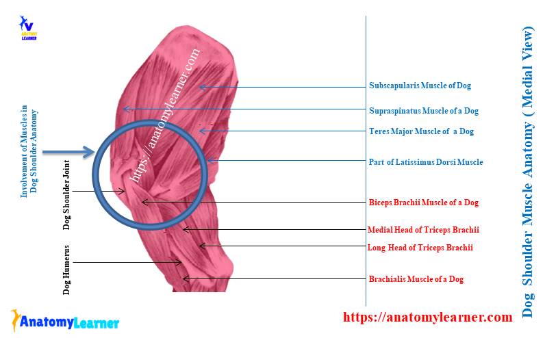
Teres major muscle of the canine shoulder
The teres major muscle also flexes the shoulder joint and draws the humerus caudally. This fleshy and slender muscle lies caudal to the subscapularis muscle.
The teres major muscle arises from the caudal angle of the scapular bone and crosses the triceps brachii and coracobrachialis muscles. It forms a short, flat tendon that blends with the latissimus dorsi muscle.
Finally, this short and flat tendon of the teres major muscle inserts on the teres major tuberosity.
This teres major muscle also rotates the shoulder joint medially. Thus it prevents the lateral rotation of the humerus bone.
Coracobrachialis muscle of dog shoulder anatomy
This is another essential muscle directly involved in the extension and adduction of the dog shoulder joint. It is a short and thick muscle in the arm of a dog.
This coracobrachialis muscle arises from the coracoid process at the distal end of the scapula. The tendon from the coracobrachialis muscle obliquely passes over the medial aspect of the shoulder joint.
Again, this arm muscle runs between the medial and accessory head of the triceps brachii muscles. Finally, the tendon of the coracobrachialis muscle inserts on the minor or lesser tubercle of the dog humerus bone.
Biceps brachii of the dog
The biceps brachii muscle is also directly attached to the canine shoulder structure. It originated from the supraglenoid tubercle of the scapular bone.
The biceps brachii muscle of the dog crosses the shoulder joint in a sharp curve. Finally, the biceps brachii muscle tendon attaches to the cranial surface of the dog humerus bone.
The tendon of the biceps brachii cranially attaches to the joint capsule or capsular ligament of the shoulder. Here, the joint capsule reflects around the tendon of the biceps brachii muscle.
At the distal end of the intertuberal groove, this tendon becomes wide and spindle-shaped and inserted into the cranial surface of the humerus. The main function of the biceps brachii (dog) muscle is to flex the elbow joint.
But, it also extends and stabilizes the dog shoulder joint during the standing condition.
Some other muscles like omotransversarium, trapezius, and log head of triceps brachii have indirect involvement on the shoulder. You may learn more about these muscles’ anatomy from another article by an anatomy learner –
Dog arm and forearm muscles anatomy with the labeled diagram,
Canine shoulder anatomy ligaments
First, let’s see the ligaments in the canine shoulder anatomy –
- Articular capsule, or joint capsule (capsular ligament),
- A transverse humeral retinaculum, and
- The medial and lateral glenohumeral ligaments,
You will also find different muscle tendons that provide the shoulder joint stability. Now, let’s know a little about these ligaments of the canine shoulder joint structure.
The articular capsule of the shoulder joint is capacious and encloses the whole joint. This capsule forms a loose connective tissue attachment just peripheral to the glenoid cavity of the scapular bone. ‘
The joint or articular capsule also attaches to the articular surface of the humeral head. Here, the joint capsule blend with the periosteum on the neck of the dog humerus bone.
A part of the joint capsule surrounds the tendon of the biceps brachii muscle. This occurs in the intertuberal groove of the proximal end of the dog humerus.
Now, the tendon locates in the groove and holds by the transverse retinaculum. Again, the capsular ligament blend with the retinaculum craniomedially. The capsular ligament also blends with the tendon of the subscapularis muscle medially.
In addition, the capsular ligament also blends with the tendon of supraspinatus and infraspinatus muscles. You will also find two fibrous capsular ligaments at the lateral and medial aspects of the shoulder joint.
These are the lateral and medial glenohumeral ligament that attaches to the glenoid cavity to the articular surface of the humeral head. You will also find some thick tendons from the subscapularis, supraspinatus, and infraspinatus muscles that provide the shoulder joint stability.
Anatomy of dog front shoulder
The anatomy of the dog front shoulder shows the bone involvements in the joint, binding materials (ligaments and tendons), and vessels. Here, the shoulder joint is mainly supplied by the different branches of the axillary artery.
You know the axillary artery is the continuation of the subclavian artery in the thoracic limb of a dog. This axillary artery extends from the cranial border of the first rib to the distal border of teres major and latissimus dorsi muscles.
Again, in the canine front shoulder anatomy, you will also find the musculocutaneous nerve (cranially), radial nerve (laterally), and the median and ulnar nerve (caudally). So, you might also describe the anatomical features of the different nerves from the brachial plexus of the dog.
The below-mentioned article might help you to get the primary idea of the different nerves from the animal brachial plexus –
Nerves of the brachial plexus with their courses
In this part, I will not describe all the courses of vessels and nerves from the dog forelimb. Rather I prefer to describe the main vessels and nerves in the shoulder joint structure.
The axillary artery at the dog shoulder joint
So, the axillary is the main artery in the shoulder structure of the dogs. You will find four primary branches of the dog axillary artery –
- The external thoracic artery of the dog,
- A lateral thoracic artery of the dog,
- A subscapular artery in the dog’s shoulder, and
- The cranial circumflex humeral artery of the canine shoulder,
Again, some other branches of the artery are also supplied to the muscles and other structures of the canine shoulder.
Let’s see the dog forelimb artery labeled diagram. Here, it shows the external thoracic artery as the first branch of the dog axillary artery. It arises lateral to the first rib and curves around the craniomedial border of the deep pectoral muscle.
The first branch of the axillary artery (external thoracic) supply to the superficial pectoral muscle of the dog.
From the diagram, let’s find out the lateral thoracic artery of the dog’s axillary. It arises from the caudal surface of the axillary artery at the level of the cranial border of the first rib.
This lateral thoracic artery crosses the lateral surface of the lymph node and runs through the fat. It supplies to the thoracic lymph node area and the latissimus dorsi muscle.
The cranial circumflex is the last branch of the dog axillary artery. Then the axillary artery of a dog continues as the brachial artery in the arm.
You will find the origin of the cranial circumflex artery of a dog at the medial surface of the axillary artery. Again, it may also originate proximal to the origin of the subscapular artery.
As there are different branches in the subscapular artery of the dog, I would like to discuss this artery separately.
Dog subscapular artery and shoulder joint
The dog subscapular artery is the larger branch from the axillary artery in the dog’s arm. This subscapular artery is responsible for supplying the different parts of the scapular region and arm.
The subscapular artery runs obliquely in a dorsocaudal direction along the caudal border of the dog scapula. It passes between the teres major and subscapularis muscles and becomes subcutaneously near the caudal angle of the dog scapular bone.
You will find three main branches (subdivision) of the subscapular artery in the dogs shoulder region –
- A thoracodorsal artery – the larger artery that supplies to the latissimus dorsi and skin,
- The caudal circumflex humeral artery, and
- Circumflex scapular arteries,
After the origin of the subscapular artery, the thoracodorsal artery arises from its caudal surface. This artery runs caudally and supplies to the teres major and latissimus dorsi muscle.
Again, the caudal circumflex humeral artery also leaves the subscapular artery at the same level as the thoracodorsal artery. It courses laterally between the head of the humerus bone and teres major muscle.
The collateral radial artery is one of the principal subdivisions of the caudal circumflex artery. This artery supplies to the teres major and medial head of the triceps brachii muscles.
The caudal part of the canine shoulder joint, infraspinatus, and coracobrachialis muscles are supplied by the branches of the collateral radial artery.
You will also find the circumflex scapular artery that arises from the cranial surface of the subscapular artery. This vessel extends obliquely between the subscapularis and the long head of the triceps brachii muscle.
Finally, it reaches the caudal border of the dog scapula and divides into the lateral and medial branches.
Dog shoulder joint nerves anatomy
Let’s try to identify the brachial plexus nerve that is supplying to the different structures near the shoulder joint –
- Axillary nerve from the brachial plexus of a dog,
- Nerves to subscapularis and teres major muscles,
- Nerve to subscapularis and joint capsule of the shoulder,
- Suprascapular nerves of the dog’s shoulder,
- Nerve to supraspinatus muscle,
- Nerve to infraspinatus muscle,
- Branches of nerve to the deltoid muscle,
- Nerve to the cranial part of the shoulder joint capsule, and
- Cranial lateral cutaneous brachial nerve,
All these nerves are identified in the dog’s shoulder joint nerve labeled diagram. But, you might learn the courses of the different nerves from the dog’s brachial plexus.
The subscapular nerve passes over the dog scapular notch and innervates the subscapularis muscle. Then this nerve continues at the neck of the scapular bone and enters the infraspinatus muscle.
The subscapular nerve in a dog may be single or double and arises from the brachial plexus. You will generally find two divisions (cranial and caudal) of the subscapular nerve that innervates the subscapularis muscle.
The axillary nerve is the larger nerve in the dog brachial plexus. It curves around the caudoventral border of the subscapular muscle near its distal end.
This axillary nerve has a wide range of innervation. Most of the muscles and structure around the dog’s shoulder joint are innervated by the axillary nerves and branches. It will also innervate to the articular capsule of the shoulder joint.
You will find the cranial lateral cutaneous brachial nerve that arises from the axillary nerve. This cutaneous brachial nerve supplies to the deltoid muscle of the shoulder region.
Again, the musculocutaneous nerve provides branches to the coracobrachialis, brachialis, and biceps brachii muscles of the dog.
Canine shoulder anatomy diagram
You have already got the different labeled diagrams on the canine shoulder structure. Again, I would like to share some of the other canine shoulder labeled diagrams so that you may understand them so quickly.
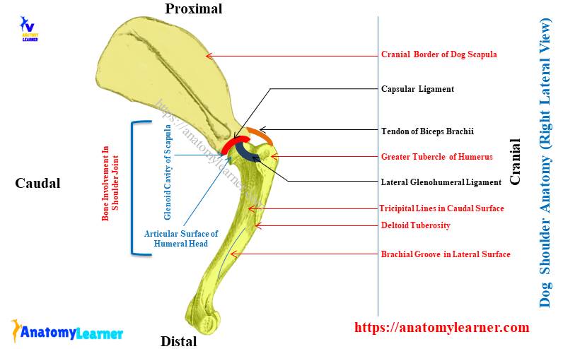
I tried to show you the different bones from the dog’s thoracic limb in the first labeled diagram. Again, I tried to focus on the different osteological features from the proximal extremity of the scapula and distal extremity of the humerus bone.
There is only one joint in the structure of a canine shoulder, and that is glenohumeral articulation. Here, the glenoid cavity of the scapula and the articular surface on the head of a dog humerus are identified.
Again, the second labeled diagram shows the different ligaments from the shoulder articulation. There the two main ligaments –capsular and lateral and medial glenohumeral ligaments are identified in the diagram.
Here, the third dog’s shoulder-labeled diagram shows the different tendons that come from the supraspinatus, infraspinatus, biceps brachii, deltoideus, and teres major muscle. The other tendons that come from the triceps brachii, subscapularis, and teres minor muscles are identified.
You may get more canine shoulder structure labeled diagrams on social media from anatomy learners. Again, you will find more details diagrams on the brachial plexus of a dog with different nerves.
You will also get videos on the canine shoulder anatomy here. There are also handmade figures available for the canine shoulder. These handmade figures may use to prepare your practical hand note.
Horse shoulder joint
The basic structure of the horse shoulder joint is almost similar to the dog shoulder anatomy. But, the joint capsule of the horse shoulder is more spacious and blends especially with the subscapular muscle.
Again, there are lateral and medial glenohumeral ligaments that provide more strength to the joint capsule of the horse shoulder. An additional band of coracohumeral ligament is present in the horse’s shoulder structure.
This coracohumeral ligament extends from the supraglenoid tubercle to the greater tubercle of the horse humerus. The lateral movement of the horse shoulder is impossible due to the unique structure of the articular surface and ligaments.
You will only find the extension and flexion movement in the horse shoulder joint.
Dog shoulder injury
Sometimes your dog may face different injuries to its shoulder. Typically, you will find intermittent lameness in case of a minor shoulder injury to your dog. But, the shoulder injury sometimes becomes more severe for the dogs.
Different forms of dog shoulder lameness lead to pain and arthritis. Let’s see the common causes and forms of dog shoulder injury –
- Supraspinatus tendinopathy – tendon may damage at the greater tubercle of the humerus,
- Biceps brachii tendonitis – the tendon of the biceps brachii muscle may be injured due to external trauma,
- Medial shoulder instability – when the inside ligaments and tendon of the canine shoulder become injured,
- Osteochondritis of the dog humerus bone – development of a loose flap of cartilage may develop on the articular surface of the humerus bone,
The most common injury to the dog is supraspinatus tendinopathy, medial shoulder instability, bicipital tenosynovitis, and shoulder luxation or dislocation. Here, I will not describe the details of all these injuries in the dogs.
Instead, I prefer to provide the leading causes and symptoms of these shoulder injuries. Let’s start with the supraspinatus tendinopathy of the dog shoulder structure.
Supraspinatus tendinopathy in dog shoulder
The working or active companion dogs are more susceptible to supraspinatus tendinopathy. It is very hard to diagnose supraspinatus tendinopathy in a dog without a laboratory examination.
Some degenerative changes may also occur in the supraspinatus tendon of the dog. There are a variety of causes of the degeneration of the supraspinatus ligament.
If any degeneration occurs in the supraspinatus tendon, it may show the discontinuation and disorganized fibers. Again, different types of strain, hitting, and jumping also may cause supraspinatus tendinopathy in the active or working dog.
Unilateral lameness is very common in the supraspinatus tendinopathy of a dog. But, some dogs show bilateral lameness in the supraspinatus tendinopathy condition.
For proper diagnosis and management of supraspinatus tendinopathy, you might contact your veterinarian as soon as possible.
Canine medial shoulder instability
You already got the idea of the structure of the dog’s shoulder. You will find different ligaments and tendons (especially glenohumeral) inside the shoulder structure. Sometimes these ligaments and also the tendons may be damaged by specific causes.
This damage to the ligaments and tendons may lead the canine shoulder joint instability. This shoulder instability of a dog may be lateral, medial, or multidirectional. But, medial shoulder instability is more common in dogs.
Sometimes the degeneration and breakage of the tissue on the shoulder joint may also lead the shoulder instability. The affected dog may show different changes in their performance.
They generally show weight-bearing lameness (unilateral or bilateral). An orthopedic evaluation and diagnostic imaging are required to diagnose this shoulder injury perfectly.
Dog shoulder dislocation or luxation
The shoulder dislocation (luxation) is widespread in all aged dogs. But, you will find two forms of dog shoulder dislocation – traumatic and congenital or degenerative.
The traumatic form of shoulder dislocation may occur in any dog which can be adequately recovered. But, congenital or degenerative shoulder luxation may happen in the small breed of dogs. If your dog has a degenerative shoulder luxation, it is dangerous to your pet.
How may the traumatic shoulder dislocation occur in the dog? To understand shoulder dislocation, you might have a good piece of knowledge on the dog shoulder anatomy. The medial side of the dog’s shoulder is weak.
So, the dog’s shoulder can dislocate in any direction when severe trauma occurs. In some cases, multiple stress on the humeral articular surface may cause the partial dislocation of the medial side of the shoulder joint.
Again, exploring the shoulder structure of the small dog breed like toy and miniature poodles, shelties, and others, you will find the less developed medial glenohumeral ligament. Again, this structure also shows the less developed capsular ligament.
So, probably these factors, you may find more shoulder dislocation or luxation cases in the small breed of dogs. Now, let’s see what you will find and what you should do in different types of dogs shoulder dislocation.
Types of dog shoulder dislocation
So, you may divide the canine shoulder dislocation into the traumatic and degenerative types. Again, according to the degree and time of luxation, it may divide into acute and chronic.
If you find any shoulder dislocation due to minor trauma, there is no need to be worried. Your veterinarian will easily correct this dislocation manually. Sometimes, a supportive bandage is needed for several weeks to recover from this situation.
In the chronic shoulder luxation of a dog, you should correct it through surgical intervention. A chronic case may occur if significant damage occurs in the articular surfaces and in the tendons or ligaments.
If you find intermittent unilateral or bilateral lameness in your dog, you should take care of it. However, there are other different symptoms of shoulder luxation.
Immediately, you should control the lifestyle of your dog and provide physical therapy (suggested by your veterinarian). These may reduce the risk in some content for your affected dog.
Biceps tendonitis and osteochondritis in dog humerus
These two injuries are not so common in the dog, but suddenly your dog may show the symptoms of these two injuries. You know, a thick tendon of biceps brachii muscle passes through the groove of the dog humerus bone.
This tendon is very much susceptible to repetitive strain-type injury. So, the biceps tendonitis may occur in the dog shoulder structure.
Again, a loose flap of cartilage develops on the articular surface of the dog humerus bone. This loose flap of cartilage will cause osteochondritis in the dog humerus.
In this case, you need the surgical correction to remove this flap of cartilage from the articular surface of the humeral head. Don’t worry; your veterinarian will perform this procedure so perfectly.
More inquiries on dog shoulder anatomy
Now, let’s see the most common questions on the dog shoulder anatomy with the different types of problems that both the learner and pet owner asks. As a veterinary student, you should know the cases and management of these problems.
But, you might know the basic structure of the dog shoulder joint to understand the following. Again, if you are a pet owner, this information might help you gather knowledge on the canine shoulder and its problems.
Can a dog pull a shoulder muscle?
Yes, a dog can pull the shoulder muscles. There are some extensor and flexor muscles in the shoulder of the dogs. So, some of the muscles help the dog extend its shoulder; again, some muscles help flex it.
You probably hear the different shoulder muscles from the dog, like trapezius, deltoideus, omotransversarius, and biceps brachii. Again, there are the supraspinatus, infraspinatus, subscapularis, rhomboideus, and tensor major and minor muscles in the shoulder region of the dogs.
You will also find a major muscle (triceps brachii) which consists of four heads and plays an essential role in the canine shoulder. All the descriptions of these shoulder muscles are discussed previously in different articles by anatomy learners.
Where is a dog’s shoulder located?
The dog’s shoulder is located between the scapula and humerus bones. Externally, you will find this shoulder at the level of the fourth to sixth ribs of the thoracic cage.
The distal extremity of the scapular bone (glenoid cavity) articulates with the proximal extremity of the humerus (articular surface of the head). And thus, they form the shoulder joint in the dogs.
In the forelimb or thoracic limb, you will also have other joints like the knee, fetlock, pastern, and coffin. Again, the scapula attaches to the axial skeleton with the help of different muscle and form the synsarcosis.
Can a dog recover from a shoulder injury?
Yes, a dog can recover from a shoulder injury. You know, shoulder injuries are more common in a dog as they used to jump.
In the small breed of dogs, this injury is widespread as they are a more active and working type of animal. Some dogs may have the typical symptoms, and some are not.
If the injuries are minimal, it is easy to recover quickly. But, in chronic cases, it needs time to recover the shoulder injuries.
But, injuries due to the degenerative changes may last for many years. The perfect recovery from these types of shoulder injuries is impossible in dogs.
What is wrong with my dog’s shoulder?
If you see the anatomy of a dog’s shoulder joint, you will find the weaker areas in the articular surface of the humerus bone. Again, fewer ligaments in the shoulder structure provide strength to this joint.
Thus, your dog may face shoulder injuries in minor or major trauma. Again, you know the active and working dogs used to jump on the land every time. Thus, they also get more stress on the shoulder joint, and injuries may occur.
If there are any injuries in the dogs shoulder, they will show intermittent unilateral or bilateral lameness. Again, you will find pain and arthritis in the shoulder joint of your dogs.
How do you tell if a dog’s shoulder is dislocated?
According to the shoulder joint structure in a dog, they are very susceptible to injuries. You may find four different forms of injuries to the dog’s shoulder – osteochondritis in the humerus bone, biceps tendonitis, supraspinatus tendinopathy, and medial shoulder instability.
Any damage in the articular surfaces of the distal part of the scapula and proximal part of the humerus may lead the shoulder dislocation. Again, any damage to the tendons and ligaments of the shoulder structure also may lead to luxation.
If there is any luxation (traumatic or degenerative), your dog will show intermittent unilateral lameness with pain. But, this is not the confirmed diagnosis for the shoulder dislocation. Immediately, you should bring your suspected dog to a professional veterinarian.
What are the ligaments and tendons in the dog shoulder anatomy?
There are three main ligaments in the dog shoulder joint structure – capsular, lateral glenohumeral, and medial glenohumeral. Again, you will find different muscles and tendons in the dogs shoulder joint structure.
You will commonly find the tendon from supraspinatus, infraspinatus, biceps brachii, subscapularis, and teres major muscles.
Conclusion
I hope you got the general idea of the dog shoulder anatomy from this article. The basic structures of the dog shoulder joint are – the glenoid cavity, articular surface on the humeral head, three primary ligaments, and different shoulder muscles with their tendons.
As there are different injuries in the shoulder joint of a dog, you might have a piece of good knowledge of their structure as a veterinarian. Again, the pet owner also knows the structure of the canine shoulder to understand why the injuries commonly occur in their dogs.
I hope all the labeled diagrams representing the dog shoulder structure help you learn it perfectly. Finally, you should practically learn the canine or dog shoulder joint structure from the actual sample at your gross veterinary anatomy teaching laboratory.