The dog elbow anatomy consists of humeroradial and humeroulnar joints. It is a typical hinge or ginglymus type of joint with the range of movement restricted to flexion and extension. You will mainly find the humerus, radius, and ulna bones in the formation of the dog elbow joint.
Here, the humeroradial joint of the dog elbow is formed by the humeral condyle with the head of the radius bone. Again, the humeroulnar joint is formed by the humeral condyle with the trochlear notch of the ulna bone.
You will also find the different muscles and ligaments in the structure of a dog’s elbow joint. I will show you everything about the dog elbow joint anatomy with the labeled diagram in this article.
So, if you want to learn the details of humeroradial and humeroulnar joints, let’s continue this article until the end.
Dog elbow anatomy
So, the dog elbow anatomy is a composite joint formed by the humeral condyle with the head of a radius and the trochlear notch of the ulnar bones. Thus, it forms the humeroradial and humeroulnar joints in the dog’s elbow.
Again, the proximal part of the radioulnar joint freely communicates with the humeroradial and humeroulnar joints. So, I will also cover the anatomy of the radioulnar joint of the dog here in this article.
Okay, first, let’s an important structure that you might know from the dog elbow joint –
- Types of joints in the dog elbow,
- Bone involvement in the elbow joints,
- Types of movement in the dog elbow joint,
- Involvement of the muscles in the elbow joint, and
- Different types of ligaments involved in the dog,
So, before learning the details and anatomical facts of the dog’s elbow joint, you might have basic knowledge of the forelimb bones. You might know the different features of the dog skeleton’s humerus, radius, and ulna bones.
Or, you may go to the below-mentioned article to review the anatomical facts of the dog humerus, radius, and ulna bones with the labeled diagrams –
- Bones from the dog skeleton with the labeled diagram, and
- Dog humerus bone anatomy with the labeled diagram,
Again, I will also show you the main structures from the dog humerus, radius, and ulna bones related to the elbow joint.
Where is a dog’s elbow
The dog elbow joint is the second true joint of the forelimb (thoracic limb). You know, the first true joint from the dog’s thoracic limb is the shoulder joint.
The dog elbow joint is between the arm (humerus) and forearm (radius and ulna) of the front leg. Again, this joint lies just below the chest on the back of the arm.
The dog’s humerus bone is directed downward and backward if you see the diagram. Again, the radius and ulna bones are directed downward and forward. Thus, the elbow joint is formed at the angle between these two bones.
You will find the olecranon process of the ulna bone at the caudal end of the dog elbow joint. This structure of the dog’s elbow joint is known as the point of the elbow.
I hope you can identify the elbow joint perfectly from the front limb (thoracic limb) of the canine skeleton.
Special features of canine or dog elbow joint
Now, let’s see the special features of the canine or dog elbow joint anatomy –
- There are two important joints (humeroradial and humeroulnar) that form the dog elbow,
- Again these two joints have a connection with the radioulnar joints,
- The distal extremity of the humerus articulate with the proximal extremity of the radius and ulna bones,
- Different important muscles of the dog’s arm like the brachialis, biceps brachii have a direct connection with the elbow joint,
- The lateral collateral, medial collateral, and annular ligaments are the most important in the dog’s elbow,
- There is also a presence of the olecranon and oblique ligaments in the dog elbow structure, and
- This is the hinge type of joint in the fore limb of the dog that possesses the extension and flexion movements,
In the dog, the range of limb movement (elbow joint) in the sagittal plane between 100 – 140 degrees of extension. But, this degree of joint movement may vary with the different breeds of the dogs. Again, in the cat, the range of this elbow movement in the sagittal plane is about 140 degrees.
The variation of the elbow movement is caused due to the eccentric proximal insertion of the collateral ligament concerning the axis of the joint. You will learn the insertion site of the collateral ligament from the details anatomical facts section of this article.
Elbow joint dog name
Sometimes you may also be asked another name or synonym of the dog elbow joint by your examiner. Another name for the dog’s elbow joint is cubiti (cubitum means the elbow).
The caudal landmark of the dog elbow includes a prominent olecranon process, the cubital fossa. This structure is also known as the elbow pit.
Again, you may be called this dog’s elbow joint the composite joint. Anatomically this composite joint comprises the humeroradial and humeroulnar joints.
So, you may also call this join the humeroradial and humeroulnar joint. Finally, these two joints (humeroradial and humeroulnar) connect with the proximal part of the radioulnar joints.
Sometimes this elbow joint is also referred to as the snap-type joint of the body. The different parts of this snap joint clip together perfectly. You may find three major types of snap joints – annular, cantilever, and torsional.
Summary of elbow joint name: you may call this dog elbow joint the cubiti joint. Anatomically, you may call this elbow the humeroradial, humeroulnar, and proximal radioulnar joints.
Dog elbow bone anatomy
Now, let’s know the dog elbow bone anatomy with the diagram. It includes three separate bones – humerus, radius, and ulna. I will focus on the osteological features from the humerus’s distal part, proximal radius, and ulna bones.
- Osteological features of the dog humerus (especially from its distal part), and
- Osteological features of the proximal radius and ulna bones of the dog thoracic limb,
Dog elbow and the humerus bone
You know the humerus is the arm’s bone that is comparatively long and less twisted in the thoracic limb of a dog. The direction of this dog’s humerus bone is important while studying the structure of the elbow joint.
This dog humerus bone is directed obliquely downward and backward from the distal extremity of the scapula (shoulder joint) to the proximal part of the radius and ulna bone (elbow joint). Anatomically, this humerus bone is divided into a long body and two distinct extremities.
These two extremities of the dog humerus include – proximal and distal. In the proximal extremity of the humerus, you will find the well-developed, rounded, and more convex head, more prominent greater tubercle (lateral), and less developed medial or lesser tubercle.
Again, the proximal extremity (upper) of the dog humerus bone shows an intertuberal groove, several small foramina, and a constricted small neck. This neck of the proximal end continues with the caudolateral surface of the humeral body.
Again, the body of the dog humerus shows four surfaces with some important osteological features. Here, the deltoid tuberosity is the most prominent osteological feature from the lateral surface of the dog humeral body. Again, you will also find the brachial groove at the lateral surface of the dog humerus (not so spiral compared to the other animal).
Again, the medial surface of the humeral body shows tuberosity of the teres major. On the cranial surface, you will see a less developed humeral crest at the distal end.
The caudal surface of the dog humerus begins at the neck and extends to the distal third of the bone, which continues with the lateral supracondylar crest. This surface of the dog humerus bone is smooth, convex, and possesses prominent nutrient foramen.
A distal end of dog humerus that forms an elbow
The osteological features from the distal part of the dog humerus bone are important as they are directly involved in the formation of the elbow joint. So, let’s know these osteological features perfectly from the distal extremity of the dog humerus bone.
The distal extremity of the dog humerus is also known as the humeral condyle. This humeral condyle of the dog divides into two parts –
- Small, lateral articular surface (known as the capitulum humeri, and
- Larger, medially located, pulley-shaped trochlear humeri,
Again, you will see the olecranon fossa, radial fossa, and medial and lateral epicondyle in the distal extremity of the humerus bone.
How do these two parts of the humeral condyle form the dog elbow joint?
- The capitulum humeri (small lateral condyle) – articulates with the head of the humerus, and
- The trochlear humeri (larger medial condyle) – articulates with the trochlear notch of the ulna bone and forms one of the most stable hinge joints in the dog’s body.
On the cranial surface of the distal end, you will see the radial fossa. Here, the head of the radius bone enters during the flexion of the elbow joint.
On the other hand, the caudal surface of the humeral distal extremity shows a deep olecranon fossa. When the dog’s elbow joint extends, this fossa receives the anconeal process of the ulna bone.
Again, you will see the supratrochlear foramen in the distal extremity of the humerus bone (foramen passes between radial and olecranon fossae). This structure is not clinically or anatomically important.
You might also perfectly identify the lateral and medial epicondyles of the dog humerus. The diagram identifies these two epicondyles from the dog humerus bone.
Canine elbow anatomy and radius bone
The proximal extremity of the canine radius and ulna bones participate in the elbow joint formation. These are two completely separated bones in the dog’s thoracic limb that possess a very narrow interosseous space. But, these two bones of a dog’s thoracic limb can not move over one another.
You know the proximal extremity of the radius and ulna are responsible for the formation of dog elbow anatomy. So, I will only focus on the osteological features from the proximal extremity of the radius and ulna bones from the dog’s thoracic limb.
But, you may learn more about the dog radius and ulna bone anatomy from the below-mentioned article –
- Dog radius and ulna bone anatomy with the labeled diagram,
Okay, let’s see and try to identify the following important structures from the dog radius and ulna bone that are involved in elbow joint formation –
- Articular circumference and fovea of the radius bone,
- Head of the radius and ulna bones of the dog,
- A trochlear notch of the ulnar bone,
- Olecranon process of the dog ulna,
- A radial notch on the ulnar dog bone, and
- Radial and ulnar tuberosities,
But, ensure you can identify the position and surfaces of the dog radius and ulna bone perfectly. I will again suggest you read the article mentioned above to know all the anatomical facts of animal radius bone perfectly (the video is there).
The proximal extremity of the dog radius
You know the radius is a weight supporting the main forearm bone, which is shorter than the ulna. You will see the proximal head, neck, long middle body, and distal trochlea.
The head of the dog radius is an irregular oval outline with a concave articular fovea. This articular fovea of the radius articulates with the capitulum and lateral part of the trochlea of the humerus.
Again, you will see the smooth osseous band on the head that articulates with the radial notch of the ulna bone. A tight body possesses two surfaces (cranial and caudal) and two borders (medial and lateral).
The cranial surface of the radius shaft is convex both transversely and vertically. Again, the caudal surface of the radius bone divides into two flat to concave areas by a verticle interosseous border.
You will see the proximally directed nutrient foramen in the middle of the caudal surface of the body. There is a radial tuberosity on the medial border and adjacent caudal surface of the shaft of the radius bone.
Again, there are carpal articular surfaces and an ulnar notch at the distal extremity of the dog radius bone. Medially, you will find the sharp, wedge-shaped styloid process that extends distally to the main articular surfaces of the carpus.
Elbow joint and dog ulna bone
The dog ulna is also the supporting bone in the forearm of the dog that contributes to the elbow joint. Here, the structures from the proximal extremity are responsible for this joint.
So, you might identify the following important osteological features from the proximal extremity of the dog ulna bone –
- Olecranon tuber and olecranon process of the dog ulna bone,
- A trochlear notch of the dog ulna bone,
- Coronoid process or articular circumference of the dog ulna,
- Ulnar tuberosity, and
- Interosseous spaces and borders in between the radius and ulna bones,
All these structures are identified in the dog radius ulnar bone labeled diagram. You may also find more related diagrams (dog bones) here.
“Ulna is the longest bone in the dog skeleton. This bone exceeds the radius bone in length.”
The olecranon process is the prominent osteological feature in the proximal extremity of the dog ulna bone. You will see the olecranon tuber and anconeal process in the olecranon process of the dog ulnar bone.
This olecranon process of the ulnar dog bone serves as the lever of the arm. It is a four-sided and laterally compressed structure. The proximal part of this olecranon is grooved cranially and enlarged and rounded caudally.
Muscles of the dog elbow joint (at olecranon process)
You will see the attachment of the major extensor muscles of the elbow joint to the olecranon process. The triceps brachii, anconeus, and tensor fascia antebrachii muscles attach to the caudal part of the olecranon process of the dog ulna bone.
Again, the flexor carpi ulnaris and flexor digitorum profundus muscles arise from the medial surface of the olecranon process of the ulna. You will find the details guide on these muscle in the below mentioned article –
- Dog leg muscle anatomy with the labeled diagram
But, you may also learn a little about these elbow joint muscles from this article (let’s see the muscles of the elbow joint section).
Other osteological features from dog ulna
There is a trochlear notch (semilunar notch) in the cranial aspect of the olecranon process of ulna bone that helps to articulate with the humerus. As well as, there is a radial notch that also articulates the ulna with the caudal part of the radius bone.
The sharp hooked anconeal process locates at the proximal end (cranially) of the olecranon process. This anconeal process of the ulna bone fits in the olecranon fossa of the dog humerus bone (during the elbow joint extension).
Let’s talk about the body and distal extremity of the dog ulna bone in a little. You will see the long and somewhat flattened body in the dog’s ulna bone. It possesses three surfaces (lateral, medial, and cranial) and three borders (caudal, lateral, and medial).
The cranial surface of the ulnar body is rough and convex. You will see the interosseous space extends proximally from the notch that separates the distal extremity, the head, from the body of the dog ulna bone.
The caudal border of the dog ulna is smooth and concave throughout its length. Again, the medial border of the ulna bone is sharper and straighter than the lateral one. The caudal border of the olecranon process continues distally and forms the lateral border of the ulna bone.
At the distal extremity of the dog ulna, you will see the oval and slightly raised articular surface (known as the articular circumference). Again, there is a pointed and enlarged styloid process at the distal extremity of the ulna bone.
Dog elbow anatomy description
Now, I will describe two major joints from the dog elbow anatomy – the humeroradial and humeroulnar joints. You will also find the description of the radioulnar articulation in the part of the article. Okay, let’s see what the special features of these two (or three) joints of the dog elbow are –
- Humeroradial articulation or joint (dog elbow front joint),
- Humeroulnar articulation (dog elbow caudal joint), and
- Radio ulnar articulation of the dog elbow,
“You might write the bone involvement, type of joint, type of movement, and different types of ligmants in both humeroradial, humeroulnar, and radio ulnar articulation of the dog elbow.”
First, let’s show the summary of these two (humeroradial and humeroulnar joints) from the dog’s elbow.
Dog front elbow anatomy (humeroradial joint)
You will clearly describe the humeroradial joint from the front view of the dog’s elbow. This is a typical hinge joint in the dog elbow that possesses extension and flexion functions.
Bone involvement: humeral condyle and humeral articular circumference of the dog radius bone (or articular fovea).
Type of joint: hinge (typical) joint,
The hinge joint is one uniaxial joint that permits movement in one plane. The main movements of the hinge joint are extension and flexion. The movement of the elbow joint is the best example of the hinge joint of the animal’s body.
You may also be called the hinge joint as the ginglymus joint.
Type of movement of the humeroradial joint: extension and flexion around an axis,
Extension and flexion are the angular type of bone movement where the angle between two adjoining bones changes. In the extension movement, the angle between two adjoining bone increase, or the segments straightens.
Again, the angle between the two bones reduces in the flexion movement, and these segments come close together.
Ligaments of the humeroradial joint: mainly, you will find the following ligaments in the humeroradial joint of the dog elbow –
Capsular ligament,
Lateral collateral ligament – proximally attaches to the depression on the lateral epicondyle of dog humerus. Again, distally this ligament attaches to the lateral tuberosity of the radius bone.
Medial collateral ligament – proximally, it attaches to the medial epicondyle of the humerus bone and divides into the superficial and deep parts. The superficial part ends at the medial border of the radius, and the deep part ends at the medial tuberosity of the radius.
You may find more information on the dog’s elbow joint ligaments in the ligament section of this article.
Humeroulnar articulation of the dog elbow
The humeroulnar articulation of the dog elbow is another best example of the typical hinge joint. It also possesses extension and flexion functions. Let’s see what the unique features of the dog humeroulnar joint are –
Bone involvement: humeral condyle of the humerus and trochlear notch of the ulna bone,
Type of joint: hinge or ginglymus joint that moves on one axis.
Type of movement: it also possesses the extension and flexion type of movement like the humeroradial joint.
Ligaments of the humeroulnar joint: you will see the following important ligament in the humeroulnar joint of the dog elbow –
- Capsular ligament or joint capsule (common),
- An annular ligament of the dog elbow,
- The olecranon ligament of the dog’s elbow, and
- Oblique ligament of the dog elbow,
Please find the information on these ligaments as mentioned earlier in this article’s (dog elbow ligament description section) (see the next part).
If you are asked to write the dog elbow structure, there is no need to write these two joints (humeroradial and humeroulnar) separately. Just write the bone involvements of these two joints separately.
Ligaments of the dog elbow joint
If you notice the dog elbow anatomy, you will find the following ligament –
- Capsular ligament or the joint capsule of the dog elbow,
- The lateral collateral ligament of the dog elbow,
- A medial collateral ligament of the dog elbow,
- An annular ligament of the dog elbow,
- The olecranon ligament of the elbow, and
- An oblique ligament of the dog elbow,
Now, let’s see the anatomical features of these ligaments from the dog elbow structure.
Joint capsule or capsular ligament of dog elbow
This is the common capsular ligament for all three components of the dog elbow joint (humeroradial, humeroulnar, and radioulnar). You will see this ligament on both sides, but it is extensive in the cranial and caudal aspects of the elbow joint.
If you see the dog front elbow structure, this joint capsule attaches proximal to the supratrochlear foramen and covers most of the radial fossa. Again, caudally this capsule forms a loose, fat-covered synovial pouch. It attaches to the distal part of the supratrochlear foramen.
The joint capsule again extends distally between the radial notch of the ulnar dog bone and the articular circumference of the radius bone. Again, medially it extends a distal pouch deep to the biceps brachii muscle.
You will also find the extension of this muscle laterally to the deep extensor carpi radialis and extensor digitorum communis muscles. Caudomedial aspect, the capsular ligament extends deep to the flexor carpi radialis and flexor digitorum profundus muscles.
Lateral collateral ligament of canine elbow joint
The anatomical name of this ligament is the collateral cubiti laterale ligament. Normally, it proximally attaches to the lateral epicondyle of the dog humerus bone.
The lateral collateral ligament of the dog elbow structure divides into two parts –
- Slightly larger cranial part, and
- A flatter small caudal part,
The larger cranial part of the lateral collateral ligament of the dog elbow attaches to the small lateral eminence of the neck of the radius bone. Again, the small flatter caudal part of the lateral collateral ligament attaches to the annular ligament at the articular circumference of the ulna bone.
Medial collateral ligament of the elbow
The anatomical name of the medial collateral ligament of the dog elbow is collateralae cubiti mediale ligament. It is smaller than the lateral collateral ligament of the elbow joint structure.
This medial collateral ligament attaches to the medial epicondyle of the humerus bone proximally and crosses the annular ligament distally. You will also find the two segments or divisions of this medial collateral ligament of the dog elbow joint –
- Thinner cranial segment, and
- A thicker caudal segment of the medial collateral ligament,
The thinner cranial part of the medial collateral attaches to the proximal part of the radial tuberosity. At the same time, the thicker caudal segment of this ligament passes deeply to the interosseous space and attaches to the ulna bone.
Annular ligament from dog elbow anatomy
The anatomical name of the annular ligament of the dog elbow joint is anulare radii. This thin band of the elbow joint structure runs transversely around the dog radius bone.
You will see the relationship among the annular, lateral, and medial collateral ligaments. This annular ligament lies deep in the collateral ligament and slightly blends with the ulnar collateral ligament.
Finally, this annular ligament attaches to the ulna bone’s lateral and medial coronoid processes.
Olecranon and oblique ligements of the dog elbow
The olecranon is an elastic ligament in the dog elbow structure. It passes between the craniomedial aspect of the olecranon process to the medial border of the olecranon fossa.
You will also see a small band but distinct band of an oblique ligament in the dog’s elbow structure. This oblique ligament arises on the proximal edge of the supratrochlear foramen.
It crosses the cranial, flexor surface of the elbow joint and distomedial to the tendon of the biceps brachii and brachialis muscles. You will see the two segments of the oblique ligament at the level of the distal annular ligament –
- A shorter segment of the oblique ligament, and
- A longer part of the oblique ligament – forms a loop around the tendon of the biceps brachi and brachialis muscle,
The shorter part of the oblique ligament attaches to the cranial part of the medial collateral ligament. At the same time, the longer segment of the oblique ligament attaches to the medial border of the dog radius bone.
A radioulnar joint of the dog
While studying the canine elbow anatomy, you might also learn the radioulnar joint (along with the humeroradial and humeroulnar joints). First, let’s see the bone involvement, type of joint, type of movement, and ligaments in the radioulnar joint in very short form –
Bone involvement in the radioulnar joint: posterolateral aspect of the radius bone and anterior aspect of the ulna bone.
Type of the joint: amphiarthrosis in young and synarthrosis in older animals. In a dog, it is a synovial type joint.
If you don’t know what the amphiarthrosis and synarthrosis joints are, then the following information is for you –
Amphiarthrosis is the cartilaginous joint that possesses restricted movement. You will see two major divisions in the amphiarthrosis joint – synchondrosis and symphysis.
When the hyaline cartilage forms the cartilaginous joint, it is a synchondrosis joint. The best example of the synchondrosis joint is a junction between the sphenoid and occipital bone.
Again, symphysis is the fibrocartilaginous joint between the symmetric bones of the skeleton. They are also known as the secondary cartilaginous joint that persists throughout life. Again, the best examples of these joints are the dogs’ mandibular and pelvic symphysis.
You may learn details about the different types of a joint from the animal body from the below-mentioned article –
- Types of a joint in the animal body with their bone involvement
- Types of movements: restricted in young and immovable in older dogs.
Ligaments of the radioulnar joint: you will see the following important ligaments in the radioulnar joint of a dog –
- Interosseous ligament of the antebrachium – attaches shaft of the ulan to radius proximally and distally, and
- Radioulnar or transverse or arciform ligament – passes proximally to the interosseous space from each border of the ulnar shaft to the caudal surface of the radius bone.
Types of radioulnar joint in the dog
The dog radius and ulna bones are articulated at the proximal and distal ends with the help of a thick interosseous ligament and thin interosseous membrane. So, you will find the following two parts in the radioulnar articulation –
- Proximal radioulnar joint, and
- Distal radioulanr joint,
The proximal radioulnar articulation is also in the part of the dog elbow joint anatomy. This joint extends distally between the articular circumference of the dog radius bone and the radial notch of the ulnar bone.
The proximal articular surface of the ulna contributes substantially to load transfer through the dog elbow joint.
The distal radioulnar articulation of the dog elbow extends between the distal part of the radius and ulna bones. If you notice at the distal end of the ulnar bone, you will see a slight convex articular surface. In contrast, the distal end of the dog’s radius bone possesses a shallow articular surface.
There is a fibrous joint capsule in the radioulnar joint of the dog’s elbow. This joint capsule of the radioulnar articulation continues with the interosseous membrane.
The fibrous joint capsule of the radioulnar articulation of the dog elbow is very short and tight cranially. It forms the radioulnar ligament at the distal part of the radioulnar joint.
Ligaments and membrane of the dog radioulnar joint
The interosseous ligament of the radioulnar articulation is a thick but short collagenous type. This ligament extends across the interosseous space from the adjacent rough areas on the radial and ulna bones.
Again, the interosseous ligament extends distally slight caudal to the middle of the ulna bone at the distal to the radioulnar joint. You will see the slightly oblique arrangement of the interosseous ligament along the long axis of the radius bone.
At the distal part, this interosseous ligament becomes wider and separated from the interosseous membrane by a small fossa. Here the interosseous membrane is narrow and thinner than the interosseous ligament of the radioulnar joint.
The interosseous membrane attaches to the radius and ulna, both proximal and distal to the interosseous ligaments. That means this membrane extends from the proximal to the distal radioulnar joint.
You will see a small pass in the distal end of the interosseous membrane of the radioulnar joint. This is due to passages of the dorsal interosseous artery and vein within it.
Dog elbow muscle anatomy
The dog elbow muscle anatomy includes the biceps brachii, brachialis, and some extensor and flexor group muscles. You will see the direct or indirect relationship of the joint capsule with the extensor and flexor group of muscles of the forearm.
Let’s see what the extensor and flexor group muscles that have a direct or indirect connection with the elbow joint capsule –
- Extensor carpi radialis muscle of the elbow,
- Extensor digitorum communis muscle of the elbow,
- Flexor carpi radialis muscle of the elbow, and
- Flexor digitorum profundus of the elbow joint,
Again, you will find the below-mentioned muscle that has a direct or indirect involvement in the structure of the dog elbow joint –
- Biceps brachi muscle of the elbow,
- Brachialis muscle of the dog elbow,
- Triceps brachii of the elbow,
- Coracobrachialis muscle of the elbow,
- Tensor fascia antebrachii muscle, and
- Anoconeus muscle of the elbow,
Let’s know the little anatomical facts of the muscles mentioned above from the dog’s elbow joint. But, you may also learn more about these muscles from the following article –
- Dog front leg bone and muscle anatomy with the labeled diagram
Brachial muscle of the dog’s elbow
You will see two groups of brachial muscle in the dog elbow structure –
- Cranial flexor muscle – includes biceps brachii, coracobrachialis, and brachialis,
- Caudal extensor muscle of the elbow – includes triceps brachii, anconeus, and tensor fascia antebrachii,
The biceps brachii is the longer and small muscle in the cranial brachial muscle. It attaches to the supraglenoid tubercle by the long tendon and the cranial surface of the humerus through the intertuberal groove.
This biceps brachii muscle flexes the elbow joint and extends the shoulder joint during locomotion. The biceps brachii of the dog elbow is innervated by the musculocutaneous nerve that comes from the brachial plexus of the dog.
The brachialis is another cranial brachial muscle that arises from the proximal part of the caudal surface of the humerus bone. You will see the extension of this muscle laterally as far as the humeral crest and medially as far as the medial surface.
This muscle becomes narrow in the middle of the humerus bone and goes over the flexor surface of the elbow joint. Again, it ends partly on the tendon of the biceps brachii muscle and is finally inserted on the radial tuberosity.
The brachialis muscle of the dog flexes the elbow joint. But, the coracobrachialis muscle (the third muscle of the cranial brachialis) has no direct action on the elbow joint.
This short and thick muscle arises from the coracoid process of the dog scapula bone. It mainly extends the shoulder joint of the dog.
Caudal brachial muscle of the dog elbow
The caudal brachial muscle of the dog elbow fills the triangular area between the scapula, humerus, and olecranon process of the ulna. These are primarily the extensor muscle of the dog elbow joint.
The principal muscle of the dog elbow extensor is the triceps brachii muscle. Again, you will also find the anconeus (caudally) and tensor fascia antebrachii muscle in the extensor of the dog elbow joint.
Let’s see the anatomical features (main) of the dog triceps brachii muscle (main extensor of the elbow).
Triceps brachii and canine elbow anatomy
There are three heads in the triceps brachii muscle of the canine elbow joint – long, lateral, and medial. The long head of the triceps brachii forms a triangular muscle belly. It lies on the caudal edge of the dog scapula and the apex on the olecranon process of the ulna bone.
The muscle arises from the distolateral two third of the caudal border of the scapula bone. Again, the tendon from the long head of the triceps brachii muscle ends on the caudal part of the olecranon tuber.
The lateral head of the dog’s triceps brachii is a large and almost triangular muscle. This muscle lies between the long head of the triceps and humerus bone.
The lateral head of the triceps brachii blends with the accessory head. A tendon from the lateral head inserts into the olecranon tuber of the dog ulna bone.
The medial head of the dog triceps brachii is spindle-shaped. It arises from the crest of the minor tubercle of the humerus.
The medial head of the triceps attaches to the olecranon process of the ulna bone medially and independently.
You will also find an irregular rectangular-shaped accessory head in the dog’s triceps brachii muscle. It lies on the caudal aspect (side) of the humerus bone between the heads of the triceps and brachialis muscles.
All these three (four) heads of the triceps brachii muscle extend the dog elbow joint.
Anoconeus and tensor fascia antebrachii of dog elbow
The anconeus muscle extends the dog elbow anatomy along with the triceps brachii. It lies on the caudal aspect (side) of the distal half of the humerus bone between the epicondyles.
The anconeus muscle of the dog elbow joint arises from the lateral epicondylar crest and lateral epicondyle. Again, it ends on the lateral surface of the proximal end of the dog ulnar bone. This muscle covers the proximal surface of the dog elbow joint.
The tensor fascia antebrachii of the dog elbow is a flat, broad, and straplike muscle. It supports the extension function of the triceps brachii. Thus, this muscle indirectly helps to extend the dog’s elbow joint.
It arises (originates) from the lateral surface of the latissimus dori muscle and ends on the olecranon process of the ulna as the common tendon with the triceps brachii muscle.
Extensor and flexor muscle of antebrachium
Except for the extensor carpi radialis and pronator teres muscles, the extensor and flexor muscles have no direct contribution to extending or flexing the dog elbow joint. But, in the structure of the dog elbow joint, you may find different extensor and flexor groups of muscles.
So, let’s discuss these two important muscles that contribute to the extension or flexion of the elbow joint –
Extensor carpi radialis muscle: it is a long, thick, and fleshy muscle that lies on the cranial surface of the dog radius bone. It arises on the lateral epicondylar crest of the dog humerus bone.
The tendon comes from the extensor carpi radialis muscle insert on the small tuberosity of the dog metacarpal bone. This muscle directly contributes to the flexion of the dog elbow joint. Again, the extensor carpi radialis extends the dog carpal joint.
The pronator teres muscle: is round in a transverse section that crosses the medial surface of the dog elbow joint. This muscle lies deep into the skin and fascia on the proximal third of the radius bone.
The pronator teres muscle arises (origin)from the medial epicondyle of the humerus bone just cranial to the flexor carpi radialis muscle. A thick tendinous band from the pronator teres muscle ends on the medial border of the dog radius bone.
This muscle is considered the flexor muscle of the dog elbow joint. Now, let’s see the other extensor and flexor muscles indirectly connecting with the dog elbow structure.
Other extensor and flexor muscles of the dog elbow
The extensor digitorum communis is the craniolateral muscle of the radius between the extensor carpi radialis and digitorum lateralis. This muscle arises (origin) from the lateral epicondyle of the dog humerus and inserts on the metacarpal bones.
This muscle extends the joints of the four principal digits of the dog.
You will find a flat and broad supinator muscle in the structure of the dog elbow joint. This muscle lies lateral to the flexor surface of the elbow joint and is covered by the extensor carpi radialis muscle.
The supinator muscle lies on the dog’s joint capsule and the radius bone. It arises on the lateral collateral ligament of the dog elbow joint and also from the lateral epicondyle.
This muscle extends obliquely distomedial of the proximal third of the dog radius bone. Again, the tendon from the supinator muscle inserts on the cranial surface of the radius bone.
Flexor carpi radialis muscle: lies in the medial part of the forearm directly deep into the skin and antebrachial fascia. This muscle arises from the medial epicondyle just caudal to the medial collateral ligament of the dog elbow joint.
It extends distally between the pronator teres and flexor digitorum superificialis. Then it forms the short, thick fusiform belly that merges into the flat tendon near the middle part of the dog’s radius bone.
Actually, this muscle possesses the flexion function for the dog’s carpal bones.
Canine elbow dysplasia
You know dysplasia means the abnormal development of the tissue or organs. So, canine elbow dysplasia means that there has been abnormal development in the structure of the elbow joint.
If there is abnormal development in the elbow joint, then the three bones of this joint (humerus, radius, and ulna) do not fit perfectly. You may find a different form of problems that may cause canine elbow dysplasia –
- Fragmentation on the medial coronoid process of the ulna bone,
- Osteoarthritis of the dog humerus bone,
- Ununited anconeal process of the dog ulnar bone, and
- Disease in the medial compartment of the dog elbow joint,
You may find different typical symptoms in canine elbow dysplasia. But, it would help if you had an x-ray of the canine elbow to confirm the dysplasia.
Normally, the dog shows lameness when it faces elbow dysplasia. The lameness of the dog may be intermittent and tends to improve with the rest. But, sometimes it may become the worse with exercise.
Again, you may find a swollen area and pain with the restricted movement of the elbow joint. A few dogs show pain, but no swollen area is found.
Management of the elbow is better than surgery to correct the dysplasia in the dog. If your dog shows the typical symptoms of elbow dysplasia, immediately bring it to a professional veterinarian.
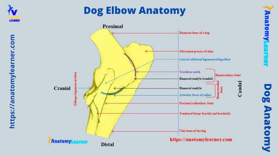
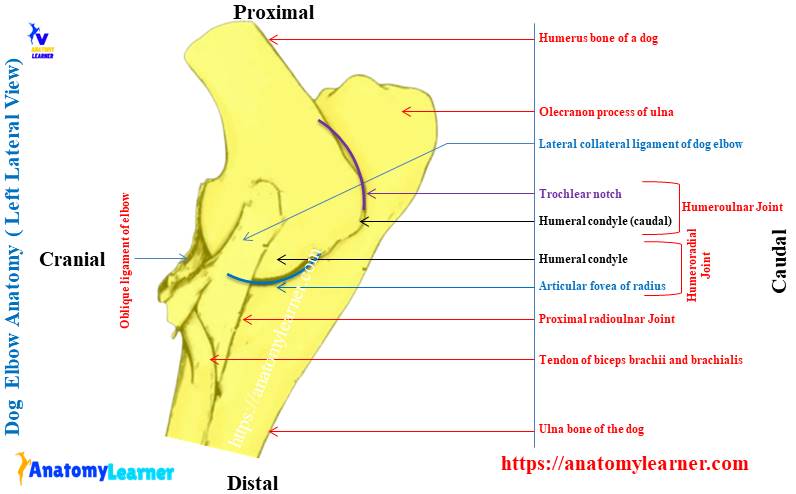
Canine elbow anatomy labeled diagram
Now, let’s see the final labeled diagram on the canine elbow anatomy. In the labeled diagram, I tried to show the different structures from the three major bones involved in the dog’s elbow joint formation.
Again, the diagram shows the ligaments and muscles from the humeroradial and humeroulanr joints of the elbow. From the front view of the dog elbow joint structure, you may easily identify the humeroradial joint and its structures.
The labeled dog elbow joint diagram shows the capsular, lateral, and medial collateral ligaments. Again, part some of the muscles are identified in the labeled diagram.
The caudal view of the dog elbow joint structure shows the annular and oblique ligaments. You may find more labeled diagrams on the dog elbow joint here.
Again, join the anatomy learner on the youtube channel to get more updates on the dog elbow structure.
Common inquiries on the dog elbow anatomy
Now, I will try to solve the common inquiries on the dog elbow joint anatomy asked by the anatomy learners. You may go through these questions and answers to gather more knowledge on the canine elbow structure. Let’s see what the common inquiries on the dog elbow structure by the anatomy learners and also by the dog owners are –
Why the lateral movement of the dog elbow joint is minimal?
Normally, the lateral movement of the dog elbow joint is minimal. This is due to the thick lateral and medial collateral ligaments in the elbow structure.
Again, the cranial extension of the anconeal process of the ulna bone logged into the deep olecranon fossa of the humerus bone. This condition also restricted the movement of the elbow joint. Thus, the collateral ligaments and the anconeal process causes minimal movement of the dog’s elbow joint.
What is the elbow joint in a dog called?
You may be called the dog elbow joint as the cubital joint. Again, this is a typical hinge or ginglymus type of joint in the thoracic limb of the dog skeleton.
The dog elbow joint is the collection of humeroradial, humeroulnar, and proximal radioulnar joints. The main function of this dog’s elbow joint is extension and flexion of this joint and thus helping in locomotion.
What does elbow dysplasia look like in dogs?
If your dog has elbow dysplasia, it will show some typical symptoms. It may show the lameness, but this lameness in the elbow dysplasia may be intermittent.
Again, the affected dog shows stiffness and reluctance to exercise. This condition may occur typically at five to ten (5 -10) months.
Again, the affected dog may show pain and swollen area on the elbow joint. But, some of the dog don’t show any external symptoms of elbow dysplasia.
Can dogs with elbow dysplasia run?
Yeah, your dog may run if they have elbow dysplasia. But, they will show intermittent lameness and stiffness and get worse in exercise.
So, if you find the typical symptoms of dog elbow dysplasia, you should immediately bring your dog to the veterinary doctor. And it would help if you had control over the dog’s running.
How do you tell if a dog has dislocated elbow?
You may suspect dysplasia when you find intermittent lameness with stiffness and swollen area on the elbow. But, this needs an x-ray of the elbow joint to confirm the dysplasia. For that, you might visit a veterinary professional with your suspected dog.
What are the common ligaments of the dog elbow anatomy?
You will see the different ligaments in the dog’s elbow structure. Commonly you will see the joint capsule (capsular ligament), lateral collateral ligament, medial collateral ligament, and the annular ligament.
Again, the dog elbow joint structure shows the olecranon and oblique ligaments.
What are the main joints that form the dog’s elbow?
The humeroradial, humeroulanr, and proximal part of the radioulnar articulation form the dog elbow structure. Some associated structures like the ligaments and muscles are involved in the dog elbow joint formation.
Conclusion
So, the dog elbow joint anatomy comprises the two major joints – humeroradial and humeroulnar. Again, the proximal part of the radioulnar joint contributes to forming the dog elbow joint structure. I hope you found the basic idea of the canine elbow joint anatomy with the labeled diagram from this article.
Six main ligaments are present in the dog elbow joint structure. Now, let’s try to identify and learn the dog elbow joint anatomy from the actual sample from your anatomy learning laboratory. You might take help from the dog elbow joint labeled diagrams I have already provided in this article.