The cat skull anatomy consists of the cranial and facial bones. These cranial and facial bones of a cat are known as the skull proper. You will find other visceral bones like the lower jaw, hyoid, and ear bone together with the cat skull proper. Here, I will share all the anatomical features of the skull bones from a cat with a diagram.
The cranial bones form the cranial cavity, and the facial bones support the face. You will find both paired and unpaired bones in the cranial and facial portion of a cat’s skull.
The osteological features of the cats or dog skull bones are somewhat different than these of the ruminant. So, if you wish to know them in detail from a domestic cat skull anatomy with a labeled diagram, continue this article till the end.
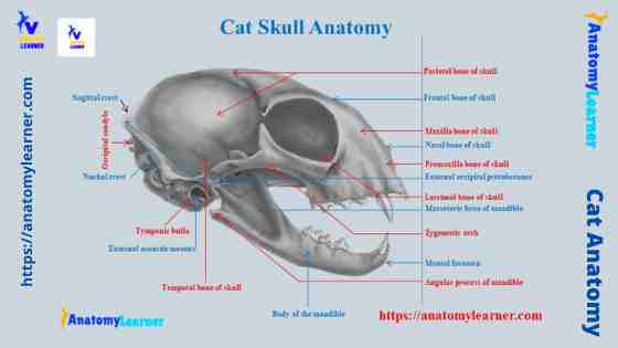
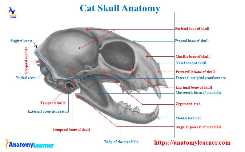
Cat skull anatomy
The cranial portion of a cat skull includes all the bones enclosing the brain cavity. First, you should know the bones in the cranial part of a cat skull. You will find the following bones in the cranial part of a cat skull anatomy –
- An occipital bone of a cat skull (single)
- The interparietal bone of a cat (single)
- A parietal bone of a cat skull (paired)
- The temporal bone of a cat skull (paired)
- Sphenoid bone (single)
- A presphenoid bone of a cat (single)
- The frontal bone of a cat skull (paired) and
- An ethmoid bone of a cat (single)
So, there are eleven bones in the cranial part of a cat skull. The occipital bone of a cat skull surrounds the foramen magnum and the interparietal bone. You will find the parietal bone at the lateral and dorsal aspects of the skull, whereas the sphenoid bone is at the ventral aspect.
The frontal bone also covers the dorsal and lateral aspects of the cat’s skull. Again, the presphenoid bone finds at the ventral part of the skull.
The facial part of a cat’s skull is much smaller than the cranial. It encloses the face and nasal cavity of a cat. You will find the below-mentioned bones in the facial part of a cat skull –
- Maxilla bone of a cat skull (paired)
- Premaxilla bone of a cat (paired)
- Lacrimal bone of a cat (paried)
- A nasal bone of a cat (paired)
- The palatine bone of a cat (paired)
- Zygomatic or malar bone (paired) and
- Vomer bone of a cat skull (single)
So, there are thirteen bones in the facial part of a cat skull. You will also find the lower jaw (mandible), hyoid bones, and ear bones in the cat skull.
Surface features of a cat skull
Now, I will show you some of the four different surfaces of a cat skull anatomy. The dorsal surface of a cat skull is relatively smooth and possesses some lines, ridges, sutures, and openings. Again, the lateral surface of a cat skull is more complicated than the dorsal and caudal.
You will find the foramen magnum and other structures; these are almost similar to the ruminant skull. But, the ventral surface of a cat skull is very complicated than those of the different surfaces.
Here, I will discuss the features of these four surfaces from a cat skull so that you may quickly identify it –
- Surface features of the dorsal aspect of the cat’s skull
- Parts of the caudal surface of the cat’s skull
- Surface features of the lateral aspect of a cat’s skull and
- Parts of the ventral surface of a cat’s skull
That’s fine, let’s start to learn and identify the features from the different surfaces of a cat skull. But, make sure you understand the detailed description of every single bone from the other aspect of a cat skull. You will find all the cranial and facial bones descriptions in the next section of this article.
Cat skull front or dorsal view
The cat’s front or dorsal view presents a smooth convex surface, a broad caudal end, and two zygomatic arches. You will also find the other structures like frontal suture, temporal line, coronal suture, sagittal suture, sagittal crest, and lambdoidal ridge in the dorsal view of a cat skull.
Again, you will also find exceptional features (incomplete orbital cavity) in the dorsal view of a cat skull. In the ruminant skull (sheep, goat, cattle), you will find the complete orbital cavity in their skull.
Now, let’s identify the bones of the cat skull that are visible from the dorsal view. Nice, you will find the following bones that are visible from the dorsal view of a cat skull anatomy –
- Maxilla and premaxilla bones
- Nasal and lacrimal bones
- Frontal and zygomatic bones
- Temporal, parietal, and interparietal bones
At the cranial part of the dorsal view, you will find a sizeable nasal opening that bounds by the premaxilla (ventrally and laterally) and nasal bone (dorsally). Again, the frontal, lacrimal, and zygomatic bones formn of the orbital cavity in the cat’s skull.
You will find a lateral prominent zygomatic process from each frontal bone of the skull. Again, the zygomatic process separates the dorsal surface of the skull from the wall of the orbital fossa and forms the supraorbital margin.
The zygomatic process of the temporal and zygomatic bones forms the zygomatic arch in a cat skull. You will find a prominent lambdoidal ridge at the caudal boundary of the dorsal surface of the skull. Again, a second ridge (sagittal crest) arises from the middle of the lambdoidal ridge of a cat skull.
The frontal and coronal sutures are the most prominent features in the dorsal surface of a cat skull.
The lateral surface of the cat skull
From the lateral view of a cat skull, you will find the different processes, sutures, foramen, and bones. The intermaxillary, nasomaxillary, sphenofrontal, coronal, squamosal, and lambdoidal sutures are more prominent at the lateral surface of a cat skull.
Again, the nasolacrimal canal, sphenopalatine foramen, optic foramen, foramen orbitorotundum, foramen ovale, and stylomastoid formen are aslo visible from the lateral aspect of the cat skull. The other essential features of the lateral view include the tympanic bulla, mastoid process, jugular process, and occipital condyles.
Now, let’s know what bones are easily visible from the lateral aspect of a cat skull. You will see the following bones at the lateral aspect of the cat skull –
- Premaxilla, maxialla, nasal, frontal, and lacrimal bones
- Palatine, parietal, temporal, interparietal, zygomatic, and occipital bones
All the lateral bones from a cat skull are identified in the labeled diagram.
The mastoid process arises from the temporal bone, just cranial to the jugular process. They rest against the side of the tympanic bulla of the cats’ skull. Again, the zygomatic arch begins as the zygomatic process of the temporal bone immediately dorsocranial to the external auditory meatus.
You will see a mandibular fossa at the base of the zygomatic process for the condyle of the mandible. This mandibular fossa is bounded caudally by the prominent post mandibular processes.
At the boundary of the orbital fossa, you will find a prominent semicircular ridge of the zygomatic arch, the zygomatic process, and the supraorbital arch of the frontal bone.
Again, at the lateral view of a cat skull, you will find teeth implanted along the alveolar border of the maxilla and premaxilla bones.
Caudal view of a cat skull
In the caudal view of a cat skull, you will find the occipital bone and its associated structures. The caudal part of a cat skull is formed by the occipital bone that surrounds the foramen magnum. There are two prominent curved occipital condyles at the side of the foramen magnum.
You know these curved occipital condyles are designed for articulation with the atlas vertebra. At the craniolateral part of the condyles, you will find a jugular process separated by a deep notch. These jugular processes attach to the caudal end of the tympanic bulla.
The lambdoidal ridges form the dorsal and dorsolateral boundaries of the caudal part of a cat skull. A dorsal external occipital crest arises at the junction of both side lambdoidal ridges. This is a faint median ridge running dorsally to the foramen magnum of a cat’s skull.
Ventral view of a cat skull
The ventral view of a cat skull anatomy is more complicated than the other views. But, I will show you some of the significant osteological features from the ventral part of a cat’s skull. You will also learn the other components from the ventral view in detail later.
At the cranial part of the ventral surface, you will find the triangular area that forms by the maxilla and premaxilla bone together with the hard palate. There are also alveolar sockets (containing teeth) at the cranial and lateral parts of the maxilla and premaxilla bone. You will also see the palatine bone with palatine groove and anterior palatine foramina.
In the middle part, you will find the pterygoid process of the palatine bone’s sphenoid and perpendicular plate. Again, some foramina and canal may easily see from the ventral view of a cat skull.
You will see the foramen ovale, foramen orbitorotundum, and pterygoid canal from the ventral view of a cat skull. The ventral view also shows you the visible part of the orbital cavity and zygomatic arch.
At the caudal part of the ventral view, there are foramen magnum, occipital condyles, and jugular processes. There are also some prominent features like the mastoid process, stylomastoid foramen, and external auditory meatus. Again, you will also find the jugular foramen and hypoglossal foramen at the ventral surface of a cat skull.
Special features of a cat skull
In this part, I will provide you with a bit of information on the unique features of a cat skull. But, make sure you will learn all the osteological features from a cat skull with the labeled diagrams.
Okay, let’s find some of the significant osteological features from the cat skull –
- The cat’s skull is narrow, slender, and more elongated than those of a ruminant skull.
- You will find a narrow frontal bone where the frontal sutures are prominent.
- There is no corneal process in the skull of a cat.
- The zygomatic processes are curved, short, and thin compared to the ruminant.
- You will get a prominent external sagittal crest in the skull of a cat.
- The orbital cavity is incomplete, and the supraorbital process is absent in the cat skull.
- There is no facial crest or tubercle in the skull of a cat.
- You will find the alveolar socket for the canine teeth in the maxilla bone of the cat’s skull.
- The body of the mandible is narrow and flatter that incompletely fuses.
- You may find two to three mental foramen at each side of the mandible (whereas you will find only one mental foramen in ruminant).
- There is an extra feature (masseteric fossa) present in the caudal part of the cat’s mandible.
These are the major osteological features from the skull of a cat. But, now, you may learn the details anatomical features of a cat skull.
Cranial bones of a cat skull anatomy
As you know, there are eleven bones in the cranial part of a skull that forms the brain cavity. You will find three paired bones and five single bones in the anatomy of a cat skull. The single and paired bones from the cranial cavity of a cat are –
- Cranial bones (paired) – include parietal, temporal, and frontal bones.
- Cranial bones (single) – include occipital, interparietal, sphenoid, presphenoid, and ethmoid bones.
Now, let’s know the details osteological features of the cranial bones from a cat skull.
An occipital bone of a cat
The occipital completes the posterior portion and base of the cat’s skull. A large opening, the foramen magnum, locates at the ventral part of the occipital bone. In a kitten, you will find four distinguished portions in their skull.
These four portions of the kitten’s occipital bones are –
- A basal part (basioccipital bone)
- Two lateral parts (exoccipital bones), and
- A dorsal part (supraoccipital bone)
But, in an older cat, these four portions fuse to form a single occipital bone. Again, you may divide this occipital bone into basal and squamous parts for convenient description.
Well, let’s know the special osteological features from the basilar and squamous parts of a cat’s occipital bone.
Basilar part of the occipital bone
The basilar part of an occipital bone is flat and elongated, articulating with cranially the basisphenoid bone. It will also communicate with the petrous portion of the temporal bone laterally.
Again, the lateral part of the basilary occipital bone articulates with the petrous and mastoid portion of the temporal bone. They form the lateral boundary of the foramen magnum and pass dorsally into the squamous part of the cat’s occipital bone.
You will find the elongated curved occipital condyle at the external surface of the basilar occipital bone. Lateral to each condyle, a triangular jugular process covers the caudal end of the tympanic bulla.
You will also find a jugular notch at the medial aspect of the jugular process of the occipital bone. There is a hypoglossal canal at the medial aspect of the jugular notch. Within this hypoglossal canal, the hypoglossal nerve passes.
A cranial opening (condyloid canal) is present at the dorsal part of the hypoglossal canal. This opening of the cat’s skull transmits a vein.
Again, you will find a longitudinal ridge at the ventral part of the basilar occipital bone.
Squamous part of cat’s occipital bone
The squamous part of the occipital bone forms an arch over the foramen magnum. There are two borders – thick and rough dorsal border and thin and smooth ventral border. The dorsal bored of the squamous part articulates with the parietal and interparietal bones of the cat’s skull.
You will find a prominent lambdoidal ridge at the external surface of the squamous part of a cat’s occipital bone. A median external occipital crest extends from the middle of the lambdoidal ridge.
You will also find an external occipital protuberance at the junction of the crest with the lambdoidal ridge.
At the inner edge of the squamous part, there is some depression for the convolution of the cerebellum.
The frontal bone of a cat
The roof, the cranial part of a cat skull, consists of paired frontal bones. These paired frontal bones articulate one another at the midline of the skull in between the nasal and parietal bones. The frontal bone forms a large part of the medial wall of the orbit and a part of the temporal fossa.
For description purposes, you may divide the cat’s frontal bone into two parts – frontal plate and orbital palate.
The dorsal surface of the frontal plate is smooth and convex. A supraorbital arch separates the cranial two-thirds of the lateral border of the frontal plate from the orbital fossa. You will find a triangular frontal spine at the cranial angle of the frontal plate of the frontal bone.
The ventral surface of the frontal plate is smooth and presents slight ridges and depressions for convolution of the cerebrum. At the medial border of the frontal plate, you will find some verticle plates that articulate with the opposite side.
The caudal border of the frontal plate is rough and articulate with the parietal bone except at its ventral end. On the other hand, the lateral boundary is smooth and articulates with the orbital plate on its right angle.
The orbital plate of the cat’s frontal bone arises from the ventral surface of the lateral border of the frontal plate. It directs ventrally and forms the dorsal portion of the medial wall of the orbital fossa. You will find an ethmoidal foramen near the ventral border of the orbital fossa.
There is a triangular spine at the cranial margin of the orbital plate of the frontal bone. Again, the ventral border articulates with the orbital plate of the palatine bone.
A temporal bone of a cat skull
This is the most complex bone in the cat skull anatomy. You will find three distinct portions in the temporal bone of a cat’s skull – squamous, petrous, and tympanic. I will teach you the osteological features from these three parts of a cat’s temporal bone with the labeled diagram.
- Squamous part of the temporal bone
- Petrous part of the temporal bone of a cat and
- Tympanic part of the cat’s temporal bone
Let’s try to identify the main osteological features from these three portions of the temporal bone of a cat.
Squamous part of the temporal bone
The squamous part of the cat’s temporal bone is oval and thin and has an equilateral triangle. You will find curved zygomatic processes that arise from the ventral border of the temporal bone.
The inner surface of the squamous part is concave and smooth, but the outer surface is convex that giving the origin of the temporal muscle.
The cranial half of the zygomatic process is beveled along the ventral border to accommodate the zygomatic process of the malar (zygomatic) bone. You will find a mandibular fossa (a groove) at the posterior part of the zygomatic process.
This mandibular fossa provides the articulating surface for the condyloid process of the cat’s mandible. Again, there is a small post mandibular process projecting from the proximal basal part of the zygomatic process.
Petrous part of the temporal bone
The petrous part of the cat’s temporal bone consists of the petrous proper and the mastoid part. You will find a very thick triangular pyramid-shaped proper petrous piece encloses the auditory labyrinth. Again, the mastoid part is less dense and flattened that attaches to its base.
The petrous proper has a base and three sides – lateral, medial, and dorsal. It forms the medial wall of the tympanic bulla of the cat. Again, the base of the petrous proper attaches to the mastoid portion of the temporal bone.
The dorsal surface of the petrous proper is triangular and represents a foramen near its apex. Again, the medial surface shows the internal auditory meatus at its middle.
You will find a fenestra cochlea (large circular foramen) at the surface of the petrous proper of the temporal bone.
The mastoid portion of the petrous is elongated and articulate with the lateral part of the occipital bone. You will find a prominent, angular mastoid process that overlaps the bulla posterior to the external auditory meatus.
Again, there is a stylomastoid foramen just anterior to the mastoid process. At the medial surface, you will find a distinct internal auditory opening.
Tympanic part of the cat’s temporal
The tympanic portion of the cat’s temporal bone is hollow and oval. You will see an irregular oval opening (external auditory meatus) that leads into a tympanic cavity. The malleus and incus are easily visible within the tympanic cavity, whereas the stapes are not visible.
The cranial part of the tympanic bone forms a short styliform spine. It lies in a horizontal groove in the ventral surface of the basisphenoid bone.
There is a short triangular spine at the ventral margin of the medial surface. This spine lies against the ventral surface of the basilar portion of the cat’s occipital bone.
You will find two or three verticles parallel grooves at the caudal end of the spine. The inner edge of the tympanic bulla is thickened.
A parietal bone of cat’s skull
The parietal bone of a cat’s skull forms the more significant part of the lateral and dorsal boundaries of the cranial cavity. Each bone is roughly rectangular with a smooth and curved outer surface. You will find a parietal tubercle at the middle part of the outer parietal bone.
Again, a curved ridge runs caudodorsal angle and indicates the boundary of the temporal muscle’s origin. You will find a smooth inner surface with some ridges and grooves. There is a shallow groove at the medial border of the ridge for the superior sagittal sinus.
The tentorium, projects anteriorly as a curved structure with a prominent medial notch. It separates the cerebellar fossa of the cranium from the cerebral fossa.
The anterior border of the parietal bone is beveled at the expense of the inner surface and articulate with the frontal bone. On the other hand, the posterior border is thick and porous medially but thin laterally. It communicates with the interparietal and mastoid parts of the temporal bone.
You will find a straight medial border in a cat’s parietal bone. In contrast, the ventral edge is concave, sharp, and articulate with the squamous part of the temporal bone.
Interparietal bone features from cat skull
The interparietal bone is a single, triangular bone locates between the parietal and occipital. You will find an apex and base in the interparietal bone. The apex of the interparietal bone directs cranially, and the base articulates with the squamous part of the occipital bone.
The dorsal surface of the interparietal bone possesses a distinguished medial projection (sagittal crest). This crest may extend anteriorly onto the parietal surface in some cats.
The sagittal crest is continuous with the lambdoidal ridge of the occipital bone posteriorly. Again, the ventral surface of the interparietal bone is irregularly triangular, smooth, and concave.
A sphenoid bone of a cat skull anatomy
The osteological features of the sphenoid bone from a cat skull anatomy are also complex. You will find two portions in the sphenoid bone – the central or base basisphenoid and two lateral alisphenoids. The basisphenoid portion of the sphenoid is the base, whereas the lateral alisphenoid is the wings.
Again, you will find a thin curved process (pterygoid process) in the structure of the sphenoid bone. So, you may divide the sphenoid bone into three parts (base, wing, and pterygoid process) for description purposes.
- Base or basisphenoid part of the sphenoid bone
- Two lateral wings or alisphenoid and
- The pterygoid processes
Let’s know the details of these three structures from the cat’s sphenoid bone.
Basisphenoid part
It is wedge-shaped and lies at the midline of the base of the cat’s skull. You will find six surfaces in the basisphenoid part of a cat’s sphenoid bone. The cranial end of the basisphenoid is square and rough. It articulates with the body of the parasphenoid bone.
The caudal end of the basisphenoid is concave and rough, articulating with the occipital bone’s basilar part. Again, the dorsal surface of the basisphenoid is distinctly saddle-shaped. You will find the tuberculum sellae, which is the anterior elevation of the saddle.
Again, there is a more prominent and rounded dorsum sellae present at the posterior part of the saddle. Between these two sellae, you will find a conspicuous depression known as the sellae turcica.
There is a small nutrient foramen near the caudal end of the sellae turcia. Again, you will find the foramen lacerum and carotid groove at the basisphenoid part of a cat’s sphenoid bone.
Wing of the cat’s sphenoid bone
The alisphenoid is a thin quadrilateral plate of the bone that attaches to the base of the sphenoid. There is a rounded groove present in the wing of the sphenoid bone. You will find three essential foramina in this rounded groove –
- Orbital fissure – a large foramen that lies among the wing, body, and pterygoid process
- Foramen orbitorotundum – small and rounded structure, and
- Foramen ovale – larger and oval structure.
Through the orbitorotundum foramen, the maxillary branch of the trigeminal nerve passes. Again, the mandibular branches of the trigeminal nerve pass through the foramen ovale.
Pterygoid process of the sphenoid
The pterygoid process is a thin plate of bone. In the pterygoid process, you will find a concave and smooth medial surface, whereas the lateral surface is convex and possesses two parallel ridges.
There is a long deep fossa (internal pterygoid fossa) between the pterygoid bone and the pterygoid process of the sphenoid bone. You will find a thin rod hamulus that extends posteriorly from the body of the pterygoid process.
A presphenoid bone of a cat
The unpaired presphenoid bone of a cat’s skull consists of a body and two wings. A rectangular body lies in the base of the skull, just cranial to the basisphenoid bone.
You will find two sphenoid sinuses which end blindly at the posterior end of the body of the presphenoid. There are also six surfaces found in the body of the presphenoid bone of a cat. The dorsal surface of the body continues with the dorsal surface of the wing.
You will find the chiasmatic groove with optic foramina at the caudal end. Again, the ventral surface of the body possesses a median ridge and continues with the ridge of basisphenoid bone. Furthermore, the caudal end of the body presents a quadrangular surface for the articulation with the body of the sphenoid bone.
The wing of the presphenoid project is posterolaterally and resembles a small triangle. You will find a smooth dorsal and ventral surface in the wing of the parasphenoid bone that continues respectively with the body’s dorsal and ventral surface.
The ethmoid bone of a cat
The ethmoid is an unpaired cranial bone of the cat’s skull but entirely associated with the nasal cavity. You will find the below-mentioned important osteological features in the cat’s ethmoid bone –
- Ethmoturbinate bone
- Perpendicular plate of the ethmoid bone, and
- Cribriform plate of the ethmoid bone.
The ethmoturbinate structure of the ethmoid bone consists of a scrolled, thin laminated plate of bone. It also comprises most of the nasal conchae and fills most of the nasal cavity.
The thin perpendicular plates of the ethmoid are separate the two lateral ethmoid turbinates. They also form the bony portion of the nasal septum and the vomer bone.
Caudal to the perpendicular plates, thin, concave, perforated cribriform plates will be found. Within these perforated cribriform plates, the olfactory nerve passes.
Facial bones of the domestic cat skull anatomy
I have already explained that there are thirteen bones in the facial region of a cat’s skull anatomy. You will find six paired bones in the facial area of a cat skull. In contrast, there is only one single bone present in the facial group of bone.
So, the paired and unpaired bones of the facial region of a cat’s skull are –
- Paired bones – includes maxilla, premaxilla, lacrimal, nasal, palatine, and zygomatic.
- Unpaired bones – vomer bone of a cat’s skull
Now, I will show you some of the essential osteological features from these above-mentioned facial bones of a cat.
Maxilla bone of a cat skull (paired)
The maxilla bone of a cat skull forms the cranial and lateral part of the face. Each maxilla bone consists of a thick ventral prismatic body and a flat frontal process.
The body of the maxilla bone is in the form of a triangular prism-shaped that helps form the nasal and orbital cavity. At the anterior border of the maxillary body, you will find the alveolar processes that accommodate the canine tooth.
You will find the most undersized three premolar teeth posterior to the canine tooth. Again, posteromedial to the third premolar is a vestigial molar.
The lateral surface of the body continues with the lateral surface of the frontal process. At the base of the frontal process on its caudal border, you will find a sizeable infraorbital foramen.
The ventral surface of the body is smooth and forms the roof of the mouth cavity. At the caudal end of the body, there is a zygomatic process.
A wing-shaped thin and flat frontal process is just dorsal to the canine and first premolar teeth. The cranial border of the frontal process articulates with the nasal bone dorsally and with the premaxillary bone ventrally.
Premaxilla bone of a cat (paired)
The premaxilla is the paired bones that bear the alveolar sockets for the incisor teeth. It also helps to form the cranial portion of the roof of the mouth. You will find irregular, horizontal palatal parts and perpendicular nasal pieces in each premaxilla bone of the cat.
You will find the anterior palatine canal at the caudal border of the palatal portion of the premaxilla bone. There is sulcus palatine in between the medial border of the premaxilla. Again, the caudal end of this medial border articulates with the maxilla laterally and vomer dorsally.
The nasal portion of the premaxilla bone possesses three elongated and triangular surfaces – dorsal, lateral, and caudal. Its dorsal border articulates with the nasal bone dorsally. Again, the caudal surface is rough and articulate with the maxillary bone.
Lacrimal bone of a cat (paried)
The lacrimal is a small wafer-like bone in the cat’s skull. It locates in the anteromedial part of the orbital cavity. You will find a significant nasolacrimal canal that is the landmark of the anterior portion of the orbit.
There is an anterior notch at the lacrimal canal which is completed by a groove in the frontal process of the maxilla bone. Again, the lacrimal bone articulates with the frontal and palatine bones of the cat’s skull.
The nasal bone of a cat (paired)
Medial to the maxilla and premaxilla and cranial to the frontal bone, you will find the paired nasal bones. These nasal bones of the cat are from the dorsal wall of the nasal cavity.
You will also find the nasal conchae nasoturbinate at the ventromedial aspect of the nasal cavity. For description purposes, you may divide the nasal lamella into verticle lamella and horizontal lamella.
The verticle lamella is curved slightly and possesses an apex that directs cranially. Again, the horizontal lamella attaches to the dorsal margin of the verticle lamella.
You will find a bony plate-like appearance at the lateral border of the horizontal lamella. These are the concha nasalis superior or nasoturbinates bone.
The palatine bone of a cat (paired)
Caudal to the palatine process of the maxilla, you will find the paired palatine bone in the cat skull anatomy. Each palatine bone of a cat comprises a horizontal plate and a curved irregular verticle plate.
Each irregular quadrilateral horizontal plate of the palatine forms the caudal and medial part of the roof of the cat’s mouth. The lateral margin of the flat palatine bone articulates over its cranial half with the maxillary bone.
You will find a short and thick maxillary spine at the middle part of the lateral margin. Again, there is a posterior nasal spine at the caudal angle of the medial margin.
The perpendicular or verticle palatine is thin and also irregular quadrilateral in shape. There is a large oval sphenopalatine foramen at the dorsal aspect of the verticle palatine.
You will also find the small ventral foramen, which is the opening of the caudal palatine canals.
Zygomatic or malar bone (paired)
The zygomatic or malar of a cat is a flat, curved bone that forms the lateral wall of the orbital cavity. It also includes the zygomatic arch and the zygomatic process of the temporal bone.
The caudal end of the zygomatic bone continues into two processes – frontal or orbital process and zygomatic process. You will find a curved orbital process surrounding the orbit with an incomplete bony ring.
Again, a ventral zygomatic process articulates with the zygomatic process of the temporal bone. There is a visible ridge on the lateral surface that marks the position of the origin of the masseter muscle.
Vomer bone of a cat skull (single)
You will find the vomer bone just dorsal to the hard palate and ventral to the ethmoid bone. This vomer bone of the cat helps to complete the nasal septum.
The vomer consists of two thin laminae of bone. You will see an articulation with the body of the presphenoid bone at its caudal end. There is a broad and rough cranial end that articulates with the palatal process of the maxillary bone.
Cavities and sinus of the cat’s skull
Here, I will discuss the nasal and cranial cavity from the cat’s skull. The nasal cavity is the most anterior space that houses the highly complex turbinates of the ethmoid, maxilla, and nasal bone. Again, the mucosa membrane of these turbinates plays an essential role in warming, humidifying, and filtering the air.
A nasal septum is present in the nasal cavity that consists of a dorsal bony perpendicular plate of the ethmoid, rostral cartilaginous part and ventrally located vomer. All these component divides the nasal cavity into the right and left portion.
You will find large external nares that open anteriorly into the nasal cavity. There are cribriform plates in between the nasal and cranial cavities.
The cranial cavity is formed by the cranial bones already described in this article.
Mandible of the cat
The mandible of a cat consists of two halves that articulate at the cranial end. Each of the cat skull anatomy mandibles consists of two portions – a horizontal body and a verticle ramus. Here, I will show you the main osteological features from the cat’s mandible’s flat body and verticle ramus.
First, let’s try to know some of the interesting anatomical facts from the cat’s mandible –
- The body of the mandible is narrow and flatter compared to that of the ruminant.
- There is two to three mental foramen present at the anterior aspect of the mandible.
- The ramus of the cat’s mandible is relatively tiny.
- You will find a pervasive and curved coronoid process in a cat’s mandible.
- There is a well-developed angular process that projects caudally from the caudal angle of the mandible.
- You will find a very deep masseteric fossa at the ramus of the mandible that creeps on the coronoid process.
- The ventral surface of the cat’s mandible is highly curved compared to that of the ruminant.
Now, let’s know some other anatomical facts of a cat’s mandible.
Body and ramus of the mandible
The body of the cat’s mandible is flatter and possesses two surfaces and two borders. You will see the mental foramen at the cranial end of the lateral surface of the body. Again, the caudal end of the ramus of the mandible consists of a deep masseteric fossa.
The medial surface of the body is smooth and bears the mandibular foramen. Again, the ventral border is soft and forms the angular process. The dorsal edge is slightly curved and accepts the alveolar sockets for the tooth. This dorsal border of the mandibular body continues with the cranial margin of the coronoid process.
The ramus of the cat’s mandible possesses the coronoid process and condyloid process. There is a smooth surface and border in the coronoid process of the mandible. The masseteric fossa occupies the outer surface of the ramus.
Again, a semicylindrical condyloid process attaches the caudal margin of the coronoid process. It articulates with the mandibular fossa of the temporal bone.
Cat skull anatomy labeled diagram
You already got the cat skull anatomy labeled diagram, but I will provide another one here. This might help you perfectly identify the essential osteological features from the cat skull.
Here, I tried to show you cranial and facial bones with unique features. But, you may find some other cat skull labeled diagrams from the social media of anatomy learners.
Frequently asked questions on cat skull.
In this part, I tried to enlist the most frequently asked questions on the cat skull. So that you may get help from these questions and get the perfect answers. Okay, let’s find the questions and answers on the cat skull.
How do you identify a cat’s skull?
This is a straightforward task to identify the cat skull if you have a good piece of knowledge of its bone. I have already described the summary of the osteological features of the cat’s skull bone. The cat’s skull is somewhat narrow, slender, and elongated than a ruminant.
The frontal bone is also narrow, and the frontal suture is more prominent in the cat’s skull. You will not see any cornual process in the cat’s skull. There is a highly curved zygomatic process in the skull of a cat.
The external sagittal crest is more prominent in the cat’s skull. Again, the orbital cavity is incomplete in a cat-like dog.
The lateral surface of the body and ramus of the mandible possess a masseteric fossa, the distinguishing feature of the cat skull.
What skull type do cats have?
Is a cat’s skull flexible?
Are cats skull strong?
If you want to get more about the cat skull bones, you may join the video tutorial offered by the anatomy learner. Again, you may learn more about the anatomical facts from the other articles of anatomy learners.
Conclusion
So, the cat skull anatomy consists of the cranial and facial bones and the mandible. All the osteological features of a cat skull’s cranial and facial bones are described with the labeled diagram. I hope you will identify these cat skull bones from the actual sample at your anatomy learning laboratory.
You will also find the same bones and almost similar osteological features in the Persian and domestic cat skull anatomy. So, it is essential to learn the normal cat skull bones first.