The cat ear anatomy consists of external, middle, and inner regions. You will find a pinna, external acoustic meatus or ear canal, and Henry’s pocket in the outer region of the cat’s ear. Again, the middle region of the cat’s ear consists of the tympanic membrane, cavity, and auditory ossicles.
The inner ear is located within the temporal bone’s petrous part and is very complicated in its structure. You will find three main parts – a membranous labyrinth, bony labyrinth, and surrounding capsule in the inner ear structure.
Here, I will provide detailed features of a cat’s external, middle, and cat inner ear anatomy with labeled diagrams. If you are serious about learning cat ear anatomy, you may continue this article till the end.
Cat ear anatomy
Do you know cats are exceedingly sensitive to sound? Yes, their hearing range extends well above and below the human hearing. They have a keener sense of hearing than that of a dog.
Except for the outer part of the ear, you will find almost similar structures in the cat ear anatomy like a dog. For description, I will tell you the anatomical facts of a cat’s ear from three different regions (external, middle, and inner) separately.
But, if you want, you may learn the basic structure of a dog’s ear anatomy before going to the deep of this article. That might help you understand the main difference between the dog and cat ears.
The ear structure of most mammals (including a cat) is very complicated and embedded within the petrous part of the temporal bone. In the external part of the cat’s ear, you will find the below-mentioned structures –
- Auricle or pinna
- External acoustic meatus
- Muscles of the cat external ear and
- Cartilages of cat’s ear
Again, the middle ear of a cat consists of the following structures –
- A tympanic membrane
- The tympanic cavity and
- Three bones of the middle ear (ossicles)
You will also find the ligaments of these three ossicles in the cat’s ear. There are two small muscles present within the ossicles of the ear. The inner ear is complicated and consists of two labyrinths – bony labyrinth and membranous labyrinth.
In the bony labyrinth of cat’s inner ear, you will find –
- Vestibule, semicircular canals, and cochlea
Again, the membranous labyrinth of the cat’s inner ear consists –
- Crista ampullary, macula, vestibular nerve, and cochlear ducts
Now, you may learn the details anatomy of these three regions of a cat’s ear.
Special features of cat’s ear
Here, I will enlist some of the special features from the cat’s ear. But, you will learn more about these features of cat’s ear with the labeled diagrams.
The outer part of the ear is cupped shaped (known as auricle or pinna) and consists of scutiform cartilage and intrinsic and extrinsic muscles.
An external auditory meatus (ear canal) extends more or less ventrally and turns essentially medially to the tympanic membrane.
You will find an oval to elliptical thin, slightly opaque membranous partition (tympanic membrane or eardrum) between a cat’s ear’s external acoustic meatus and tympanic cavity.
The tympanic cavity of a cat ear is an air-filled cavity that communicates with the nasopharynx through an auditory tube (present in the inner ear).
Three movable auditory ossicles (malleus, incus, and stapes) are present within the tympanic cavity of a cat’s ear.
You will also find the ligaments, nerves, and auditory muscles in the middle ear.
There is a very spacious cavity in the bony labyrinth of the cat’s inner ear. You will find three bony semicircular canals in the inner ear embedded in the petrous part of the temporal bone.
You will also find the cochlea, scala vestibule, and scala tympani in the structure of a cat’s inner ear.
All these features from a cat ear will be discussed in detail in the specific part of this article. So, you might continue this article to know the details facts of a cat ear.
Cat external ear anatomy
So, in the cat external ear anatomy, you will find a sound collecting part (auricle or pinna) and a short sound conducting portion (ear canal). The auricle is the externally visible part that possesses important features like Henry’s pocket. Again, the external acoustic meatus (ear canal) ends at the tympanic membrane of the middle ear.
First, let’s see the diagram and try to identify the following structures from the cat’s external ear –
- Clubbed shaped pinna
- Auricular cartilage of cat’s ear
- The apex of the cat’s pinna
- Conchal cavity of the ear
- Helix (medial and lateral) and scapha of cat’s ear
- Targus and antitragus (lateral and medial)
- Annular and scutiform cartilages of cat’s ear
- The Henry’s pocket of cat’s ear
- An external acoustic meatus (ear canal)
You will find the list of muscles from the external ear in the next part of this article. I already described all the above-mention terms or structures in the external ear anatomy (of a dog). If you are interested in knowing about these structures with the labeled diagram, please read that article.
Fine, let’s get into the details description of the different parts from a cat’s external ear with diagrams.
Cat ear pinna or auricle
The sound collecting part of the external ear is pinna or auricle. Most cats have an erect ear, but an exception is found in the Scottish fold. The most distal part of their auricle blent rostroventrally begins early.
The whole auricle is covered by a thin curved plate of auricular cartilage. You will also find the auricular muscles between the auricular cartilage and the outer part. The size and shape of the cat’s ear sometimes depend on the auricular cartilage.
“There are three types of cartilage in the structure of a cat’s ear – auricular, annular, and scutiform.”
Again, the auricle of a cat covers by the skin. Sometimes, the ear may be traumatized and rupture the blood vessels, and develop hematomas between the skin and cartilage. In this case, you need surgical intervention to solve this problem.
The concave surface of a cat’s auricle directs rostrally, and the convex surface directs caudally. Again, you will find a conchal cavity at the proximal part of the auricle. This conchal cavity is funnel-shaped at the entrance into the external acoustic meatus (ear canal).
“It is very difficult to describe the pinna or auricle without the description of cartilage. But, for you easy understanding, I will discuss the cat’s ear cartilage separately. Please combine these topics (auricle and cartilages) when you will describe or write on your exam paper.”
So, let’s know about the cartilages from the cat’s external ear.
Cat ear cartilage anatomy
The cartilage of a cat ear anatomy plays an important role as they maintain the shape and size of ears. Like the dog ear, you will also find three different types of cartilage in a cat’s ear structure. They are –
- Auricular cartilage of the cat’s ear (main cartilage)
- The annular cartilage of cat’s ear, and
- Scutiform cartilage (long and narrow)
From the cat ear diagram, you will find this three cartilage easily.
Auricular cartilage of cat’s ear
The auricular cartilage is thin, elastic, and pliable in a cat. It thickens proximally where it rolls into the external acoustic tube. You may hear about the term helix of a cat’s ear. It is the entire free margin of the auricle and passes over the apex.
There is a spine of a helix that is the medial projection on the proximal part of the helix medially. Again, the proximal lateral part of the helix forms a fold supported by the cartilage. This structure of the cat’s ear is a marginal cutaneous sac.
There is a transverse fold (anthelix) of auricular cartilage found on the concave surface of the auricle. Again, you will find a flat large concave structure (scapha) at the inner side of the auricle or pina of a cat’s ear.
You will also find some cartilaginous projections at the base of the external acoustic meatus. The thick blunt irregular quadrangular plate of the cartilage that projects from the rostral border of the tube is Targus. Lateral to the Targus, you will find the antitragus, a thin elongated projection of the cartilage.
Annular cartilage of a cat
The annular cartilage of a cat’s ear is a short and tubular-like structure. You will also get the same cartilage in the ear of a dog. The external acoustic meatus of a cat’s ear extends ventrally and bends medially until it meets this small annular cartilage.
Again, the proximal part of the annular cartilage overlaps and attaches to the bony portion of the external acoustic meatus.
Scutiform cartilage of the ear
The scutiform is a small, narrow plate-like cartilage that is closely attached to the outer part of the ear. It lies in the temporal fossa between the skin and temporalis muscle, beneath the rostromedial part of the auricle.
The several muscles that move the cat ear attach to the scutiform cartilage. In addition, some short auricular muscles form its attachment on the dorsomedial border of the scutiform cartilage.
Cat ear Henry’s pocket
In most cats, you will find Henry’s pocket on the lower posterior part of the external ear. This is a marginal cutaneous pouch (a fold of skin) of a cat ear.
The Henry’s pocket is commonly found in the ear of a domestic and big breed of cats. You may also find this marginal cutaneous pouch in other mammalian species.
Cat external ear muscles
You will find one group of muscles that connects the cartilages of the external ear with the other part of the head. This group of muscles is the extrinsic auricular muscles of a cat. Again, another group of muscles mostly interconnects the cartilages of the external ear.
So, what are the extrinsic muscles of the cat’s external ear? Well, I will enlist the extrinsic auricular group of muscles from the cat below –
- Rostral auricular muscles – include scutuloauricularis superficialis, scutuloauricularis profundus, frontoscutularis, zygomaticoscutularis, and zygomaticoauricularis muscles.
- Dorsal auricular muscles – include interscutularis, parietoscutularis, and parietoauricularis muscle.
- Caudal auricular muscles – include cervicoscutularis, cervicoauricularis superficialis, medius, and profundus muscles.
- Ventral auricular muscles – include parotidoauricularis and styloauricularis muscles.
Most of the rostral auricular muscles of a cat’s ear pull their external ear rostrally. The zygomaticoauricularis muscle pulls the external ear of a cat rostrodorsally. Again, the scutuloauricularis profundus muscle pulls the concha rostrodorsally.
The interscutularis muscle draws the ear dorsally toward the midline. Again, the parietiscutularis and parietoauricularis muscles pull the external ear dorsally.
The cervicoscutularis, cervicoauricularis superficialis muscles pull the external ear dorsocaudally. Inconstrat, the cervicoauricularis medius and profundus muscles pull the external ear caudally.
The parotidoauricularis muscle of the cat’s ear draws the external ear ventrally. At the same time, the styloauricularis muscle pulls the external ear ventrally and rotates it outward.
Intrinsic muscles of cat’s ear
Now, I will enlist some of the intrinsic muscles from the cat ear. These intrinsic muscles of the cat flex or constrict their ear or associated structures. Okay, let’s find the intrinsic muscles from a cat ear –
- Helicis and helicis minor – draws rostral margin of auricle proximally
- Tragicus – flexes the concha
- Antitragicus muscle – constricts the external auditory opening
- Caudoantitragicus muscle – constricts the concha
- Transversus auriculae muscle – flexes scapha medially on the concha
I think you got the main idea on the cat’s ear intrinsic and extrinsic muscles. You may learn the anatomy of these ear muscles from the myology part.
Cat ear canal anatomy
The cat ear canal (external acoustic meatus) extends from the base of the auricle to the tympanic membrane. This ear canal surrounds the annular cartilage and tubular part of the auricular cartilage.
In a cat’s ear canal, you will find a long lateral cartilaginous part and a short bony external acoustic meatus. The long lateral cartilaginous part of the cat ear canal is formed by the deeper part of the auricular and annular cartilage.
The external (outer) auditory meatus is lined by the lateral surface of the tympanic membrane. You will find the ceruminous glands in the subcutaneous tissue of the cat’s ear canal.
The parotid glands closely bound the lateral and ventral walls of the cat’s ear canal.
Nerve and blood supply to the external ear
The auricular branch of the vagus, facial, and second cervical nerves supply to the external ear. Most of the external auricle muscles are innervated by the facial nerve.
The caudal auricular and superficial temporal arteries supply the external auricle. Both the caudal auricular and superficial temporal arteries are the external carotid artery branches.
Cat middle ear anatomy
In the cat middle ear anatomy, I will show you the features of the tympanic membrane, tympanic cavity, and three ossicles. You will also find some information on the ligaments and muscles related to the ossicles.
The air-filled tympanic cavity connects with the nasopharynx via the auditory tube. It closes outside by the tympanic membrane at the level of the external acoustic meatus.
Do you know the exact location of the middle ear in a cat? It locates within the large, hollow, oval-shaped tympanic bulla of the temporal bone. The tympanic bulla develops from two bones – pars tympanic and pars endo-tympanic.
The pars tympanic makes the lateral wall of the middle ear and surrounds the opening of the external acoustic meatus. An incomplete bony septum bulla in a cat divides the middle ear cavity into small dorsolateral and large ventrolateral compartments.
Again, the middle ear’s two-compartments communicate with each other by an opening at the caudodorsal margin of the septum bullae near the oval window.
Now, let’s discuss the anatomical facts of the tympanic membrane and tympanic cavity with contents from a cat’s middle ear with labeled diagrams.
The tympanic membrane of the middle ear
The tympanic membrane (eardrum) is a thin, semitransparent, three-layered, somewhat oval shape. It is a deep border of the external acoustic meatus and an external border of the tympanic cavity.
The lateral surface of the tympanic membrane (cat) is lined with a layer of skin that continues with the skin of the external auditory meatus. Again, the medial surface lines with the mucous membrane that also lines the tympanic cavity.
The tympanic membrane of a cat’s middle ear divides into pars flaccida and pars tensa.
A small dorsal triangular portion lies between the short lateral process of the malleus and the right margin of the tympanic incisures. This small dorsal triangular portion of the tympanic membrane is the pars flaccida.
On the other hand, the pars tensa constitute the remainder of the membrane that attaches peripherally to the fibrocartilaginous annulus.
The external surface of the tympanic membrane is somewhat concave due to the traction on the medial surface by the manubrium of the malleus. You will find a more depressing point (umbo membrane tympani) opposite the manubrium’s distal end.
The manubrium embeds in the tunica propria and covers with the epithelium of the tympanic cavity. Again, the epithelium of the external surface of the tympanic membrane originates around the site of attachment of manubrium of the malleus to the membrane.
The tympanic cavity of a cat’s middle ear
The tympanic cavity is the oblique air-filled space between the petrous and tympanic parts of the temporal bone. It communicates via the auditory tube with the nasopharynx to maintain equal pressure on either side of the tympanic membrane.
If you remove the thin, ventrolateral surface of the tympanic bullae, you will find the septum bullae and ventromedial compartment of the tympanic cavity of the middle ear.
The lateral wall of the cats tympanic cavity is an irregular quadrangular shape in a cat. You will find a cochlear window (round window). Just within the cochlear window is the cochlear canaliculus.
The ventral part of the tympanic cavity within the tympanic bulla looks like an eggshell. You will find an elliptical opening at the dorsal aspect of the cavity. With the help of this opening, it communicates with the tympanic cavity properly.
You will find a bony prominence (promontory) on the medial wall of the tympanic cavity. This medial wall houses the cochlea of the ear.
There is a vestibular window on the dorsolateral surface of the promontory, just medial to the pars flaccida. You will find the ostium of the auditory tube at the rostral extremity of the tympanic cavity proper.
Auditory ossicles
In the cat middle ear anatomy, you will find three major bones (malleus, incus, and stapes) within the tympanic cavity. These are the small bones that transmit air vibrations from the tympanic membrane across the middle ear cavity to the inner ear.
So, they extend like a chain across the tympanic cavity and connect the tympanic membrane with the vestibular window. The malleus is the most lateral ossicle that attaché to the tympanic membrane. Again, the stapes is the most medial ossicle that directly attaches with the perilymph fluid.
On the other hand, the incus is small, located between and articulating these two ossicles. Thus, the vibration of the tympanic membrane transmits through the chain of ossicles to the perilymph fluid with the vestibule.
Malleus of cat’s ear
The malleus of a cat’s middle ear consists of a head, a wide and thin neck, a manubrium, and three processes. You will see the rostral, muscular, and lateral processes in the cat’s malleus bone.
The head of the cat’s malleus bone is smooth and rounded that articulates with the incus. A long, curved, wide, and thin neck is present in the malleus bone.
You will find asymmetric manubrium in the malleus bone of a cat’s ear that directly attaches to the tympanic membrane.
The muscular process of the malleus serves as the attachment of the tendon of the tensor tympani muscle. Again, the rostral process is long that attaches to the tympanic membrane. It extends directly rostral from the neck of the malleus. At the same level, you will also find the muscular process.
There is a very short lateral process on the dorsal aspect of the manubrium by which it attaches to the tympanic membrane.
Incus bone of a cat’s ear
The incus of a cat has a similar shape to premolar teeth. You will find a head, two unequal crus (short and long), and a process. Another name of the short crus is crus breve, and long crus is crus longum. The body of the incus (bone) articulates with the malleus bone.
The short crus directs caudally and attaches at its tip by a strong ligament to the caudal wall of the cat’s middle ear. On the other hand, crus longum extends ventrally and medially, nearly parallel to the neck of the malleus bone.
There is a lenticular process at the distal extremity of the long crus (crus longum). This lenticular process articulates with the cupped head of the stapes bone of the middle ear.
In some cat breeds, you will find separated lenticular processes in the incus bone. Then it is considered the lenticular bone of the middle ear of a cat.
Stapes bone of a cat’s middle ear
The stapes of a cat’s middle ear consist of a head, two crus, a base, and a muscular process. It is the innermost ossicle and the smallest bone of a cat’s body.
These stapes of the cat ear lie horizontal plane where the base faces medially. The head of the stapes (bone) articulates with the incus bone through the lenticular process or bone.
Again, the base of the stapes articulates with the fibrocartilaginous ring that covers the edge of the vestibular window. Both the rostral and caudal crus are hollow on their concave sides.
You will find a thin stapedial membrane that connects one crus. The rostral crus is slightly longer than the caudal crus in a cat.
A muscular process arises from the caudal crus near the head of the stapes. This muscular process provides the attachment for the stapedius muscle.
Ligaments of the ear ossicles
I will not go into a details discussion on the ligaments of the cat’s middle ear ossicles. Several ligaments attach the ossicles to the wall of the tympanic membrane. First, let’s enlist these ligaments that bind the ossicles with the tympanic membrane –
- Lateral ligament of the malleus bone
- Dorsal ligament of the malleus bone
- Rostral ligament of the malleus bone
- Dorsal and caudal ligaments of the incus bone
- An annular ligament of the ossicles
The lateral ligament of the cat’s malleus bone is short that connects the lateral process of the malleus to the margin of the tympanic notch. Again, the dorsal ligament of the malleus bone connects the head of the malleus to a small area on the roof of the epitympanic recess.
You will find a short rostral ligament that attaches the rostral process of the malleus bone to the bony tympanic ring. There is an attachment of incus bone to the roof of the epitympanic recess. They bind with each other by the dorsal ligament of the incus.
With the help of the incus’s caudal ligament, the incus’s short crus attaches to the fossa incudis. Again, an annular ligament joins the base of the stapes to the cartilage that lines the vestibular window.
Muscles of the middle ear of a cat
You will find two small muscles in the middle ear that are associated with the two ossicles. These two muscles of the middle ear of a cat are –
- Tensor tympani muscles and
- Stapedius muscle of the middle ear
The tensor tympani muscle is spherical, with the base of the fossa tensor tympani. This tensor tympani muscle attaches to the neck of the malleus and a small fossa in the lateral surface of the petrous bone, just near the vestibular window.
The tensor tympani muscle draws the manubrium medially and tightens the tympanic membrane. You will find the branch of the mandibular nerve that innervates the tensor tympani muscle of the cat.
The stapedius muscle is the smallest muscle of the cat’s body. It attaches to the head of the stapes and a narrow fossa on the lateral surface of the petrous bone.
The body of the stapedius muscle largely lies medial to the facial nerve. Again, the tendon of this muscle inserts into the muscular process of the stapes bone. Contraction of the stapedius muscle moves the rostral end of the base of the stapes caudolaterally.
Nerves of the middle ear of a cat
You will find two types of nerves in the middle ear anatomy of a cat. One group of nerves plays the normal functions within the middle ear. Again, another group of nerves transmits the impulse from the middle ear to the distant location.
Here, I will enlist the essential nerves pf the middle ear of a cat –
- Postganglionic sympathetic nerves
- Chorda tympani nerve
- Tympanic nerve and tympanic plexus
- Minor petrosal nerve
- Tensor tympani nerve
- Stapedius nerve of the middle ear
The postganglionic fibers run via the tympano-occipital fissure. And out from beneath the medial portion of the tympanic bullae. Again, the chorda tympani nerve arises from the facial nerve within the petrous temporal bone. Then it enters the cavity of the middle ear of a cat and passes medial to the malleus to join the lingual and trigeminal nerves ultimately.
The tympanic nerve enters the tympanic cavity of the ear and from the tympanic plexus on the promontory of the petrous bone. In a cat, you will find the tympanic plexus on the ventral and rostral to the tympanic tensor muscle.
The minor petrosal nerve arises from the tympanic plexus on the promontory of the petrous temporal bone. This nerve passes dorsal to the tensor tympani muscle and medial aspect of the auditory tube.
The tensor tympani nerve arises from the mandibular nerve near the location of otic ganglion. It enters the tympanic cavity below the auditory tube to innervate the tensor tympani muscle.
You will find three to four stapedial nerves that arise from the facial nerve. These stapedial nerves of the middle ear innervate the stapedius muscle.
The auditory tube
The auditory tube (Eustachian tube) is a short canal that extends from the rostral part of the tympanic cavity to the nasopharynx of a cat. You will find a small slit pharyngeal opening in the nasopharyngeal wall.
An elliptical lumen in the tube possesses the cartilaginous medial wall and thin connective tissue lateral wall. You will find the mucous membrane in the lining of the auditory tube of a cat that continues with the tympanic cavity.
In addition, the auditory tube of the ear leaves the skull through the muscular canal near the rostromedial tympanic bullae.
Cat inner ear anatomy
In the cat inner ear anatomy, you will find two primary parts – bony labyrinth (vestibule, semicircular canal, and cochlea) and membranous labyrinth. Again, you will also find an otic capsule surrounding the bony or osseous labyrinth.
The internal ear of a cat consists of the sensory organs of the hearing and equilibrium. Again, the otic capsule is the innermost layer of the bone bordering the osseous labyrinth within the petrous temporal bone.
Now, I will discuss the different parts of the bony labyrinth (osseous) and membranous labyrinth with the labeled diagrams.
Bony labyrinth (osseous) of a cat’s inner ear
The bony labyrinth of a cat’s inner ear is the petrous part of the temporal bone that consists of three fluid-fill portions. And these portions of the bony labyrinth are the large vestibule, three semicircular canals, and the cochlea.
All these bony component contains the perilymph (similar cerebrospinal fluid). So, you may say, the perilymph locates internal to the bony labyrinth and external to the membranous labyrinth.
Vestibule of the bony labyrinth
The vestibule is the most spacious cavity of the bony labyrinth. It is an irregular, oval space that communicates with the cochlea rostrally and with the semicircular caudal caudally.
You will find various depressions and ridges on the surface of a vestibule. At the middle wall of the vestibule, there are two depressions –
- Caudodorsal depression (elliptical recess) and
- Rostroventral depression (spherical recess)
There is a vestibular crus that separate the two depression. The caudodorsal depression contains utricle, whereas the rostroventral depression contains saccule. You will find several groups of small openings for the vestibular nerves.
Again, you will also find two major openings in the vestibule – one is the vestibular window, and another is a cochlear window. The vestibular window is more dorsal and inserts to the footplate of the stapes bone. In addition, the ventrolateral cochlear window is located at the end of the cochlea and is closed by a membrane.
Semicircular canals of the inner ear
You will find three bony semicircular canals in the inner ear anatomy of a cat. These three bony semicircular canals are – anterior, lateral, and posterior.
All these osseous canals embed petrous temporal bone caudodorsal to the vestibule.
The segment of the canal that communicates with the vesituble is the crus. Each canal has two crus that communicate with the vestibule. You will find the osseous ampulla (dilation) at the crus of each canal.
Five ampullae (aperture) from the semicircular canals open into the vestibule. At the same time, the nonampullated ends of the two canals fuse to form the common crus.
The anterior and posterior semicircular canals lie near the verticle planes, with the right angle between them opening laterally. Which one is the shortest and longest semicircular canal in cat’s ear? The lateral semicircular canal is the shortest, and the anterior canal is the longest in a cat’s ear.
The cochlea of the inner ear of a cat
The cochlea of the inner ear of a cat is conical shaped structure. It locates in the petrous temporal bone’s promontory, ventral, barrel-shaped elevation. This cochlea also extends ventrorostarlly from the cochlear window.
The base of this cochlea lies upon the internal acoustic meatus, and the apex directed ventrorostral and slightly lateral. You will find the spiral cochlear canals or tube that makes three turns about the central axis (modiolus).
There is a thin, osseous spiral lamina that projects from the modiolus. So, the osseous spiral lamina begins within the vestibule and ends at the apex in a free hook-like process (hamulus).
The osseous spiral canal divides into an upper passage (scala vestibule) and a lower passage (scala tympani). Again, the two scala joins at the apex of the cochlea, the helicotrema, where the spiral lamina ends freely at the hamulus.
The scala vestibule is the extension of the space of the vestibule along one surface of the cochlea that contains the perilymph. A vestibular membrane forms by the continuation of the lining epithelium of the scala vestibule with the epithelium of the membranous cochlear duct.
The scala tympani lies opposite of the cochlea from the scala vestibule. This scala tympani of the inner ear also contains the perilymph. The lining epithelium of the scala tympani fuses with the epithelium lining of the cochlear ducts and forms a basilar membrane.
Again, the scala tympani opens into the tympanic cavity through the cochlear window. You will also find a secondary tympanic membrane that closes the cochlear window.
Membranous labyrinth of cat’s inner ear
The membranous labyrinth of a cat’s inner ear is an interconnecting system of epithelium-lined tubes and spaces filled with endolymph. It continues with the bony labyrinth and very complicated structure in the inner ear of a cat.
The membranous compartment of the bony vestibule form two sacs – utricle and saccule. Both the sacs connect indirectly with each other and the cochlea and semicircular canals.
Utricle and saccule of the ear
You will find the utricle at the caudodorsal region of the bony vestibule. The macula acustica utriculi mark the location of the sensory receptor’s response to the endolymph’s movement.
You will find the opening of semicircular ducts at the caudal end of the utricle. Again, there are slender utriculosaccular ducts on the rostral wall that communicate with the saccule.
The saccule is smaller than that of the utricle and locates the rostroventral part of the bony vestibule. There is a macula sacculi which is the location of sensory receptors that respond to the movement of the surrounding endolymph.
You will find an opening that connects the saccule with the cochlear duct.
Semicircular ducts of the membranous labyrinth
You will find three membranous tubes within the bony semicircular canals that open into the utricle. Each semicircular duct possesses less than one-third the diameter of the bony canal.
There is a large perilymph space within the membranous semicircular tubes.
You will find the dilated ends at each membranous tube (known as the membranous ampulla). Each ampulla contains a crista ampullary, where you will find the sensory receptor.
You know this sensory receptor response to movement of the perilymph.
The semicircular ducts with their crista ampullary, utricle and saccule constitute the organ of equilibrium. Do you know what the crista ampullary are? The crista ampullary is the receptor organ associated with each semicircular duct.
There is a highly developed and differentiated part in the membranous labyrinth: the cochlear duct. This is a triangular shape structure adjacent to the peripheral wall of the cochlea. You will find a thin vestibular membrane that forms the roof of the cochlear duct. Again, a thicker basilar membrane forms the floor of the cochlear duct.
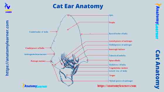
Cat ear pinna diagram
Here, I will show you the different parts of the cat ear pinna with a labeled diagram. Again, in another diagram, I will show you the different parts of a cat’s middle ear and inner ear.
So, the labeled diagram shows the different parts of the auricle, external acoustic meatus, and three different types of cartilages of the cat’s ear. Again, the diagram also shows the tympanic membrane, tympanic cavity, and auricular ossicles of the middle ear.
You may also get more labeled diagrams on the cat ear (including the external, middle, and internal parts).
Frequently asked questions on cat ear anatomy.
In this part of the article, I will provide information on the frequently asked questions on cat ear anatomy. I hope you will get your questions and answers on cat’s ear below.
What is the little pocket on a cat’s ear for?
This is the Henry’s pocket on a cat’s ear. Henry’s pocket of the cat’s ear locates on the lower portion of the external ear. This is the marginal cutaneous fold of the ear of a cat.
Do all cats have Henry’s pocket?
Probably, you will not find Henry’s pocket in all cat’s breeds. This Henry’s pocket is more common in the domestic and big cat breed. You may also find this pocket in dogs and other mammalian species.
What is inside a cat’s ear?
Inside the cat’s ear, you will find the middle and internal parts. The outer part is the only visible portion of the cat ear. In the middle and inner ear, you will find the important structures described previously in this article.
You may also read the other related articles (on cat anatomy).
Conclusion
So, the information provided in this article might help you to enrich your knowledge of the cat ear anatomy. The cat ear consists of the visible external part, middle part, and internal part. You might know the different parts of the cat ear pinna (auricle) and three basic cartilages.
All these structures from the cat’s external ear are described with the labeled diagram.
Again, from the middle and inner cat ear anatomy, you might have good knowledge of the basic structures. The tympanic membrane, cavity, and the three small bones are the main component of the cat’s middle ear structure. Again, in the cat’s inner ear, it consists of osseous and membranous labyrinths.