The cat muscle anatomy includes the origin, insertion, and fiber direction of every single muscle from the different regions of the body. Here, I will show you the essential muscles from the face, neck, forelimb, abdomen, and hindlimb. You will also find the description of these muscles from the different regions.
I am sure that this article might help you to identify the clinically significant muscle from the cat’s body. So, if you are a newbie in learning cat muscle anatomy, you may continue this article until the end.
Cat muscle anatomy
The cat muscle anatomy is somewhat different than those of a ruminant. You will find some particular muscles in a cat compared to other animals. But, the structure of a cat’s muscle (composition and parts) is almost similar to other species.
Okay, first, let’s know the most important muscles in a cat. There are many clinically essential muscles in the different regions of a cat’s body. You know, the cat’s body may divide into several areas – head, neck, thorax, abdomen, pelvic, fore, and hindlimb.
So, here I will show you these clinically significant muscles from the below-mentioned regions of a cat –
- Muscles of the cat faces or head region
- Neck muscles of a cat
- Superficial and deep muscles from the back of a cat
- Cat forelimb muscles
- The muscles of the cats’ forelimb
- Hindlimb muscles of a cat (leg)
- Muscles of the abdomen of a cat
- The pelvic and tail muscles of a cat
You will find the list of the muscles from the face, neck, forelimb, hindlimb, abdomen, and tail region of a cat in the particular part. Again, all these muscles from different regions of the body will be identified in the cat-labeled diagram.
But, for now, I will tell you some of the muscles from the cat’s body with a labeled diagram. Make sure you will identify and learn the anatomical facts of the other muscles of a cat from the specific part of this article.
Now, let’s know the list of muscles and their anatomical facts from the different regions of a cat’s body.
Cat face and head muscle anatomy
There are both superficial and deep muscles in the head of a cat. Most superficial muscles derive from the platysma and involve facial expression.
First, let’s know some of the essential muscles from the head region of a cat –
- Muscles of facial expression
- The muscles of the eye, forehead, and ear
- Muscles of the mastication
- Cat tongue muscles
- Muscles of pharynx and larynx
- Muscles of the soft palate and hyoid apparatus
Fine, you will learn and identify all the muscles from the head one by one.
Muscles of the fascial expression
The cat’s facial expression muscles divide into superficial and deep groups. The superficial group of fascial expression muscles includes –
- Sphincter colli superficialis muscle
- Platysma muscle and
- Sphincter colli profundus muscle
The sphincter Colli superficial muscle of a cat is well-developed, and fiber may reach the thorax over the shoulder joint. Again, this muscle may blend with the cervical part of the platysma.
You will find a well-developed platysma muscle sheet that originates from the mid-dorsal tendinous raphe of the neck and skin. It helps to draw the commissure of the cat’s lip caudally.
The sphincter Colli profundus muscle extends dorsoventrally from the base of the ear, lateral to the masseter muscle and parotid gland.
Again, the deep muscles of the facial expression will find in the nose, lip, eyelid, forehead, and ear. These muscles include –
- Zygomaticus
- Orbicularis oris muscle of a cat
- Superior and inferior incisive
- Levator labii superiroris
- Caninus, buccinators, and mentalis muscles
- Levator nasolabialis muscle
The orbicularis oris is the main muscle of the cat’s lip. It extends from the commissural region into the lips near their free borders. The zygomaticus is a straplike, long muscle that extends from the rostral angle of the scutiform cartilage to the edge of the superior lip and cheek.
You will find the superior and inferior incisive deep to the orbicularis muscle. The buccinator is the thick, flat, and broad muscle that forms the foundation of the cheek. Again, the mentalis muscle of a cat arises from the alveolar border of the mandible.
The levator nasolabial is a flat, thin muscle lying deep to the skin on the lateral surface of the nasal and maxillary bones. Again, the caninus lies ventral to levator nasolabial and extends rostrally.
Muscles of the eyelid, forehead, and ear
From the eyelid and forehead of a cat, I will enlist the most important muscles. Again, in the ear of a cat, you will find both the extrinsic and intrinsic muscles.
Let’s try to identify the following muscles from the eyelid and forehead of a cat –
- Orbicularis oculi muscles
- Retractor anguli oculi lateralis
- Levator anguli oculi medialis
- Levator palpebrae superioris
- Orbitalis and occipitalis muscles
The orbicularis oculi muscle surrounds the palpebral fissure of a cat. Again, the retractor anguli oculi lateralis muscle arises beside the frontalis muscle from the temporal fossa. The levator anguli oculi medialis is a small, thick muscle that arises from the median line on the frontal bone.
The extrinsic muscles (rostral) of the cat’s ear are –
- Superficial scutuloauricularis
- Deep scutuloauricularis
- Frontalis and frontoscutularis muscles
- Zygomaticoauricularis
- Again, the dorsal extrinsic muscles of a cat include –
- Interscutularis
- Parietoscutularis and
- Parietoauricularis muscle
At the caudal aspect, you will also find the following muscles –
- Cervicoscutularis
- Superificial, middle, and deep cervicoscutularis muscles
- Parotidoauricularis and styloauricularis muscles
- The intrinsic muscles of the cat’s ear include the following –
- Helicis and helicis minor muscles
- Tragicus muscle
- Transverse and oblique auriculae muscles
Again, in the middle ear of a cat, you will find two essential muscles – stapedius and tensor tympani muscles.
The zygomaticoauricularis muscle turns the auricular concha rostrally. You will learn more about all the extrinsic and intrinsic muscles of the cat’s ear in detail from another article of anatomy learner.
Muscles of mastication
Do you know what the muscles of mastication are in a cat? The muscles of mastication include masseter, temporal, lateral, and medial pterygoid, and the digastricus. All of these mastication muscles are innervated by the mandibular nerve except the caudal part of the digastricus.
- Masseter muscle of a cat
- Temporal muscle of a cat
- Lateral and medial pterygoid muscles
- Digastricus muscle of a cat
The masseter muscle of a cat lies on the lateral surface of the ramus of the mandible ventral to the zygomatic arch. You will find three layers in the masseter muscle of a cat – superficial, deep, and middle.
The superficial layer is a large part that arises from the ventral border of the rostral half of the zygomatic arch. Again, the middle layer is thinnest, and the deep layer attaches to the temporalis muscles.
The temporalis is the most significant muscle of the cat head anatomy that occupies the temporal fossa. Again, the lateral pterygoid is a small and short muscle in a cat than the medial pterygoid. It arises from the sphenoid bone in a small fossa that lies ventral to the alar canal and orbital fissure.
The medial pterygoid muscle of a cat arises from the lateral surface of the pterygoid, palatine, and sphenoid bones. These both medial and lateral pterygoid muscles help to raise the mandible.
In the digastricus muscle of a cat, you will find two parts – rostral and caudal belly. This muscle runs from the paracondylar process to the ventral border of the mandible.
Muscles of the cat’s tongue
The muscles of the cat’s tongue include styloglossus, hyoglossus, genioglossus, and intrinsic lingual muscles. All these muscles of a cat’s tongue are suppling by the hypoglossal nerve.
- Styloglossus muscle of a cats tongue
- Hyoglossus muscle of tongue
- Genioglossus muscle of the cat
- Intrinsic lingual muscles of a cat
The styloglossus muscle of a cat extends from the stylohyoid bone to the tongue. You will find three heads in the structure of this muscle, and it helps to draw the tongue caudally.
The hyoglossus muscle locates at the root of the cat’s tongue. It arises from the ventrolateral surface of the basihyoid and thyrohyoid bones.
You will find a thin triangular genioglossus muscle in the intermandibular space and ventral to the tongue. The action of this muscle is to depress the cat’s tongue.
Muscles of the pharynx, larynx, and hyoid apparatus
The muscles of the pharynx are associated with the laryngopharynx. Here, I will only enlist the muscles from the larynx, pharynx, and hyoid apparatus of a cat with a labeled diagram. But, you may learn the detailed anatomy of these laryngopharynx muscles from another article of anatomy learner.
So, what are the muscles found in the larynx of a cat? Well, you will find the below-mentioned muscles in the cat larynx –
- Hyopharyngeus muscle
- Thyropharyngeus muscle
- Cricopharyngeus muscle
- Stylopharyngeus muscle
- Palatopharyngeus and pterygopharyngeus muscle of a cat
Again, in the cats’ larynx, you will find the following muscles –
- Cricothyroideus muscle
- Cricothyroideus dorsalis and lateralis muscle
- Thyroarytenoideus muscle
- Vocalis and ventricularis muscles
- Arytenoideus transverse muscle of a cat
- Muscles of the cat hyoid apparatus include –
- Sternohyoideus muscle
- Thyrohyoideus muscle
- Mylohyoideus muscle
- Ceratohyoideus muscle
- Occipitohyoideus muscle
- Geniohyoideus and stylohyoideus muscles of a cat
The sternohyoideus is a straplike muscle that arises from the dorsal surface of the manubrium and the cranial edge of the costal cartilage. This muscle pulls the basihyoid bone and tongue caudally.
You will find sterothyroideus muscle dorsal to the sterohyoideus that consists of two parts – cranial and caudal. Again, the thyrohyoideus muscle of a cat originates from the lamina of the thyroid cartilage. You will find the mylohyoideus muscle that lies ventrally in the intermandibular spaces.
Ceratohyoideus is the small triangular palate-like muscles attached to the rostral border of the thyrohyoid bone. Again, the genioglossus muscle is a fusiform muscle in a cat. It remains in the intermandibular space to the mid-ventral of the basisphenoid bone.
Cat neck muscle anatomy
From the cat neck muscle anatomy, you might have a good piece of knowledge on these muscles that are primarily located in the neck and attaches to the head or thoracic limb. You will find some clinically essential muscles in the neck of a cat.
The neck of a cat includes the following muscles –
- Brachiocephalic muscle
- Sterocephalicus muscle
- Omontransversarius muscle
- Splenius muscle
- Longus capitis muscle
- Longus colli muscle of a cat
- Scalneus muscle
- Serratus ventralis cervicis muscle
Again, at the deep part of the cat’s neck, you will find rhomboideus capitis, splenius, longissimus capitis, semispinalis capitis and cervicis, and longus colli muscles.
The brachiocephalic is a long, flat muscle that extends between the brachium and head and neck. It lies ventral to the sphincter colli superficilis and platysma of the cat.
You will find two main parts in the brachiocephalicus muscle of a cat – cleidobrachialis and cleidocephalicus. Again, the cleidocephalicus of a cat divides into cervical and mastoid parts.
The sterocephalicus is another large and flat cat muscle that arises from the manubrium. You will also find two parts in the strnocephalicus muscle of a cat – mastoid and occipital regions. This muscle draws the neck and head of the cat.
The omotransversarius is a flat but narrow muscle that lies lateral to the cervical vertebrae. This muscle helps to draw the forelimb cranially.
The splenius capitis is a flat, fleshy, triangular muscle in the dorsolateral part of the cat’s neck. You will find a flat, long longus capitis muscle at the ventrolateral aspect of the longus colli muscle of a cat.
The longus colli is the long muscle in the cat’s neck that comprises two separate bundles. Again, the serratus ventralis cover the caudal half of the lateral neck.
Deep muscle of the neck
Rhomboideus is a very complex muscle in the cat consisting of capitis, cervicis and thoracic. The rhomboideus capitis is the cranial part that consists of narrow, flat, and thin bundles. This part of the rhomboideus muscle inserts at the dorsal border of the scapula.
The other parts of the rhomboideus muscle insert to the scapula’s vertebral border and outer surface. You will find a large flat splenius muscle on the dorsolateral aspect of the neck just beneath the rhomboideus muscle.
The longissimus capitis is a narrow, straplike muscle that is a cranial continuation of the longissimus dorsi. It lies along the ventral edge of the splenius muscle of the cats’ neck.
The insertion of the longissimus muscle is in the mastoid process of the temporal bone. Again, it helps to lateral flexion of the cat’s head.
Both semispinalis capitis and cervicis muscles are inserted at the median third of the lambdoidal crest. They both help to elevate the head of a cat.
Lumbar, thoracic, and back muscles of a cat
You will find lots of muscle in the lumbar, thoracic and back region of cat anatomy. So, it is complicated to enlist and describe all the muscles from these respective regions of the cat’s body. Instead, I prefer to enlist some of the essential muscles from these regions of a cat.
But, you may know the detailed anatomical facts of these lumbar, thoracic, and back muscles of a cat from another article of anatomy learner. Here, I will show you the muscles from the following regions of a cat –
- Superficial thoracic muscles of the cat
- Deep thoracic muscles of the cat
- Superficial back muscles of the cat and
- Lower back muscles of a cat
- Superficial thoracic muscle of a cat
You will find many superficial muscles in the thoracic region of a cat. But, I will enlist and describe some of the essential muscles from the superficial muscles. These superficial muscles of a cat remain to adhere tightly with one another.
Superficial muscles of thorax
Okay, let’s identify the below-mentioned superficial muscles from the thoracic region of a cat –
- Pectoantebrachialis muscle of the cat
- Pectoralis major (superficial and deep parts)
- Pectoralis minor muscle of a cat
- Xiphihumeralis muscle of a cat
The pectoantebrachialis is the most external muscle of a cat’s chest or pectoral region. It is a narrow and thin band of muscle in a cat.
This extends from the midline of the body to the upper part of the forelimb of a cat.
The origin of the pectoantebrachialis muscle is from the manubrium of the sternum. This muscle helps to draw the forelimb toward the midline.
You will find a superficial and deep part in the pectoralis major muscle of a cat. The outer part of the pectoralis major muscle is the flat and thin band that is partially covered by the pectoantebrachialis muscle.
Again, the deep part of the pectoralis major is a flat band three times wider than that of the superficial aspect. The pectoral muscles help draw the forelimb toward the midline and turn the manus forward.
Xiphihumeralis is a long, thin, and narrow band of the muscle of a cat that lies along the posterior border of the minor pectoral muscle. This muscle has a synergistic action with the pectoral minor in drawing the forelimb toward the midline.
Deep thoracic muscles of the cat
First, I would like to enlist the deep thoracic muscles from the cat’s body. The name, origin, insertion, and the fibers direction of these deep thoracic muscles are almost similar to the cat.
Okay, let’s try to identify and learn details about the deep thoracic muscles of a cat –
- Serratus ventralis muscle
- Scalenus (cranial, caudal, and medius part)
- Transverse costarum and thoracic muscles
- Intercostalis internus and externus muscles
- Serratus dorsalis cranial and caudal muscles
Serratus ventralis is a large, fan-shaped muscle in a cat that extends between the thorax and scapula. This muscle helps draw the scapula towards the thoracic wall and support the scapula.
The three parts of the scalenus muscle are a bandlike structure that lies along the lateral aspect of the thorax. These muscles help to bend the neck and pull the ribs cranially.
The transverse costarum is a thin, bandlike muscle that extends from the sternum and covers the cranial part of the rectus abdominus muscle. It also pulls the ribs cranially.
The intercostalis externus of a cat originates from the cranial rib and inserts to an adjacent caudal rib. Again, the intercostalis internus lies directly below the externus part and originates from the caudal rib. The externus part protracts the ribs, whereas the internus part retracts the ribs of the cat skeleton.
The transverse thoracic muscle originates from the dorsolateral border of the sternum between the third to eight ribs. It inserts at the costal cartilage near the junction of the ribs.
Serratus dorsalis cranialis is a thin layer of muscle in a cat that extends along the dorsal part of the thorax and neck. Again, the serratus dorsalis ventralis is also a thin muscle that extends from the caudal end of the serratus dorsalis cranialis muscle.
Superficial back muscle of a cat
In the superficial back region of a cat, you will find some essential muscles. Here, I will show you these essential superficial back muscles from a cat.
First, I would like to enlist the superficial back muscles from a cat –
- Clavotrapezius muscle of a cat
- Clavobrachialis muscle of a cat
- Acromiotrapezius muscle of a cat
- Spinotrapezious muscle of a cat and
- Latissimus dorsi muscle of a cat
Clavotrapezius is a flat, broad muscle of a cat that covers the lateral part of the neck. It originates from the lambdoidal ridge, middorsal raphe over the spine, and inserts clavicle. This muscle helps to the protraction of the humerus.
The clavobrachialis muscle of a cat originates from the clavicle and inserts on the medial surface of the ulna distal to semilunar notch. You will get a thin acromiotrapezius muscle in a cat that covers the scapula.
Spinotrapezious of a cat is a triangular muscle and most posterior part of the trapezious group. It originates from the spinous process of most of the thoracic vertebrae. It helps to pull the scapula dorsally and caudally.
The latissimus dorsi is a thick, flat, and large triangular muscle posterior to the trapezius group.
The spinotrapezious muscle craniodorsally covers this muscle. It pulls the forelimb of a cat dorsocaudally.
Other muscles of the thorax and lumbar region
You will also find some other muscles in a cat’s thorax and lumbar region. These muscles include – multifidus spinae, longissimus dorsi, spinalis dorsi, and iliocostalis.
The multifidus spinae is an extensive muscle that primarily originates from the transverse processes of the vertebrae. Again, the longissimus dorsi is another extensive muscle in a cat that lies in the space between the neural spine and transverse processes.
You will find the insertion of this longissimus dorsi muscle on the various processes of the anterior vertebrae. This muscle help to extend the vertebral column of a cat.
Spinalis dorsi is the medial sepearation of the longissimus dorsi in the thoracic region of a cat. It originates from the neural spines of more posterior thoracic vertebrae. Again, it inserts to the transverse processes of the more cranial vertebrae. This muscle also helps to extend the vertebral column of a cat.
Iliocostalis is a thin muscle that attaches to the thoracic region and originates from the lateral surface of the ribs. This iliocostalis help to pull the ribs together.
Cat abdomen muscle anatomy
This is very important to learn the cat abdomen muscle anatomy as they are clinically significant. You will find the obliques externus, obliques internus, transverse abdominis, and rectus abdominis muscles in the wall of a cat’s abdomen.
These muscles are layered and adhere closely to one another using abdominal fascia. You will find distinctive fibers direction in each muscle, and this feature is used to identify these muscles perfectly.
So, the muscles of the cat’s abdomen are –
- External oblique abdominal muscle
- Internal oblique abdominal muscle
- Transverse abdominal muscle and
- Rectus abdominis muscle of a cat
Let’s know the detailed anatomical facts of these abdominal muscles of a cat.
External oblique muscle
This is the most superficial oblique muscle of the abdomen of a cat. The direction of the fibers of this muscle extends craniodorsally.
It originates from the lumbodorsal fascia and the last ninth or tenth rib. Again, it inserts at the median raphe of the distal part of the sternum, linea alba, and pubis. The action of this external muscle is to compress the abdominal region.
Internal oblique abdominal muscle
The internal oblique abdominal muscle of a cat lies directly beneath the external oblique muscle. In this muscle, the direction of the fibers extends caudodorsally.
It originates from the lumbodorsal fascia with the external oblique muscle and iliac crest. Again, this muscle inserts at the linea alba by a thin aponeurosis in common with the external oblique and transverse abdominal muscles.
This internal oblique abdominal muscle of a cat also compresses the abdominal region.
Transverse abdominus muscle of a cat
The transverse abdominis muscle of a cat lies directly beneath the internal oblique muscle. Fibers of this muscle extend nearly transversely between the origin and insertion.
This muscle originates from the aponeurosis of the costal cartilage, the transverse lumbar vertebrae, and the ventral border of the ilium bone. You will find the insertion of this transverse muscle at linea alba in common with the two obliques muscles of the cat’s abdomen.
Again, this transverse abdominal muscle compresses the abdomen of a cat.
Rectus abdominal muscle of a cat
This is a ventral muscle of a cat’s abdomen that forms by the aponeurosis of the other three abdominal muscles. The fibers of this muscle are directed longitudinally on either side of the linea alba of a cat.
Again, this rectus abdominal muscle of a cat originates from the tubercle of the pubic bone. It inserts the first and second costal cartilage and the proximal end of the sternum. This rectus abdominal muscle of a cat also helps to compress the abdomen. Again, it pulls the sternum and ribs caudally and causes flexion of the trunk.
Cat forelimb muscle anatomy
I will show you all the muscles from the different regions with the labeled diagram from the cat forelimb muscle anatomy. You know the forelimb of a cat divides into shoulder region, arm region, forearm region, and manus region. Here, I will provide you with a list of muscles from the different areas of a cat’s forelimb.
Fine, let’s try to identify the below-mentioned muscles from the different regions of the forelimb of a cat –
- Shoulder muscles of a cat’s forelimb
- Brachium or arm muscles of a cat
- Antebrachium or forearm muscles of a cat and
- Muscles of the manus
Though I have another article here in anatomy learner where I have described the cat leg muscles briefly. You may also follow that guide to get a basic idea of the cat leg muscles (both front leg and hind leg).
Shoulder muscles of a cat
The shoulder muscles are attached to the scapular region of a cat. You will find the following essential shoulder muscles in a cat –
- Supraspinatus, infraspinatus, and subscapularis muscles
- The teres major and minor muscles
- A levator scapulae ventralis muscle
- The acromiodeltoid muscle of the cats shoulder
- A spinodeltoid muscle of a cat
The supraspinatus is a thick muscle in the cat that lies in the supraspinatus fossa of the scapula. This muscle originates from the entire surface of the supraspinous fossa. Again, this muscle inserts at the greater tuberosity of the humerus bone. The action of this muscle is to protract the humerus bone.
The infraspinatus is also a thick muscle that originates from the surface of the infraspinous fossa. This muscle inserts to the lateral surface of the greater tuberosity of the cat’s humerus.
The subscapularis is a triangular muscle that occupies the subscapular fossa. Again, the subscapular muscle arises from the subscapular fossa of the scapula. It inserts to the dorsal border of the lateral tuberosity of the cat’s humerus.
The teres major is a thick and triangular muscle that arises from the cranial border of the scapula. It inserts with the latissimus dorsi muscle on the medial surface of the proximal end of the humerus bone.
Again, the teres minor is a small muscle that originates from the cranial border of the scapula that inserts into the greater tuberosity of the humerus.
You will find a bandlike levator scapulae ventralis muscle that possesses two heads. The acromiodeltoid muscle is a flat muscle that arises from the acromion process of the scapula bone. Again, the spiodeltoid muscle arises from the scapular spine and inserts at the deltoid ridge of the cat’s humerus bone.
Cat arm muscle anatomy
Cat arm muscle anatomy includes coracobrachialis, biceps brachii, triceps brachii, brachialis, anconeus, and epithrochlearis muscles. First, let’s identify these muscles from the arm region of the cat’s forelimb.
- The coracobrachialis – is a very short, bandlike muscle that arises from the coracoid process of the scapula. The insertion site of the coracobrachialis muscle is at the proximal end of the humerus.
- Biceps brachii – is a thick muscle of the cat’s arm that arises from the glenoid fossa and inserts on the radial tuberosity.
- Triceps brachii – is a considerable lateral muscle of the cat’s arm consisting of three heads (lateral, medial, and long). The lateral head of the triceps arises from the deltoid ridge of the proximal end of the humerus. Again, the long head originates from the glenoid fossa of the scapula. The medial head of the triceps arises from the humerus of the cat. These three parts of the triceps muscle form a strong tendon that inserts into the olecranon process of the ulna.
- Brachialis – is a lateral muscle of the arm that covers the lateral head of the triceps brachii muscle. This brachialis originates from the lateral surface of the shaft of the humerus bone.
- Anconeus – this is a small and triangular muscle in the arm of a cat. It covers the lateral surface of the cat’s arm. You will find the origin of this anconeus muscle at the dorsal surface of the lateral epicondyle of the humerus. The insertion site of this anconeus muscle is at the lateral surface of the ulna bone.
- Epitrochlearis – is a thin and flat muscle on the medial aspect of the cat’s arm. It arises from the lateral border of the latissimus dorsi muscle and inserts into the olecranon process of the ulna bone.
Muscles of the cat’s forearm and manus
The forearm of a cat consists of some of the particular muscles and the extensor-flexor groups of muscle. First, I will provide the list of forearm muscles of a cat. Then, you will also find the cat forearm muscle labeled diagram.
Okay, the forearm of a cat consists of the following muscles –
- Brachioradialis muscle of a cat’s forearm
- The entensor carpi radialis longus and brevis msucels
- The extensor digitorum communis and lateralis muscles
- Extensor carpi ulnaris muscle of a cat
- Supinator muscle of a cat
- Abductor pollicis longus of a cat
- Extensor indicus muscle of a cat
- The pronator teres and quadratus muscles
- Flexor carpi radialis and ulnaris muscles
- The flexor digitorum superficial and profundus muscles of a cat
- Lumbrical muscles of the cat manus
Now, let’s try to identify these forearm muscles from the labeled diagram.
The brachioradialis is a narrow and bandlike muscle that arises from the mid-shaft of the humerus bone. You will find the extensor carpi radialis at the radial side of the forearm of a cat. Again, the extensor carpi brevis lies medial to the extensor carpi radialis muscle.
Both the extensor digitorum communis and lateralis msucles helps to extend the digits of the cats. You will find a flat supinator muscle at the proximal end of the radius bone.
The abductor pollicis longus arises from the ventrolateral surface of the ulnar shaft. And this abductor inserts at the radial side of the first metacarpal.
You will find five heads in the structure of a cat’s flexor digitorum profundus muscle.
On the other hand, flexor digitorum superificialis two heads – superficial and deep. The flexor carpi ulnaris arises from the epicondyle of the humerus and inserts the second carpal bone.
Cat hindlimb muscle anatomy
In the cat hindlimb muscle anatomy, I will discuss the muscles from the different regions with proper diagrams. You will find four other areas (pelvic, thigh, leg, and pes) in the hindlimb of a cat.
Okay, let’s enlist the muscles from the following regions of a cat’s hindlimb –
- Muscles of the hip or pelvic region of a cat’s hindlimb
- Thigh muscles of a cat
- Muscles of the cat’s leg and
- Pes muscles of a cat
You might know the muscles of these different regions of the cat’s hindlimb. Then, you may go with the detailed anatomical facts of these hindlimb muscles.
Cat hip muscles
In the cat hip region, you will find the following muscles –
- Gluteus muscles (three parts – maximus, medius, and minimus)
- Pyriformis muscle of the cat’s hip
- Articularis coxae muscle of cat’s hip
- Gemellus msucles (two parts – cranialis and caudalis)
- Obturator muscles (two parts – interus and externus)
- Quadratus femoris muscles of the cat’s hip
The gluteus maximus is a thin muscle that arises from the transverse process of the last sacral and first caudal vertebrae. This gluteus maximus inserts to the greater trochanter of the femur bone.
The gluteus medius is a thick muscle in the cat’s hip that arises from the crest and lateral border of the ilium bone. It (gluteus medius) inserts on the proximal end of the greater trochanter of the femur bone.
The gluteus minimus possesses a spindle-shaped cranial part and fan-shaped posterior part. This gluteus minimus originates from the lateral surface of the ilium and inserts the greater trochanter of the femur.
Pyriformis is a fan-shaped muscle in the cat’s hip that arises from the transverse processes of the last two sacral and first caudal vertebrae. Again, you will find the insertion of this muscle at the proximal end of the greater trochanter of the femur bone.
The articularis coxae is a small and flat muscle that arises from the cranial end of the ilium bone. You will find a fan-shaped gemellus cranialis muscle just below the pyriformis muscle. Again, the gemellus caudalis is a flat muscle that originates from the dorsolateral surface of the ischium bone.
The obturator internus (of a cat) arises from the medial surface of the ramus of the ischium bone. Again, it inserts into the trochanteric fossa of the femur. On the other hand, the obturator externus originates from the lateral surface of the pubis and ischium bones of the cat’s hip.
Cat thigh muscle anatomy
I will show you the cat thigh muscle anatomy with a labeled diagram. Again, you will find a short description of the thigh muscles of a cat. The thigh muscles of a cat include –
- Sartorius muscle of a cat’s thigh
- Gracilis muscle of a cat
- Biceps femoris of a cat’s thigh
- Semitendinosus and semimembranosus muscles of a cat
- The adductor longus and femoris muscles
- Pectineus muscle of a cat
- Quadriceps muscle (vastus medialis, lateralis, intermediate, and rectus femoris parts)
You may identify these cat’s thigh muscles with the help of the labeled diagram.
Sartorius is a narrow and bandlike muscle that arises from the crest and ventral border of the ilium bone. You will find a thin gracilis muscle at the caudal half of the medial surface of the cat’s thigh.
The biceps femoris is the large muscle covering most parts of the cat’s thigh. This biceps femoris of a cat originates from the ischial tuberosity and inserts proximal one-third of the tibia bone.
There is a long semitendinosus muscle at the caudal border of the cat’s thigh. Again, you will find a thick semimembranosus muscle on the medial aspect of the thigh. They arise from the ischial tuberosity and ramus of the ischium bone.
The abductor femoris arises from the ramus of the pubis and ischium bones. Again, the thin adductor longus arises from the craniomedial border of the pubic bone. Both the adductor femoris and longus muscles of the cat helps to adduct their thighs.
You will find a small and triangular pectineus muscle in the thigh of a cat. This pectineus muscle arises from the cranial border of the pubic bone and inserts the proximal shaft of the femur bone.
Quadriceps muscle of a cat
The quadriceps are complex muscles in the cat thigh anatomy that consist of four components. It lies on the craniolateral and craniomedial aspect of the thigh of a cat.
The four components of the quadriceps muscle of a cat are –
- Vastus medius muscle
- The vastus lateralis muscle
- A vestus intermedius muscle and
- The rectus femoris muscle
The vastus medius is the most medial muscle of these four parts of the quadriceps. This medius part of the quadriceps arises from the shaft of the femur bone and inserts on the tibial tuberosity.
The vastus lateralis is a large and flat muscle that covers the cranial and lateral surface of the cat’s thigh. This vastus lateralis originates from the shaft and greater trochanter of the femur bone. Again, this muscle inserts in common with the vastus medialis and rectus femoris muscles.
The vastus intermedius arises from the entire shaft of the femur bone and inserts with the other three parts of the quadriceps muscle. In contrast, the rectus femoris is a spindle-shaped muscle in the cat’s thigh that arises from the ilium bone.
This rectus femoris muscle of the cat inserts in common with the vastus medius and lateralis muscles. All these four parts of the quadriceps muscles of the cat help to extend their thighs.
Cat leg muscle
In the cat leg muscle anatomy, you will find some particular muscles and the extensor and flexor groups of muscle. Here, I will show the cat leg muscles from both extensor and flexor groups with the labeled diagrams.
So, in the cat leg, you will find the following essential muscles –
- Tibialis cranialis and caudalis muscles of a cat
- The extensor digitorum longus of a cat leg
- An extensor hallucis longus of a cat’s leg
- The peroneus longus and tertius muscles
- Flexor digitorum longus muscle of a cat’s leg
- The flexor hallucis longus muscle of a cat
- Flexor digitorum brevis muscle of a cat
- Popliteus muscle of the cat’s leg
- Gastrocnemius muscle of the leg
- Soleus muscle of the leg
- Planters and lumbrical muscle of the pes
Fine, now, you may identify these cat leg muscles with the help of the labeled diagrams.
You will see a flat, sizeable tibialis cranialis muscle on the craniolateral aspect of the tibia bone.
You will find three parts of the peroneus muscle in the cat’s leg. Peroneus longus is the most superficial muscle of the peroneal group.
This peroneus longus arises from the head and lateral surface of the shaft of the fibula bone.
Peroneus Tertius is a slender muscle in the cat’s leg beneath the peroneus longus muscle. This part of the peroneus muscle arises from the lateral surface of the fibula bone.
You will find a short peroneus brevis muscle at the posterior aspect of the other two parts. There is a triangular popliteus muscle that surrounds the posterior aspect of the knee joint of a cat.
The soleus muscle originates from the proximal part of the fibula bone and inserts with the tendon of the gastrocnemius muscle.
Gastrocnemius muscle of a cat
The gastrocnemius is a more heavy muscle in the posterior aspect of the lower part of a cat’s leg. You will find lateral and medial heads in the structure of the gastrocnemius muscle of a cat.
The lateral head of the cat’s gastrocnemius muscle arises from the patella’s lateral border and the leg’s superficial fascia. Again, the medial head of the cat’s gastrocnemius muscle originates from the sesamoid bone above the medial epicondyle of the femur and its distal adjacent shaft.
All the heads, along with some other thigh muscles, form a common tendon (known as Achilles tendon) that inserts on the proximal end of the calcaneus bone.
The gastrocnemius muscle of a cat leg helps to extend the pes.
Other extensor muscles of cat’s leg
The extensor digitroum longus is a large muscle of the cat’s leg beneath the tibialis cranialis muscle. This extensor muscle originates from the lateral epicondyle of the femur bone and helps extend the digits and flexes the pes of the cat.
You will see the extensor hallucis longus deep to the tibialis cranialis muscle in the cat leg. It (extensor hallucis longus) arises from the anterior surface of the fibula bone and is inserted on the first metatarsal bone. This extensor muscle joins with the common tendon with the tibialis cranialis muscle.
The extensor digitorum brevis of a cat is a thin muscle of their leg. It covers the dorsolateral surface of the tarsus and metatarsus of the cat. Again, this extensor muscle of the cat leg arises from the proximal end of metatarsus bones III to V.
The three tendons from the III to V metatarsus split into medial and lateral and terminated on the dorsal and lateral surface of the first phalanges.
This extensor digitorum brevis muscle of a cat leg helps extend the toes.
Other flexor muscles
In the cat leg muscle anatomy, you will find some essential flexor muscles like – the flexor digitorum longus, flexor digitorum brevis, and flexor hallucis longus. I will show you these flexor muscles from the cat’s leg with a labeled diagram.
The flexor digitorum longus is a long muscle that lies in the medial aspect of the cat’s leg. This muscle arises from the head of the fibula and shaft of the tibia bone. Again, the muscle forms the common tendon that extends over the plantar surface of the foot and divides into four small tendons.
These four small tendons of the flexor digitorum longus muscle insert on the base of the terminal phalanges of each toe.
The flexor hallucus longus is a large muscle of the posterior aspect of the cat’s leg. It arises from the fibula’s shaft and the tibia bone shaft.
This flexor muscle will form a common tendon with the flexor digitorum longus muscle.
This flexor muscle of the cat’s leg helps to flex the toes and pes.
The flexor digitorum brevis is a small muscle that lies on the plantar surface of the cat’s foot. This muscle originates from the extension of the planteris tendon. Again, this flexor muscle of the cat leg inserts to the second phalanx of the digits II to V.
This flexor digitorum brevis muscle of the cat leg also helps to flex the toe.
Tail muscle of a cat
The muscles of cat’s tail are almost similar to the dog. Here, I will provide the list of cattail muscles with a labeled diagram.
Let’s see the tail muscles of a cat –
- Sacrocaudalis dorsalis lateralis muscle of a cat
- Sacrocaudalis dorsalis medialis muscle of a cat
- The muscle sacrocaudalis ventralis lateralis of a cat’s tail
- A muscle sacrocaudalis ventralis medialis of a cat
- Intertransversarius dorsale caudae muscle of a cat
- Intertransversarius ventral caudal muscle of a cat’s tail
- Levator ani and sphincter ani externus muscle of a cat
You may find the anatomical description of these tail muscles of the cat in another article of anatomy learner.
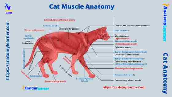
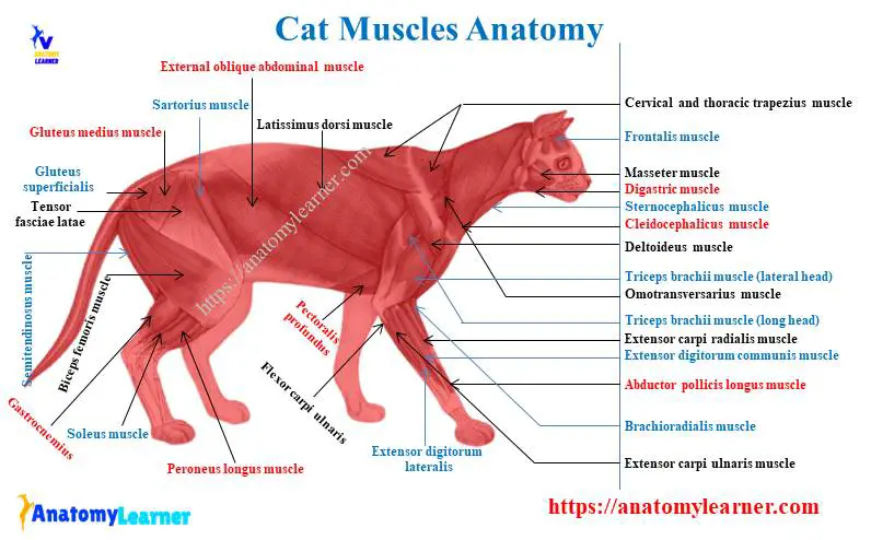
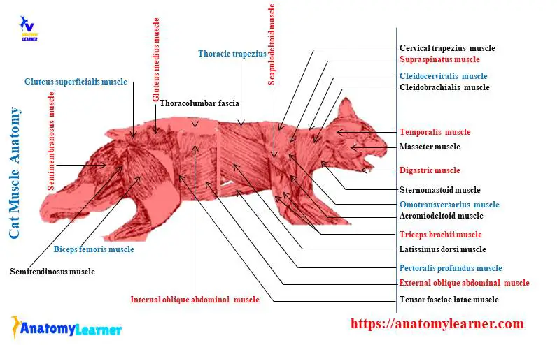
Cat muscle anatomy diagram
Again, I will show you the cat’s tail muscle with a labeled diagram. In the diagram, I tried to show you all the muscles from the different regions of the cats’ bodies.
Here, some of the essential muscles are identified from the different body parts. You will only get the basic idea of important muscles from a cat.
You may get more cat muscle anatomy labeled diagrams on social media of anatomy learners.
Frequently asked questions on cat muscle.
Most of the information on cat muscles is already described in short. But, again, I will provide some frequently asked questions on cat muscles with their answers.
What are the essential muscles in a cats neck?
Do you found any differences in the cat muscles from the ruminant?
Which one is the vital muscle in the back of a cat?
If you wish to learn cat anatomy or dog anatomy, you may read other articles from anatomy learners.
Conclusion
I think you learn the basic idea on the cat leg muscle anatomy from this article. For the field practice, you might have a good piece of knowledge on the back, forelimb, hindlimb, and abdomen muscle anatomy from a cat. That’s not mean you will not learn the other muscle from a cat; of course, you should have basic knowledge of cat muscle if you are a veterinary student.
All the labeled diagrams related to the cat muscle anatomy might be helpful for you. You may use these diagrams to identify all the muscles from the cat practically at your laboratory.