The colon histology slide possesses the typical four layers of a tubular organ – mucosa, submucosa, muscularis, and serosa. But, there are no permanent plica circularis and villi in the colon slide as found in the different segments of the small intestine. You will also see a thicker tunica mucosa in the colon slide because of the increased length of the intestinal glands.
There is a distended tunica submucosa in the colon histology slide that possesses lymphatic tissue. Again, you will see a disrupted lamina muscularis layer in the colon slide. Here, I will show you the different unique histological features from the four layers of a colon slide with the labeled diagram.
I will also provide the useful identifying features of the colon slide under the light microscope. These might help you differentiate it from different segments of the small and large intestine.
Again, I will help you draw the colon slide image at the end of this article. So, if you intend to know the details of histological features of an animal colon, let’s continue this article till the end.
Colon histology slide
First, I would like to summarize the histological features from the different layers of a colon slide. You will see the following four layers in the wall of the animal colon as the typical gastrointestinal tract have –
- Tunica mucosa of the animal colon,
- The tunica submucosa of the colon,
- Tunica muscularis layer of the colon, and
- The tunica serosa of the animal colon
The tunica mucosa of the colon histology slide possesses the simple columnar epithelium. But, you will not find any permanent plica circularis and villi on the mucosal surface of the colon. As there are no villi, the mucosal surface becomes smooth.
Again, the tunica mucosa of the colon slide is thicker than the small intestine. This is because of the increased length of the intestinal glands. The tunica mucosa layer of the colon also shows the numerous goblet cells.
You will also find the lamina propria and lamina muscularis layers within this tunica mucosa layer of the colon slide. But, the lamina muscularis of the colon may be disrupted.
Due to the disrupted lamina muscularis, the tunica mucosa’s longer intestinal glands may extend to the tunica submucosa layer. Again, the colon’s tunica submucosa is also distended compared to the different parts of the small intestine.
You will find the lymphatic tissue in the tunica submucosa of the colon microscopic slide. Again, the tunica submucosa of the colon slide also contains the blood vessels.
The tunica muscularis layer of the colon microscopic slide shows inner circular and outer longitudinal muscles. But, in some animals like the pig and horses, you will find the taenia coli. If you want to know what taenia coli is, read the details guide from the description section.
Identifying features of a colon histology slide
The main goal of this article is to identify the colon histology slide under a light microscope with its useful identifying features. First, it will be better to find the following histological features from the colon slide under the light microscope.
From the tunica mucosa of the colon slide, let’s try to find the following features –
- Temporary mucosal folds of the colon,
- The lining epithelium (simple columnar) of the tunica mucosa,
- Lamina propria and intestinal glands of the colon,
- Muscularis mucosa of the colon slide (under the tunica muscosa layer),
From the tunica submucosa of the colon slide, let’s try to find the following histological features –
- Lymphatic nodules in the tunica submucosa layer of the colon slide,
- Dense connective tissue fibers, cells, and ground substances in the tunica submucosa,
Again, in the tunica muscularis layer of a colon microscope slide, try to find out the following features –
- The inner circular muscle layer of the colon,
- The outer longitudinal smooth muscle layer of the colon slide,
- Myenteric nerve plexus of the colon slide (within the muscle layers),
- The taeniae coli (smooth muscle band) in the muscularis layer of the colon (in some species only like pigs and horses),
In addition, the tunica serosa of the colon microscope slide consists of loose connective tissue, numerous blood vessels, and adipose tissue. You will see a simple squamous epithelium lining in the tunica serosa layer of the colon slide.
Identification points of a colon microscope slide
Now, it will be easy for you to write the identification points of the colon microscope slide. The unique identification points of the colon slide under a light microscope are –
- The wall (total) of the sample tissue section shows four distinct layers – tunica mucosa, tunica submucosa, tunica muscularis, and tunica serosa layers,
- There is a simple columnar epithelium lining on the tunica mucosa layer. But, the sample tissue section did not show any plica circularis and villi on the tunica mucosa layer,
- The presence of numerous longer intestinal glands in the tunica mucosa layer of the provided sample tissue,
- Again, the submucosa layer of the provided tissue sample shows dense connective tissue, blood vessels, and lymphatic nodules,
- The tunica muscularis layer (muscular externa) shows an inner circular and an outer longitudinal layer of smooth muscle,
- In addition, the external layer (tunica serosa) shows the loose connective tissue layer that surrounds by a simple squamous epithelium lining,
So, this is a colon histology slide under the light microscope. If you find the taeniae coli (three longitudinal bands of smooth muscle) in the muscularis externa layer, then this is the colon microscope slide of a pig or horse.
I think this information is enough for identifying the different microscopic features of an animal colon. Now, you may know more about the different layers of the colon microscopic slide with labeled diagrams.
Colon histology layers
There are four different tunica layers in the wall of a colon microscope slide – mucosa, submucosa, muscular, and serosa. The structure of the four different layers of the colon microscope slide is almost similar to the structure of a tubular organ.
So, it will be better if you read the following article (general structural pattern of a tubular organ or gastrointestinal tract –
- The general structural pattern of a tubular organ with the labeled diagram
I suggest that the article as different labeled diagrams show various features of a typical tubular organ. Okay, now let’s know the unique microscopic features of the animal colon slide.
So, what I will cover in this part of this article –
- Microscopic features of the tunica mucosa layer of the colon slide,
- Features of the tunica submucosa of the colon microscope slide,
- Microscopic features of the tunica muscular layer of the colon slide, and
- Microscopie features of the tunica serosa layer of the animal colon
That’s fine; let’s start to know the details of the colon microscope slide with the labeled diagrams.
Features of the tunica mucosa of colon slide
The tunica mucosa layer of the colon slide is next to the lumen. You may also be called this layer the mucous membrane or mucosa of the colon.
In the tunica mucosa of the colon microscope slide, you will find the following features –
- An epithelium lining (simple columnar epithelium),
- A lamina propria layer, and
- A lamina muscularis layer,
The tunica mucosa of the colon shows numerous crescent-shaped folds. But, there are no plica circularis or any villi present on the mucosal surface of the animal colon.
If you don’t know what the plica circularis or villi are, then you may read the below-mentioned article –
Histological features of the different segments of the small intestine like – the duodenum, jejunum, and ileum with the labeled diagrams.
Now, let’s describe the features of the lining epithelium, lamina propria, and lamina muscularis layer of the colon slide.
Epithelium lining of the colon
You know that the mucosal surface of the colon microscope slide is lined predominantly by the simple columnar epithelium. I hope you have a great idea about the simple columnar cells. These are the long tall cells that have an elongated nucleus.
Under the light microscope, you will only see the elongated nuclei placed vertically towards the colon’s lumen. Do you know the main functions of these epithelium cells of the colon?
The simple columnar epithelium of the colon has the main function of absorbing excess water and electrolytes from the contents within the colon. Again, the columnar cells of the colon mucosa secrete mucous and antibodies (immunoglobulin A).
You know the antibody protects the pathogenic organism.
Under the light microscope, you may also find goblet cells on the mucosal surface of the colon. The number of goblet cells on the colon mucosal surface increases at its caudal portion.
The goblet cells of the colon secrete mucous that serves as a lubricant that facilitates the passage of the semisolid content through the lumen of the colon.
You will not see any paneth cells on the mucosal surface of the colon microscope slide. But, sometimes, you may see some endocrine cells and some stem cells in the mucosa of the colon.
Lamina propria of the colon slide
The lamina propria is a layer of dense connective tissue immediately beneath the lining epithelium. In the lamina propria of the colon histology slide, you will find fine collagen, elastic and reticular fibers, and all of the cells of the typical connective tissue.
You will also find two important structures in the lamina propria of the colon microscope slide – intestinal glands and lymphatic nodules. The intestinal glands (mucosal glands) are confined to the mucosal surface, an elongated structure in the colon slide.
Do you know the epithelium lining of these intestinal glands of the colon? The intestinal glands of the colon are lined with the simple columnar epithelium.
Because of the increased length of the intestinal or mucosal gland, you will see a thicker lamina propria (mucosa) in the colon compared to the different parts of the small intestine.
Sometimes, the colon slide’s intestinal glands may extend to the tunica submucosa layer. This figure will see in that area of the submucosa where the lamina muscularis is disrupted.
But why is the lamina muscularis layer disrupted in the colon? Let’s jump to the next section to know why the lamina muscularis layer of the colon slide is disrupted.
The lymphatic nodules in the lamina propria are not common in the colon slide. In some species, you may find diffuse lymphatic tissue in the lamina propria of their colon. These diffuse lymphatic nodes of the colon contain the immunocompetent T and B lymphocytes that initiate the immune response to the injury agents.
Again, the lamina propria of the colon slide also shows blood vessels and nerve fiber.
Lamina muscularis layer of the colon slide
The lamina muscularis layer in the colon microscope slide is inconstantly present. You will see a thin layer of smooth muscle just beneath the lamina propria of the colon slide.
But, due to the lymphatic nodes in the lamina propria of the colon microscope slide, it becomes distended. Thus this enlargement of the lamina propria may disrupt the lamina muscularis layer of the colon.
In other different tubular organs or different parts of the intestine, you may see the two or three layers of smooth muscle in their lamina muscualris layer. The lamina muscualris layer of the colon slide may play important roles in the following ways –
- They allow independent movement of the tunica mucosa,
- Facilitates the easy movement of the content within the colon, and
- Assists in the expression of secretions from the mucosal glands of the colon
I hope you can understand all the histological features from the lamina muscularis of the colon microscope slide.
Tunica submucosa of colon microscope slide
The structure of the tunica submucosa of the colon microscope slide is similar to the structure of a typical tubular organ. It is a layer of dense connective tissue where you will see different cells, fibers, submucosal glands, and lymphatic nodules.
But, in the tunica submucosa of the colon, you will find dense connective tissue with no submucosal glands. This layer is just beneath the lamina propria of the colon structure. So, how will you differentiate the lamina propria from the tunica submucosa layer?
Fine, the connective tissue of the tunica submucosa layer of a colon is denser than the lamina propria layer. You will also find different blood vessels, lymph vessels, and nerve fibers in the tunica submucosa of the colon microscope slide.
The tunica submucosa of the colon slide also contains the lymphatic nodules. Again, you may also find fat cells in the tunica submucosa of the colon.
Some cells in the tunica submucosa of the colon are PAS-positive. These cells are the muciphage in the tunica submucosa of the colon slide. But, these cells do not constantly occur in the tunica submucosa of the colon slide.
Tunica muscular of a colon histology slide
The tunica muscular layer of a colon histology slide may possess a unique feature (taenia coli) in some specific species like pigs and horses. But, normally, you will find the inner circular and outer longitudinal smooth muscle layers in the tunica muscular of a colon microscope slide.
If you compare the tunica muscular layer of the small and large intestine, you will find a great variation under the light microscope. The inner layer’s smooth muscle fibers (cells) are oriented circularly or in a tightly coiled pattern.
On the other hand, the smooth muscle fibers of the outer layer of the tunica muscularis are arranged longitudinally or in a loosely coiled pattern. The parasympathetic ganglionic cells of the myenteric nerve plexus are found between the two layers of the smooth muscle fibers.
What is taeniae coli of a colon?
As I told you before, the colon’s tunica muscularis is somewhat different from these the different parts of the small intestine. The inner circular layer of the smooth muscle remains continuous in the colon, but you may see the difference in the outer layer.
The outer smooth muscle layer of the muscular externus is condensed into three broad, longitudinal bands. These three broad and longitudinal bands of the smooth muscle are the taeniae coli of the colon.
You will not find the taeniae coli in all the animals’ tunica muscular layer. The taeniae coli may be evident in the tunica muscular layer of pigs’ and horses’ colons.
The taeniae coli of the colon is shorter in length than another layer of the wall and contains numerous elastic fibers. This structure helps to produce the sacculation on the wall of a colon.
Tunica serosa of colon slide
The tunica serosa of the colon microscope slide is the outermost layer that lines with the mesothelium (simple squamous lining epithelium). You will also find numerous blood vessels and adipose tissue in the tunica serosa of the colon microscope slide.
But, in some cases, the tunica serosa may be absent in the caudal aspect of the ascending or descending part of the colon. In this case, the peritoneum forms a small pouch-like structure that fills with yellow fat.
Colon histology slide labeled diagram
Now, I will show you the colon histology slide with a labeled diagram so that you may easily understand. There are two more labeled diagrams where I identify all of the structures from the different layers of a colon. Let’s see and learn more from the colon-labeled diagrams.
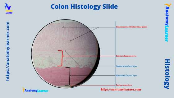
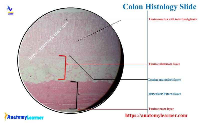
Let’s see the first diagram of the colon microscope slide. Here, I identify the four different layers of the colon (tunica mucosa, tunica submucosa, tunica muscular, and tunica serosa) from the diagram.
The lumen of the colon is identified here in the diagram. Different temporal folds (plica circularis in the small intestine) are shown in the colon microscope slide labeled diagram.
In these temporary folds of the colon mucosa, you will see the lining epithelium (simple columnar) and mucosal gland in the lamina propria. I will show the different structures from the tunica mucosa in the next labeled diagram.
Let’s see the thicker tunica submucosa of the colon from the labeled diagram. Some lymphatic nodules and blood vessels are shown on the colon labeled diagram.
Again, the muscularis externa of the colon are identified in the labeled diagram. There is an extra flat band of smooth muscle in the tunica muscualris layer of the colon microscope labeled diagram. This is the taeniae coli of the colon.
In addition, the colon labeled diagram also shows the bundle of nerve fibers (not seen under the binocular microscope). Finally, the labeled diagram shows a thin layer of tunica serosa that lines with a single layer of squamous cells.
The mucosa of a colon labeled diagram
Let’s see the second labeled diagram of the animal colon. Here, I will show you all the structures from the tunica mucosa layer of a colon microscope slide.
The diagram shows some temporary folds of the colon. Let’s see the absorptive cells from the surface epithelium of the colon mucosa. The diagram also shows the crypts (lumen of the intestinal glands) of the colon.
The lamina propria is also identified from the different temporary folds of the colon. Here, the lamina propria of the colon shows numerous blood vessels (capillaries) and numerous intestinal glands.
The colon labeled diagram also shows the other cells like – endocrine cells (basal granular cells), goblet cells, macrophages, lymphocytes, and plasma cells. Again, the colon labeled diagram also shows some smooth muscle cells in the lamina propria.
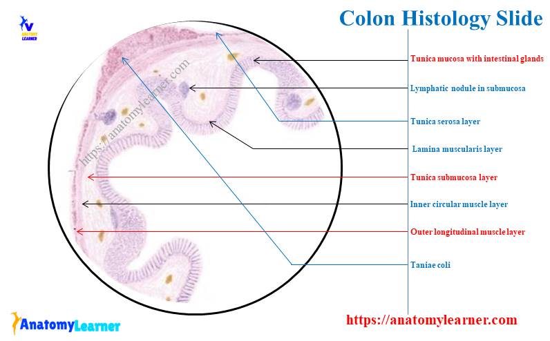
Again, let’s see the third colon microscope slide labeled diagram. It shows the lymphatic nodules in the lamina propria of the colon that disrupts the lamina muscularis.
It also shows the inner circular and outer longitudinal smooth muscle layers of the colon microscope slide. In addition, you will find a great difference in the connective tissue between the lamina propria and the tunica submucosa of the colon from the diagram.
For more colon microscope slide labeled diagrams, you may join with social media of anatomy learners.
How do you differentiate colon slides from others?
You may be asked to differentiate the histology slides of the different parts of small and large intestines. Now, I will show how you will differentiate the colon histology slide from the other slides like the duodenum, jejunum, ileum, cecum, and end part of the large intestine.
First, let’s know what the major differences between the large and small intestine (in the point of microscopic view) are –
Different segments of the small intestine present –
- Permanent circular folds of the tunica mucosa and tunica submucosa – known as the plica circularis,
- There are minutes finger-like projections that contain a central core of the lamina propria,
- The surface of the columnar epithelium cells presents very small finger-like projection (villi) of the plasma membrane,
- Again, the tunica submucosa of the large intestine shows Brunner’s gland and Peyer’s patches,
In addition, the different segments of the large intestine show the following peculiar features –
- Present small angular lumen compare to the thicker wall,
- There are few short crypts but no plica circularis and villi,
- Present more goblet cells and longer, straight, and more compact intestinal glands,
- May present the disrupted lamina muscularis in some segments, and
- The outer longitudinal muscle layer of the tunica muscualris may show three flat bands of taenia coli
But, both the small and large intestine shows the typical features of a tubular organ.
Duodenum and colon slides
So, let’s see the duodenum and colon slides under the light microscope. You will easily identify and differentiate the duodenum slide from the colon slide.
In the duodenum histology slide, you will find the short leaf-like villi lined with the simple columnar epithelium cells. You will see the different crypts of Liberkuhn in the duodenum microscope slide.
But, in the colon histology slide, you will not find any villi. You will only see some of the temporary mucosal folds with lines with the simple columnar epithelium.
Again, the crypts of the colon are shorter and not so distinct under the light microscope. The duodenum slide has continuous muscualris mucosae (consists of two or three layers), but the colon may show the disrupted muscularis mucosae.
In addition, the tunica submucosa of the duodenum histology slide shows Brunner’s glands. But, you will not see any glands in the tunica submucosa of the colon microscope slide.
The jejunum and colon microscope slides
You already have a piece of great knowledge on the microscopic feature of a colon slide. So, now see the specific features found in the jejunum histology slide that helps you differentiate it from the colon.
In the jejunum histology slide under the light microscope, you will get the longer club-shaped intestinal villi that line with the simple columnar epithelium and goblet cells. The crypts of Liberkuhn are also distinct in the microscope slide of jejunum.
The muscularis mucosae are also distinct in the jejunum histology slide. But, the tunica submucosa of the jejunum lacks Brunner’s glands.
Again, there are no Peyer’s patches in the tunica submucosa of the jejunum histology slide.
Ileum and colon microscope slides
Let’s see the special microscopic features of the ileum slide under the light microscope. There are short finger-like intestinal villi on the mucosal surface of the ileum histology slide.
The lining epithelium of the ileum mucosa is similar to the duodenum or jejunum (simple columnar epithelium). There is a thick lamina propria in the ileum histology slide that possesses some crypt of Liberkuhn.
The muscularis mucosae are also distinct in the ileum histology slide compared to the colon slide. You will see a special and unique feature in the tunica submucosa of the ileum histology slide, Peyer’s patches.
I hope you can understand the basic difference between the duodenum, jejunum, and colon microscope slides. Now, let’s see how you differentiate the colon slide from the cecum and the end part of the large intestine.
How to differentiate colon slide from cecum?
In the cecum histology slide, you will find four different layers in the typical tubular organs. The tunica mucosa of the cecum shows simple columnar epithelium lining like the colon.
Again, you will not see any villi and plica circularis as you find in the colon histology slide. So, you may be confused about differentiating the colon microscope slide from the cecum slide.
Don’t worry; you will easily differentiate the colon microscope slide from the cecum slide. The tunica mucosa of the colon microscope slide presents a thicker layer than the cecum due to longer intestinal glands.
Again, the other histological features of different layers of a cecum are almost similar. But, in the colon of a pig and horse, you will see a taenia coli, whereas you will also see taeniae Ceci in their cecum.
You will get the details of histological features of the cecum histology slide in another article by anatomy learner –
- Cecum microscope slide with the labeled diagram
I hope this article will help you understand every single feature of the cecum perfectly.
Colon microscope slide and end part of the large intestine
How you may differentiate the colon microscope slide from the end part of the large intestine. The end part of the large intestine possesses some unique histological features. You will see both permanent and temporary mucosal folds in the end part of the large intestine.
The permanent mucosal folds are present in the lower part of the end portion of the large intestine. Again, the temporary mucosal folds are present in the upper part of the end portion of the large intestine.
These folds of the end part of the large intestine consist of a tunica submucosa core surrounded by the tunica mucosa. You will see the developed muscualris mucosae and lamina propria in the end part of the large intestine.
The lamina propria of the end part of the large intestine consists of numerous large intestinal glands, adipose tissues, lymphatic nodules, and numerous goblet cells.
Again, the tunica submucosa of the end part of the large intestine possesses numerous blood vessels and adipose tissue. You will find more elastic fibers in the horse and cow’s tunica submucosa and adventitia layers.
Colon histology slide drawing
The drawing of a colon histology slide image is not such a hard task for the learners. I will show you the simple process of drawing a colon microscope slide image. Again, I will provide the colon microscope slide drawing image for you.
Let’s try to draw the colon microscope image by following the below-mentioned simple procedure –
So, you should draw the different histological features of a colon from its four different layers. First, let’s try with the tunica mucosa of the colon microscope slide.
To draw the tunica mucosa of the colon, you may put some wave (temporary folds) that surrounds the lumen. Now, you should draw the simple columnar epithelium on the tunica musical surface of the temporary folds (shown in the diagram).
You may draw some other cells on the tunica mucosal surface of the colon, like goblet cells and endocrine cells.
Now, you should provide the loose connective tissue fibers and cells in the lamina propria of the colon microscope image. Here in the colon microscope image, I draw some blood vessels (capillaries).
You know there are numerous longer intestinal glands present in the lamina propria of the colon. So, let’s draw the lumen and lining epithelium of the intestinal glands of the colon image.
You may also place some macrophages, lymphocytes, and plasma cells in the lamina propria of the colon microscope image. Finally, you should draw the muscularis mucosa in the colon slide image.
Here, I have not drawn any lymphatic nodules in the lamina propria of the colon microscope slide image. But, you may draw the diffuse lymphatic tissue in the lamina propria of the colon slide.
Drawing other layers of colon slide image
Now, let’s draw the thicker tunica submucosa of the colon microscope image. It would be best to place the dense connective tissue with numerous blood vessels here. Again, you know this layer of the colon also contains the lymphatic nodules.
So, now, I will draw some lymphatic nodules in the tunica submucosa of the colon microscope slide image.
It’s time to draw the thin muscularis externa of the colon slide image. First, draw the inner circular layer of the smooth muscle. Then try to draw the outer longitudinal smooth muscle layer of the colon.
Here in the image, I tried to draw the taenia coli of the horse colon. If you draw the cow or goat colon, there is no need to draw the taeniae coli.
Again, if you want, you may draw the myenteric nerve plexus between the smooth muscle’s inner and outer layers.
Finally, let’s draw a thin layer of loose connective tissue surrounding the tunica muscular layer. You should provide a single layer of flattened epithelium (mesothelium or simple squamous epithelium) at the outer surface of the thin connective tissue.
Frequently asked questions on a colon histology slide
I will try to make answers to all the questions on the colon histology slide asked by the learners. If you read the full article on the colon, you may skip this part. Let’s see the common questions on the colon microscope slide that the learners ask.
What is the histology of the colon?
The wall of the colon microscope slide contains four tunics – mucosa, submucosa, muscular, and serosa. In the mucosa of the colon, you will see simple columnar epithelium lining, lamina propria, and a well-defined lamina muscularis layer.
The mucosa of the colon does not have any plica circularis and villi. Rather they possess temporary mucosal folds whose surface is smooth.
The morse characteristics features of the colon mucosa is the presence of larger, compact intestinal glands that lines with simple columnar and goblet cells. This layer of the colon also contains a few lymphatic tissues.
The submucosa of the colon consists of dense connective tissue with numerous blood vessels and lymphatic nodules. Again, the muscularis externa consists of an inner circular and outer longitudinal smooth muscle layer.
The outer longitudinal muscle shows three broad, longitudinal bands known as the taeniae coli. This is another characteristic feature of the colon microscope slide in the case of pig and horse.
What type of tissue is the colon?
So, if you see the histology slide of a colon, you will see different types of tissue like connective tissue, epithelial tissue, adipose tissue, and muscular tissue. You will also find the glandular tissue in the structure of the colon.
The tunica mucosa is made of loose connective tissue fibers and cells. Again, the tunica submucosa of the colon slide consists of dense connective tissue. Finally, the tunica serosa of the colon slide possesses a thin layer of the loose connective tissue layer.
The colon microscope slide also shows the diffuse lymphatic tissue and nodules in the lamina propria and tunica submucosa layer.
What are the layers of the colon?
Like the structure of a typical gastrointestinal tract, the colon also possesses four different layers. They are – tunica mucosa or mucous membrane, tunica submucosa, tunica muscularis or muscularis externa, and tunica serosa.
I have already described different histological features from these four layers of the colon microscope slide with the labeled diagrams.
What are the major histological features of the large intestine?
Here, I will only provide the major histological features of the large intestine of an animal. The three different segments of the large intestine (like the cecum, colon, and end part) show longitudinal folds, but they do not possess any plica circulairs.
There are no villi on the mucosal surface of the cecum, colon, and end part of the large intestine; they present a smooth surface. All the parts of the large intestine contain longer, straight, and compact intestinal glands.
The goblet cells and lymphatic nodules increase from the first part to the end part of the animal’s large intestine.
Conclusion
So, you got the basic idea of the different layers of the colon histology slide with the labeled diagram. The colon microscope slide’s tunica mucosa and tunica submucosa are thicker compared to the other parts of the large intestine. Again, most of the unique features are present in the tunica mucosa layer of the colon.
In addition, the tunica muscular layer contains the inner circular, outer longitudinal smooth muscle and the taeniae coli in some species. You should practice the colon microscope slide under the light microscope at your histology learning laboratory.