The cow udder anatomy consists of the glandular mass (body) and the papilla (teat). It is a modified cutaneous gland, also called the cow’s mammary gland.
You will find 4 mammary glands (udder) in a cow that develop in puberty and become more marked in the later stage of pregnancy (especially after parturition). In the structure of the cow udder, you will find the base, milk cistern, and apex in the glandular mass or body.
Again, the teat of a cow udder possesses a single lactiferous duct with some surrounding structure. In this short guide, I will show you the anatomical facts from the teat and body of the cow udder with the labeled diagram.
In addition, I will show you the significant difference in the udder anatomy of a mare, pig, bitch, and rabbit compared to a cow. So, if you want to get the details structure of the cow udders or mammary glands anatomy, let’s continue this article till the end.
Cow udder anatomy
The 4 mammary glands of the cow are popularly known as the udder. So, you may have a question – how many udders does a cow have?
- Number of cow udder – 1,
- Compartment of cow udder – 4,
- Mammary glands of a cow – 2 pairs (4),
So, a cow udder is a single structure that hangs beneath the inguinal region of the cow. The term udder is used in ruminants and horses. Sometimes, it is used for the sow’s mammary glands.
While reading the cow udder anatomy, you will find another term – quarters. These are the 4 parts of the cow udder, each associated with one teat. There is an intermammary groove which indicates the separation of the 2 halves of the cow udder.
The cow udder of a cow is very much larger than in the mare. Again, the body of each mammary gland (upper glandular mass) is somewhat ellipsoidal and flattened transversely.
The upper part of each mammary gland of the cow (called the base) is slightly concave and slopes obliquely ventrally. This part of the glands is attached to the ventral abdominal wall with the well-developed suspensory apparatus.
In addition, the lower narrow part of the cow’s mammary gland (called apex) possesses the teat with a lactiferous sinus. So, from the cow udder structure, you should complete the below-mentioned anatomical facts –
- Base and apex of the lobes or mammary glands,
- Internal features of the body of the mammary glands or udder,
- Structure of the teat or papillae with the lactiferous ducts and sinus,
- Features of the teat canal and papillary duct,
- Teat opening and the sphincter muscle of the teat,
- Component of the suspensory apparatus of the cow udder, and
- Blood and nerve supply of the cow udder,
Where is the cow udder – cow udder location
The cow udder is located in the ventral part of the inguinal region along the midline of the body. As there are 4 mammary glands in the structure of a cow udder, each 2 of them remains on either side of the median line between the thighs.
In the male and immature female, the udder is rudimentary. You will also find the udder in the small ruminant like sheep and goats, but only 2 mammary glands are in their udder.
You know, the udder or mammary glands of the cow develop in puberty and become more marked in the later stage of the pregnancy. This enlargement of the cow udder is due to the action of estrogen and progesterone.
This action causes fat deposition, stromal development, and growth of the lobules, ducts, and alveoli of the cow’s udder.
If you want to know the location of the mammary glands of other animal species like horses, dogs, cats, pigs, elephants, and rabbits with their unique features, let’s find them at the end of this article.
Cow mammary glands or udder identification
Before going into the details anatomy of the cow mammary gland, let’s try to identify the below-mentioned features –
- Mamma or mammae (mammary glands) – 4,
- Base and apex of the mammary glands,
- Alveoli of the gland and lobular ducts,
- Lactiferous ducts and sinus,
- Teat with the teat canals,
- Teat opening of the mammary glands,
- Intermammary groove from the cow udder,
- Suspensory apparatus (primary, lateral, medial, and secondary laminae,
- Symphyseal tendon and pelvic symphysis,
Again, you will find another vital feature in the internal structures like the annular fold and venous ring of the cow udder. Most of these anatomical features from the cow udder are identified in the labeled diagram.
Unique features of cow udder anatomy
The unique features of the cow udder anatomy are enlisted below. These features might provide the basic concept of the cow udder structure.
- In the cow udder, there are 4 mammary glands structure that is associated with the teat,
- There are different lobes and lobules in the internal compartment of the cow udder,
- The lobule of the mammary glands consists of connective tissue alveoli and a cluster of milk-secreting cells,
- Different larger lactiferous or milk ducts convey milk from the alveoli of the mammary glands to the lactiferous sinus,
- The lactiferous sinus is the larger milk storage cavity located in the body of the mammary gland as well as in the teat of the udder,
- There is a teat or papilla that projected from the apex of the body of each mammary gland of the cow udder,
- There is a teat canal in each teat from which milk comes from the lactiferous sinus into the opening,
- You will find stratified squamous epithelium in the structure of the teat canal (longitudinal fold) of the cow udder,
- The sphincter muscle fibers remain around the teat opening, which prevents the milk flow,
- In a Furstenberg’s rosette, the fold of the teat canal of the cow udder extends into the teat sinus,
- There is also a constricted annular fold present between the glandular and teat part of the lactiferous sinus,
- The suspensory ligament of the cow udder is well-developed and attaches the udder to the symphyseal tendon and also to the ventral abdominal wall,
There are also large caudal superficial epigastric vein, external pudendal artery, mammary lymph node, and nerves (iliohypogastric, ilioinguinal, and genitofemoral) find in the structure of a cow udder.
Cow mammary gland anatomy
In the anatomy of the cow mammary glands, you will find an intermammary groove (between the right and left glands). From this groove, a connective tissue septum develops in between the right and left side mammary glands.
But, the boundary between the right and left side’s mammary glands is not so distinct. Based on the external appearance of the cow mammary glands, each of these is called the quarters.
Each mammary gland of the cow’s udder possesses a concave base and a convex apex. Here, the concave base of each of these mammary glands attaches to the convex abdominal wall with the help of the suspensory apparatus.
In addition, the apex of each mammary gland of the cow directs ventrally and possesses a teat in each. The medial surface of each gland of the cow udder is almost flat, whereas the lateral surfaces are convex.
Suspensory apparatus of cow udder
The suspensory apparatus of the cow udder is the unique binding material of the udder. It attaches to the cow udder with the symphyseal tendon and the ventral convex abdominal wall.
It runs below the pelvic symphysis of the cow and is well-developed in the cow. In the structure of the suspensory apparatus of the cow udder, you will find the thick membrane of the fibrous tissue that invests over the surface of the mammary glands.
You will typically find 4 sheets of yellow elastic tissue in the structure of the suspensory apparatus of the cow udder. Two of which are well-developed and median in position and chiefly yellow elastic tissue.
Again, there is a lateral sheet or lamina in the structure of the suspensory apparatus of the cow udder. In this lateral lamina of the cow’s suspensory apparatus, you will find 2 layers – superficial and deep.
In addition, a secondary lamina is also found in the structure of the suspensory apparatus of the cow udder. Now, let’s see the details anatomical facts of these laminae from the cow udder’s suspensory apparatus –
Medial laminae of cow udder
The right and left mammary glands of the cow udder anatomy are separated by the medial laminae (double-layered septum). This double-layered septum attaches to the flat medial surface of each mammary gland of the cow.
These 2 medial laminae arise from the ventral abdominal wall near the linea alba. Now, it extends ventrally between the two halves of the cow udder.
As there is elastic tissue in the structure of the medial laminae of the cow udder, it stretches more than the lateral lamina. It helps to prevent the cow udder from dragging on the ground.
Lateral lamina of the cow udder
The lateral lamina of the suspensory apparatus arises from the symphysial tendon caudal to the udder. In the structure of the lateral lamina of the suspensory apparatus, you will find less yellow elastic tissue.
It passes laterally and covers the lateral surface of the mammary gland of the cow. In addition, this lamina reaches the abdominal floor. It diverges and passes laterally to the superficial inguinal ring of the cow.
Now, the lateral lamina extends ventrally over the cow udder and divides into superficial and deep layers. The superficial layer of the lateral lamina attaches to the skin of the udder to the medial face of the thighs.
Again, the deep layer of this lamina is thicker and attaches to the convex lateral surface of the udder. Here, you will find the larger mammary lymph node (caudal to the lamina) and more fat.
Internal features of cow udder anatomy
The internal features of the cow udder anatomy (mammary gland) are well understood under the histology slide. I have already described the microscopic features of the mammary gland structure previously. You may get that information from the below-mentioned article –
In the internal structure of the cow udder, you will find the parenchyma (epithelial component) and a connective tissue stroma. And you know, these 2 components are common in every single gland. The epithelial parenchyma of the mammary gland of a cow varies with the active and inactive condition (proliferating and lactating mammary glands).
Under the skin of the mammary gland of a cow, you will find 2 layers of connective tissue fascia. A fibroelastic tissue, along with the smooth muscle fibers, covers the mammary gland substance.
This fibro elastic tissue layer of the mammary gland sends numerous septa into the structure of the glands. Thus, they divide the gland parenchyma into different lobes and lobules.
Alveoli in the udder of a cow
In each lobule of the cow udder structure, you will find numerous alveoli (the smallest unit of the parenchyma). There are numerous alveolar cells (simple cuboidal) in the structure of the mammary gland alveoli.
But, the form and connections of the alveolar cell vary in the stages of activities. You will find a significant difference in the microscopic figure between the proliferative and lactating phages of the cow’s mammary gland.
In the lactating phage of the cow mammary gland, you will see the distended alveoli, which are lined with a single layer of cuboidal epithelium. These lining cells of the alveoli also vary in lactation and resting conditions.
You will also find the myoepithelial cells in the structure of the alveoli and the ducts. You know these myoepithelial cells of the cow mammary glands locates between the basement membrane and the epithelial cells.
There are also alveolar ducts in the structure of the mammary gland parenchyma. These alveolar ducts from each lobule of the cow udder join together to form the excretory lobular ducts.
Now, the numerous lobular ducts of the mammary gland parenchyma join to form the larger ducts. Again, many larger ducts of the parenchyma unite to form the lobular ducts.
Finally, the lobular ducts join in the center and form the sinus. This is the milk or lactiferous sinus of the cow udder structure (here is the diagram).
Teat cistern in cow udder anatomy
Externally you will find 4 well-developed teats in the cow udder anatomy. In the structure of the cow teat, you will find the below-mentioned features –
- A lactiferous duct,
- Teat cistern or sinus,
- Different layers of the teat,
- Teat canal and opening,
- Furstenberg’s rosette, and
- An annular fold of the teat,
Each teat of the cow udder structure possesses a single lactiferous duct. It widens dorsally into a roomy lactiferous sinus, formally known as the teat cistern.
The lower part of the lactiferous duct is narrow and closed by a sphincter (which comprises the smooth muscle and elastic tissue). Again, the lactiferous duct lines with the stratified squamous epithelium, which change into the cuboidal epithelium.
The wall of the cow teat consists of distinct 5 layers –
- The outer skin,
- Outer fibrous layer accompanying the skin,
- An intermediate layer of the teat wall,
- The inner fibrous layer of the teat wall, and
- Mucosa of the teat canal,
The Furstenberg’s rosette is the fold of the teat canal mucosa of the cow udder that extends into the teat sinus or cistern. Again, at the base of the teat, you will find Furstenberg’s venous ring.
An annular fold is a constriction between the glandular and teat part of the lactiferous cistern. You will find muscle fibers, connective tissue, and a circular venous ring in this annular fold structure.
Blood supply to a cow udder
The external pudendal artery, which continues as the mammary artery, is the main arterial supply to the cow udder. But, let’s know how the pudendal artery forms and supplies to the cow udder.
You know an external iliac artery (terminal branch of the abdominal aorta) gives off the chief branch – the pudendoepigastric trunk. The terminal branch of the pudendoepigastric trunk is the caudal (deep) epigastric and external pudendal artery.
Now, the external pudendal artery of the cow divides into –
- Cranial mammary artery (caudal deep epigastric artery), and
- Caudal mammary artery (cranial superficial epigastric artery),
These 2 branches of the external pudendal artery (cranial and caudal mammary arteries) anastomoses form a ring around the base of the cow udder.
The cranial mammary artery runs cranially on the ventral abdominal wall to anastomoses with the caudal mammary artery. On the other hand, the internal pudendal branch of the pudendoepigastric trunk enters into the caudal part of the ring at the base of the cow’s udder.
Mammary vein
The venous drainage of the cow udder runs along with the arterial supply. Here, the main vein is mammary vein in the cow’s udder.
The vein forms a circle at the base of the cow udder. From this circle, blood drain by three major trunk –
- Very large subcutaneous abdominal vein,
- The external pudendal vein, and
- Perineal vein,
The milk vein from the cow’s udder disappears in the dorsal recumbency. So, if you want to perform any surgery in the cow udder, you might mark it before the lateral recumbency.
Nerve supply to cow udder structure
The nerves that supply the cow udder are derived from the inguinal nerves and the caudal mesenteric plexus of the sympathetic. Here, the iliohypogastric and ilioinguinal nerves to the skin of the cranial udder of the cow.
Again, the genitofemoral nerve supply to the skin of the caudal udder.
Mammary lymph node
The lymph vessels are numerous and pass chiefly to the mammary lymph nodes. There are 2 lymph nodes above the caudal border of the base of the cow’s udder.
The length and thickness of the mammary lymph nodes may vary. You may know more about the different lymph nodes in the cow’s body from another article by anatomy learner.
Udder anatomy in other animals compare to cow
You will find a great variation in the udder anatomy in different animals compared to the cow. First, let’s see the location and number of the mammary glands in the udder anatomy of different animals –
Location of the mammary glands:
- Thoracic region: elephant and monkey,
- In the thoracoabdominal region: cat,
- Thoracoabdominoinguinal region: dog and pig,
- Inguinal region: horse and ruminant (cow, sheep, goat),
It is also essential to know the number of mammary glands in the udder structure of any animal. Let’s see the number of mammary glands from the different animal species (Table 1) –
| Animal | Location | Number of Mammae |
| Cow | Inguinal | 4 |
| Sheep, Goat | Inguinal | 2 |
| Horse | Inguinal | 2 |
| Dog | Thoraco-abdomino-inguinal | up to 10 |
| Cat | Thoraco abdominal | 8 |
| Pig | Thoraco-abdomino-inguinal | 14 – 18 |
So, Table 1 shows the 4 mammary glands (2 on each side) altogether bound to form the cow’s udder. But, in the horse, you will find only 2 mammary glands that form a small udder compared to the cow.
In the small ruminant like sheep and goats, you will also find variations in the udder structure compared to the cow. Here, you will also find the 2 mammary glands in their udder, but the udder is pendulous compared to the cows.
The number of mammary glands in the dog varies – usually 10. A distinct intermammary groove separates every 5 mammary glands on both the right and left side. On the other hand, there are 8 mammary glands in the cat that locates in the ventral abdominal wall.
Again, in the pig, you will find a great variation in the number of mammary glands compared to the cow. Typically you will find 14 mammary glands (7 on each side). But, the number may increase up to 18 – 20 in the pig.
Unique features of horse udder
- There are 2 smaller size mammary glands in the horse udder compared to the cow,
- Each mammary gland of the horse udder possesses 1 teat with 2 lactiferous ducts,
- The lactiferous sinus is very small in the horse udder compared to the cow,
- The teat of the horse udder is flattened transversely and comparatively shorter in length,
Special features of dog mammary gland
- There usually are 10 mammary glands in the dog, 5 on each side that arrange in parallel longitudinal rows,
- Among the 5 pairs of the mammary gland, 2 pairs are pectoral, 2 pairs are abdominal, and 1 pair is inguinal in position,
- There are no distinct lactiferous sinuses in the structure of the dog mammary gland,
- You will find 8 – 10 lactiferous ducts in every single teat of the dog’s mammary gland,
Unique features of pig mammary gland
- There are 10 mammary glands in the thoraco abdominal inguinal region in the pig,
- The lactiferous sinus is not so developed in the structure of the pig’s mammary gland,
- There are 2 lactiferous ducts in the structure of each teat of the pig’s mammary gland,
In the rabbit, you will find 6 – 8 mammary glands in the pectoral, abdominal, and inguinal regions. The lactiferous sinus is not developed in the rabbit’s mammary glands.
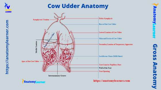
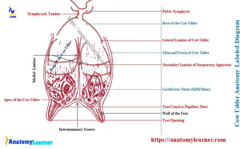
Cow udder labeled diagram
Now, I will show you all the structures from the cow udder with the labeled diagram. Here, in the diagram, I tried to show you the glandular mass and teat of the cow udder.
The 4 mammary glands from the cow udder are identified in the labeled diagram. A wider base, a rounded body, and a narrow apex from the glandular mass of a cow udder are identified in the labeled diagram.
Now, the lactiferous sinus, both from the apex of the body and the base of the teat, is identified. The teat canal with its opening is also identified in the labeled diagram.
The cow udder labeled diagram also shows the intermammary groove. In the other diagram, I tried to show you the suspensory apparatus of the cow udder. The lateral, medial, and secondary laminas are identified in the cow udder labeled diagram.
Again, the symphyseal tendon and pelvic symphysis are also shown with the cow udder labeled diagram.
Other inquiries on cow udder anatomy
Now, I will try to provide concise answers to these questions on cow udder structure that learner commonly asks. But, it is highly recommended to read the whole article to get a basic idea of the cow udder anatomy.
Let’s see the other different inquiries on the cow udder –
What part of the cow is the udder?
As you know, there are 4 mammary glands on the ventral part of the inguinal region of a cow. These 4 mammary glands bound all together and form the udder in the cow.
That means you will find only 1 udder below the inguinal region of a cow. But, there is a distinct intermammary groove between the right and left mammary glands.
In a single udder of a cow, there are 4 compartments (actually 4 mammary glands).
How does a cow’s udder work?
Though the udder’s action is complex, I will try to make it simple here. Estrogen stimulates the growth and branching of the duct system of the mammary gland structure.
Again, progesterone stimulates the formation of alveoli in the inner structure of the cow’s mammary gland. During pregnancy, the placental estrogen and progesterone stimulate the true secretory alveoli.
Now, the prolactin and GH main the lactation in the cow. Finally, oxytocin helps in milk ejection from the cow udder.
This is a straightforward way that I described. But, you should know more about the working system of a cow’s udder.
What are the parts of a cow’s udder?
There are 2 main parts in the cow udder structure – the glandular mass and the teat. Again, in the glandular mass of the cow udder, you will see a wider base, a rounded body, and a narrow apex externally.
Internally, you will find the different structures I have already described in this article in the glandular mass. You will see the parenchyma and stroma (connective tissue) in the internal structure of the cow’s mammary gland.
There are also developed lobular and interlobular ducts system in the structure of the glandular body of the cow’s udder.
At the apex of the glandular mass, there are lactiferous sinuses. The teat of the cow udder possesses some anatomical facts that were also described in this article previously. Here, the most important features of the cow teat are the lactiferous canal, opening, and wall.
What is udder anatomy?
In the cow udder anatomy, you might describe the anatomical facts of the glandular mass or body and also the teat. All the anatomical facts of both the 2 parts of the cow udder have been described with the labeled diagrams.
The cow udder’s external and internal anatomical facts are essential to understand this organ’s action perfectly. If you want to understand the duct system of the cow udder, you might learn it from the macroscopic view of the organ.
Conclusion
So, this article gives you the basic idea of the cow udder anatomy. The 2 main parts of the cow udder – glandular mass and teat- possess peculiar anatomical features both externally and internally.
The internal duct system and lactiferous sinus are the most important to know for the veterinarian. You should practice all the internal and external features from the actual sample of the cow udder at your anatomy learning laboratory. The provided sample labeled diagram on cow udder structure might help you identify these features from the actual sample.