Most first-year vet learners like you have a common question – how to study veterinary anatomy? As a vast veterinary anatomy syllabus, it seems very hard to learn or study for the beginner. But, I will show you my most straightforward and interesting way to study veterinary anatomy so that you never think it so hard.
Quick answer: combining systemic, surface, and comparative anatomy studying methods will make your veterinary anatomy learning easy. To study veterinary anatomy, you might use the labeled diagram, video lectures, and actual live samples.
I believe if you follow the ways I will describe throughout this article, it might help you find veterinary anatomy learning so easy.
But, I should be clear that these methods of veterinary anatomy learning worked for me when I was a first-year vet student. You may follow these methods or modify them in your ways to make anatomy learning easier.
So, if you wish to follow my strategy to learn veterinary anatomy quickly, continue this article till the end.
How to study veterinary anatomy
When I was a first-year student like you, I also felt learning veterinary anatomy was so hard. I was searching for a solution for it but failed to get the proper one.
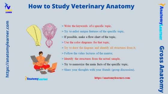
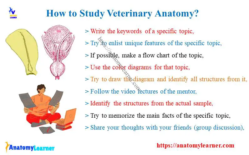
Then I tried to find out the perfect ways of veterinary anatomy learning. I tried several methods to make veterinary anatomy easy. Finally, I found the combination of the following methods that make my anatomy learning so easy –
- Write the keywords of a specific topic,
- Try to enlist unique features of the specific topic,
- If possible, make a flow chart of the topic,
- Use the color diagrams for that topic,
- Try to draw the diagram and identify all structures from it,
- Follow the video lectures of the mentor,
- Identify the structures from the actual sample,
- Try to memorize the main facts of the specific topic,
- Share your thoughts with your friends (group discussion),
Don’t worry; I will provide an example and discuss all these ways so that you may understand how to study veterinary anatomy (any topic) perfectly.
But make sure you know the basics of the different anatomical terms and planes of the body. You know the anatomical terms and plane of the body used to indicate precisely the position, direction, and surface of the different parts of the animal’s body.
I have already described different anatomical terms and planes of the animal’s body in the below-mentioned article –
- Surface anatomy – a vital learning method of veterinary anatomy,
You will find some basic terms like anterior, posterior, lateral, medial, proximal, distal, and so on from that article. So, I think you should check that simple guide to learn the basics of the different anatomical terms to start your veterinary anatomy.
Now, let’s discuss how to study anatomy with an example.
Write the keywords of a specific topic
This way of learning veterinary anatomy work best for me. I used to read the whole topic carefully and try to understand the main facts of that topic.
Then I enlist the keywords that I think are more appropriate to express these topics. I usually used to prefer these methods for every single line of any anatomical description of any organs or structures of the animal body.
Let’s provide an example – how I write the keyword from any anatomical description of organs, bones, or structures.
Suppose you are going to learn the anatomical facts of a dog scapula. You may read the lecture notes provided by your mentor or from the book. Then, you may follow the following I have shown in the picture.
Here, from the first paragraph of the dog’s scapular description, I enlist the below-mentioned keywords –
- Appearance – flat triangular bone,
- Direction – downward and forward,
- Possess – 2 surfaces, 3 angles, and 3 borders,
In this way, you may write some other essential keywords from the dog’s scapular anatomy. You may write the key features from the lateral and medial aspects. Again, you may write the structures from the different borders and angles very shortly.
I hope you can understand how you may write the keywords from any anatomical description of any organs or structures, or bones.
Let’s provide a second example – how to write the keywords for any organ anatomy.
Example of an organ (keywords)
Suppose you are going to learn the anatomy of a cow kidney. Then, you may write the following key features from the whole kidney anatomy –
- Shape – bean-shaped organs, lobulated,
- Location – below the proximal end of the last rib and first three lumbar transverse processes (for right), below and behind the right kidney and 3 – 4 lumbar vertebrae bodies,
- Surface – right (2), left (3 – dorsal, ventral, ruminal)
- Hilus – right (ventral and craniomedial), left (dorsocranial),
- Structure – capsule, cortex, medulla, renal pyramid, calyx, and pelvis,
These features are enough to get the basic structure of a cow kidney. As you read the whole lecture note or book, you may easily memorize if you see your keywords (key features) from your note or guide.
Try to enlist unique features ( summarize) of the specific topic
Writing the key features of the anatomy of any organs, structures, and bones will make a short summary for you. Or, you may enlist the unique features of the specific bones, organs, and structures.
That will help you to get a better idea of this topic. Suppose you are learning the anatomical facts of the horse femur and want to compare it with the cow’s or dog’s femur bone.
In that case, you should enlist the unique features of the horse’s femur that make them different from other animals. So, you may write the below-mentioned anatomical facts of the horse’s femur bone –
There are deep supracondyloid fossa and a third trochanter in the horse femur (whereas you will not find these features in a dog’s femur),
The fovea capitis femoris is deep and notch in the head of the horse femur bone, and
Trochanteric ridge (see here) is directed along the length of the bone (verticle), but in cows and dogs, this structure directs obliquely,
So, you find 3 essential and unique features (third trochanter, deep supracondyloid fossa, and verticle arranged trochanteric ridge) in the horse femur. In this way, you might write the unique anatomical facts of any organs and structure.
Internal organ’s unique features
Let’s take another example from the internal organ of a dog. Suppose you are learning the anatomy of the dog liver, which possesses somewhat different features compared to other animals (cow or sheep, goat).
So, you may write the unique features of the dog liver anatomy in this way –
- There are four well-separated lobes in the dog liver anatomy – left, right, caudate, and quadrate,
- In the caudate lobe of the dog liver, you will find the 2 processes – caudate (right side) and papillary processes (left side),
- The diaphragmatic surface of the dog liver anatomy is highly convex, whereas the visceral surface is irregularly concave,
- In the visceral surface of the dog liver, you will find only the impression of the stomach and duodenum. In contrast, you will find more impressions on different organs in the visceral surface of the ruminant liver.
While reading the dog liver anatomy, you might point – out these unique features on your note (blank space on the note). This will help you a lot while you again read or memorize the basic idea of the dog liver structure.
By the way, if you want to know more about the anatomical features of the dog liver anatomy with their unique features, you may read the below-mentioned article from anatomy learner –
- Dog liver anatomy – lobes, surfaces, processes with the labeled diagram,
Writing the unique features on your note is one of the best methods to learn veterinary anatomy ideally. I hope you have got an essential point to your question – how to study veterinary anatomy.
Let’s move to the third strategy you may follow to learn veterinary anatomy quickly.
If possible, make a flow chart of the topic
This is topic specific but so important to learn veterinary anatomy so easily. If possible, I always try to make a flow chart of the courses of veins, arteries, nerves, and some others.
I also love to make the flow chart of different types of circulation and fluid transportation like – general blood circulation, lymph circulation, and cerebrospinal fluid flow. This made veterinary anatomy so easy for me.
I have already shown different flow charts and short forms of the anatomical topics in the different articles for anatomy learners. In my previous article, I have shown the flow chart of lymph formation and transportation.
Again, I have also shown the flow chart for the general systemic and pulmonary circulation of the blood in the animal’s body. You may follow the same flow chart for cerebrospinal fluid (CSF) formation and transportation.
Let’s see the below-mentioned flow chart on the cerebrospinal fluid, where I show the formation and pathway of the CSF. You may follow this procedure to make your veterinary anatomy learning easy.
This is better than traditional learning (I mean reading in the form of a long paragraph) on the specific topic. It would help if you made the flow chart or sketch of the organ’s anatomy in your way. But make sure you read the details information on the specific topic (organ anatomy) first.
However, you will find more sample flow charts from the below-mentioned articles –
- What is the difference between general systemic and pulmonary blood circulation?
- Fetal circulation flow chart,
- Brachial plexus formation and distribution of nerves in ox,
- Lumbosacral plexus in the ox – courses, and distribution of pelvic limb nerves,
List of topics where you may follow a flow chart
Here, I will enlist some topics (anatomy of different organs, structures, and fluid) where you may follow the flow chart to make them easy. But, this list will not cover the whole veterinary anatomy, so you might search and think about the topics that may go under the flow chart.
So, what are the common anatomical topics where the learner should follow the flow chart or sketch form –
- Courses of the major blood vessels (veins and arteries) of an animal’s body – for example, axillary artery, femoral artery, cephalic vein, and saphenous vein,
- Courses of major peripheral nerves – example: radial, median, ulnar, ischiatic (tibia and fibula) of animals like dogs, cows, and sheep,
- General and systemic blood flow in the animals,
- Fetal blood circulation,
- Formation and transportation of lymph, and
- Formation and transportation of cerebrospinal fluid (CSF),
You may follow the flow chart or sketch diagram in these sample topics. So, this flow chart of sketch diagram might help learn the anatomical course of animals’ organs or structures.
Use the color diagrams for that topic
Using color diagrams is considered the most helpful way to learn veterinary anatomy. I always tried to follow the colored veterinary anatomy books to learn the basic things about any organs from the animal’s body.
This color diagram of any organs will provide almost the reality of the features you will find in the live samples. But the question is where to find the color diagrams to learn veterinary anatomy.
Well, you may follow some of the suggested books where you will find the color diagrams to learn the anatomical features of any organs from the animals –
- Veterinary anatomy coloring books,
- Saunders veterinary anatomy coloring books,
- Dog anatomy coloring books,
- Introduction to veterinary anatomy,
- Color atlas of ruminant anatomy,
- Animal anatomy coloring book, and
- Other different suggested books (let’s check here),
All these suggested books are great for learning gross veterinary anatomy with the color-labeled diagram. Again, you will find the different color diagrams here on anatomy learner (in different species like dogs, cats, rabbits, horses, pigs, cows, sheep, and goats).
I have prepared the gross anatomy coloring books based on the ruminant, which will provide the basic concepts on the different features of bones, internal organs, and other structures.
The color diagrams on the internal organs of the animals will help you to identify the distributed area of the specific organ. Again, you will clearly understand the relationship of different organs in the same regions or area (topographic anatomy).
In addition, you might also use color diagrams and accurate pictures to study microscopic anatomy (histology). You will find the complete guide on the color diagrams and real microscopic pictures here.
Where should you use color diagrams?
If possible, you may use the color-labeled diagrams for all the structures and organs of the animals. But, using the color-labeled diagram is mandatory for the internal organs.
Let’s see where you should use the color diagrams (mandatory) –
- Internal organs – digestive, respiratory, urogenital, and nervous system organs,
- In the courses of significant veins like the cephalic and saphenous,
- In the courses of peripheral nerves (like radial, median, ulnar, ischiatic, tibia, and fibula) that supply up to the digits of the animals,
- Different bones of the animal’s skull (to identify the area and extension of the skull bones),
- Using color diagrams of the structure of the different essential joints to understand the ligaments and bone involvement,
The books I have suggested before containing labeled diagrams and genuine pictures. So, you will get an idea of the actual color of the specific organ or structure from the animal’s body.
I have also found the best book to learn dog anatomy, where you will find both labeled diagrams and real pictures –
- Anatomy of the dog, and
- Color atlas of the dog and cat anatomy,
You may also follow these books to learn dog and cat anatomy. But, I think you should always follow the ruminant or horse anatomy books first, as some organs possess the ideal features.
On the other hand, you will not find the ideal anatomical features in some of the organs of dogs and cats. So, if you have a clear concept of horse anatomy, you may compare these features with other species. But it entirely depends on you whether you follow this strategy or not.
Try to draw the diagram and identify all structure
This is also an excellent practice for learning gross veterinary anatomy perfectly. When I was a doctor of veterinary medicine student (first year), I used to draw the diagram and label all the features from that diagrams.
This provides the best result in learning veterinary anatomy. So, you may follow this strategy (drawing the diagram on your own) to improve your anatomy learning.
But, no need to draw the perfect diagrams on the specific topics of veterinary anatomy. You are drawing these diagrams for your own use, so it is enough if you understand the structure and features of these diagrams.
Let’s share my personal experience of drawing anatomical diagrams. I am not very good at drawing, so I always try sketching the organ I read.
I used a separate notebook to draw these organ’s shapes and identified all the possible features from it. You may see the drawing (anatomical diagram) of what I have done in my student life. This is not so good, but it was so helpful for me when I tried to memorize the main features of the specific organs.
I hope this method of learning gross veterinary anatomy might also work for me. But you may design this method in your way to make things simple and easy.
Should I use a colored pencil on the anatomical diagram?
It depends on you, but my reply is – no. It would help if you did not use the colored pen or pencil as it will take more time to draw every single feature from the specific organs.
It is better to use the standard pencil or pen to draw or sketch the shape of the organs and other features. Then you should identify the anatomical features (unique and general) with the same pen or pencil. This might save you time and boost your gross anatomy learning.
But what should you do (learn) in the case of microscopic anatomy (histology)? Should you use the color pen to draw the diagram for histology slides?
Okay, drawing the microscopic figure using the color pen is obviously an excellent practice. But I wouldn’t say I like to draw the microscopic figure of any organ using a colored pen or pencil. Instead, I prefer to use color diagrams and actual microscope figures to learn the microscopic anatomy of any internal organs (histology of internal organs).
Follow the video lectures of the mentor
You may follow the video lectures of your mentor if they provide these resources. Or you may take a video of your mentor’s lectures with their permission.
Most mentors now have online resources like a blog, youtube, Facebook page, Instagram page, and other social media. So, you may join these online platforms to get access to the video lectures or other different resources provided by your mentor.
But, if your mentor does not have an online platform, you may take a video (with permission) while they show you the different anatomical features of different organs.
Here, in anatomy learner, you will find most of the articles that might help you get a basic idea of the specific organs from the first few sections. Then you will learn the anatomical details of that specific organ with different-level diagrams.
Again, you will find a youtube channel where anatomy learners provide short videos on the different organs or structures of animal bodies. So, you may also follow this channel to get an update on different anatomical topics.
You may also make short videos by yourself on the different anatomical facts of different animal organs. Let’s see how you may create a short video on different organs and structures.
How to create a short video on veterinary anatomy?
I will not tell you to spend more time creating a short video on gross veterinary anatomy. Instead, you may follow the procedure to make a short video on different animal organs.
Process – 1
It would help if you kept notes when your mentor demonstrated the different anatomical facts from the different organs or systems.
After that, you may summarize the anatomical facts within 1 minute of that specific organ or system,
Now, you should identify and tell these features and ask your friend to capture them with your phone,
That’s enough, and watch the recorded video when you read that specific topic in your room. Make sure the video length will not exceed 3 or 4 minutes.
Process – 2
This is time-consuming, but it works smoothly to learn gross veterinary anatomy. Let’s see the process – 2 to make a short video on different organs from the animal body –
You have already taken notes and summarized the anatomical facts that your mentors provided earlier,
Now, you should take different pictures from different views (dorsal, ventral, cranial, and caudal views) and different planes (longitudinal and transverse plane),
You should identify the different features from these pictures instantly with your handphone,
Now, you may add these pictures and make an awesome video. But, if you don’t want to spend more time, it is better to follow the process – 1 to make a video on gross veterinary anatomy.
You may think creating a video on a different animal’s organs is a waste of time while studying as a first-year veterinary student. But this simple video will make great value and make your gross veterinary anatomy learning easy.
Identify the structures from the actual sample
This is true that your mentor will not demonstrate all the anatomical features of the different organs from the different animal species. The gross veterinary anatomy syllabus is large and complicated to cover within a brief period.
So, you need your own effort to get an idea of some extra anatomical features from different animal species. In all conditions, identifying the anatomical features from the real or live samples will work fine to gather knowledge on veterinary anatomy.
Suppose your mentor or teacher taught you the anatomy of a horse; now, you want to know the anatomical features of different organs from the dog. You have great resources for learning dog anatomy – books, online platforms, and liver samples in your laboratory.
In this situation, you should read the essential and exceptional anatomical features of the different organs from the book. You may also get help from the video lectures from the other mentors (social media platforms).
Then you will get a basic concept of the specific anatomical features of the animal’s organ. Just note these key features and try to identify them from the live sample at your veterinary anatomy learning laboratory.
Veterinary anatomy education is incomplete without observing and practicing from live anatomical samples. So, in all situations, you should have a plan to practice all the anatomical facts from the actual samples.
You should also palpate different peripheral organs or structures (like lymph nodes and blood vessels) from a live animal. As this strategy of learning veterinary anatomy is under the surface anatomy section, I will discuss it later in this article.
Again, you may read the below-mentioned article –
- Surface anatomy – another best learning method of veterinary anatomy,
Try to memorize the main facts of the specific topic
This is another best way to learn gross veterinary anatomy perfectly. It would help if you made a routine on a daily or weekly basis where you might provide at least 5 minutes for each topic you have learned previously.
This is the best practice for learning and scoring well in the veterinary gross anatomy examination. Again, this procedure will reduce your studying load before the anatomy examination.
While practicing the anatomical features of any specific organs, you may forget some points. Immediately, it would help if you went through the videos you have created before by yourself.
Or, if you didn’t create any video, you may use your short note (key points you have listed before in the class or your room).
I am not telling you to practice the same topics (anatomy of any specific organ) weekly. You may practice this only for 2 weeks and see your improvement. If it works for you, you may follow the same procedure and compete with other anatomical features on different organs.
If you follow my previous strategy (especially no – 1, 2, and 6) of learning gross veterinary anatomy, then this method will work smoothly.
I hope you also got the best method in this section for your inquiry – how to study veterinary anatomy. Let’s see the following method that will also play an essential role in learning veterinary anatomy perfectly.
Share your thoughts with your friends (group discussion),
This is another effective method for learning gross veterinary anatomy perfectly. I think 70% of first-year veterinary students follow this method to learn veterinary anatomy, which is very effective.
This group study method is not only for the first-year veterinary student; you may follow this method throughout your DVM (doctor of veterinary medicine) life.
But, some students are not interested to learn in a group. And if you are under this condition, you may avoid this veterinary anatomy learning strategy. Again, if you love to learn anatomy in the group, then let’s continue this article section.
You may make a group of 4 – 5 friends who will share their learning on a specific day. Sometimes listening to others may make the topics easier than before.
Sharing your anatomical knowledge may help you to improve your expression quality. As well as you may also get different techniques to memorize complex topics easily. Thus, you may sharpen your veterinary anatomy learning quickly.
If you are not interested in creating a discussion group physically, you may join different social media groups (like Facebook and whatsapp), forums, and QnA (Questions and Answers) groups.
- Learning anatomy online and sharing your thoughts with others is very enjoyable. You may join different platform to learn and share your veterinary anatomy knowledge –
- Facebook page and group like – anatomy learner page, and anatomy learner group,
- Veterinary discussion forums – there are different veterinary discussion forums where you may ask or answer the inquiries of anatomy learners,
- Blog website and Youtube channel – like anatomy learner, where you may also share your idea or ask any questions on specific topics under a single specific article or video,
Best way to study veterinary anatomy
The strategy of learning veterinary anatomy that I described combines some effective methods. If you want to follow these points, you might have a good concept of effective anatomy learning methods like systemic anatomy, comparative anatomy, topographic anatomy, etc.
First, let’s see the different major methods by which gross veterinary anatomy can be studied. Here, I will enlist the significant methods of learning veterinary anatomy perfectly –
- Systemic anatomy learning method (my first choice),
- Topographic and surface anatomy learning method (needs to fulfill the systemic anatomy learning methods),
- Applied or surgical anatomy learning method (practically related to field practice),
- Comparative anatomy learning method (effective for learning anatomy among different species),
- Regional anatomy learning method,
- Functional anatomy learning method, and
- Radiological anatomy learning method,
Methods of learning veterinary anatomy may vary from person to person. But, let’s see which is the best method to learn veterinary anatomy perfectly and efficiently.
What is the best way to study veterinary anatomy?
You may already understand the best methods or ways of learning veterinary anatomy from the list I provided above.
In my sense, the combination of systemic, surface, and comparative anatomy learning ways are very effective for learning veterinary anatomy. You may go with the surgical or applied anatomy when you get the basic concept of the different organ systems from different animal species and can identify them from an external approach (most essential organs).
Without the systemic anatomy learning method, no other methods are completed. So, I think you might start with the systemic anatomy of any ideal animal species (you may choose a horse or ruminant). Then, you should compare your acquired knowledge with the other different species.
Now, these will give you a clear concept of the location of different organs from different animal species. Let’s give an example – you know the location of a dog and cow’s kidneys (both right and left) are not located in the same area.
So, the surface anatomy of the dog’s and cow’s kidneys will not be the same. Thus, you should first know the comparative anatomy of every structure and organ from the most essential domesticated and pet animals.
Then, you might apply your knowledge practically under the course of veterinary applied anatomy. So, this combination of three major learning methods will make your veterinary anatomy easy.
Let’s learn about the different major ways of studying veterinary anatomy.
Systemic veterinary anatomy learning method
In this primary learning method, you will learn the anatomical facts of the particular organ system of an animal’s body serially. Here, you will not learn the anatomical facts of the surrounding organs.
In this method, the organs or apparatus of the specific system have a common origin and structure. They associate with one particular function (sometimes more than one) of the animal’s body.
Most anatomy mentors or teachers follow this systemic veterinary anatomy learning method to teach their students. So, under this method, you might learn the organs from the different body systems of an animal –
- Osteology – deals with the bones, skeleton, ligaments, tendons,
- Syndesmology or orthology – deals with the joints of the animal’s body (with ligaments),
- Myology – deals with the origin, insertion, and fibers direction of the muscles from different regions of an animal’s body,
- Digestive system – deals with the different parts of the digestive tract and some accessory organs,
- Respiratory system – deals with the lung, trachea, and some associated organs,
- The cardiovascular system of animals – deals with the anatomical features of the heart, and blood vessels (veins and arteries),
- Urogenital organs system of the animals,
- Endocrine system – deals with the anatomy of the different endocrine glands,
- Special sense organ – deal with the anatomical facts of the eye, ear, and skin,
- Common integumentary system – deal with the anatomical features of the animal’s horn, hoof, and nails,
- Now, let’s see what you mean by the topographic and surface veterinary anatomy learning methods.
Topographic and surface anatomy learning methods
Some authors sometimes use these 2 terms (topographic and surface) to mean the same things. But, you will find a little difference between these 2 terms – topographic and surface anatomy.
When you want to accurately learn the relative position of the various parts of an animal’s body, this is under the topographic anatomy learning method. That means the anatomy of any specific organ will be studied with the associated organs of the animal’s body.
Let’s see the example – suppose you want to learn the course of a cow’s trachea. You know, the trachea of a cow starts from the larynx at the level of the base of the heart and ends in the lungs as a separated primary bronchus.
If you notice, here I have related another organ (heart) to express the exact starting point of the cow’s trachea.
You will find 2 distinct parts in the cow’s trachea – cervical and thoracic. The cervical part of the cow’s trachea extends at the level of the thoracic inlet. Here, this cow’s trachea is dorsally related to the esophagus (up to the third cervical vertebrae) and the longus coli muscle.
Ventrally to this cow’s trachea, you will find the Sterno-thyrohyoideus muscle. In addition, you will see the carotid artery, vagosympathetic trunk, jugular vein, and recurrent laryngeal nerve on the lateral aspect of the cow’s trachea.
So, to express the location or course of the cervical part of the cow’s trachea, here I have used some other organs, and parts or structures. When you also express the location, course, and formation of any organ with the relative positions of other organs, it will go under the topographic anatomy.
What is surface anatomy?
Surface anatomy is one type of topographic anatomy where you should identify the organ from the animal body with the surface approach. Here in the surface anatomy, you don’t need to point out the relative position of the surrounding organs of an animal’s body.
Let’s see the example of the surface anatomy – suppose you want to know the surface anatomy of the right lung of a cow. Then you may follow the below-mentioned process to identify the cow’s right lung from the surface approach –
Let’s draw 3 lines that I have shown in the diagram (one line from the caudal border of the scapula to the tuber coxae, the second line from the caudal border of the scapula to the olecranon process of the ulna, and a third line from the olecranon process of ulna to the last ribs of the thoracic cage),
You will make a triangular area in the thoracic region of the cow where you will find the right lung,
In the same way, you may find the surface anatomy of the cow’s heart from an external approach. You should count the intercostal spaces from the thorax and identify the third or fourth intercostal spaces.
Now, you may divide the thoracic cavity (vertically) into three equal parts and identify the exact location of the cow’s heart, as I have shown on the diagram.
Or, you may consider the olecranon process as the landmark to identify the cow’s heart from the external approach.
Comparative anatomy learning method
This is another best way to learn veterinary anatomy from different animal species. You know comparative anatomy is the description and comparison of the structure of animals and forms the basis of their classification.
So, first, you might have a good piece of knowledge of the basic anatomical features of any organs, structures, or parts of the animal’s body. That means you might follow one ideal structure from any animal. Then, it would help if you went with the comparison of the specific organs from different animals.
It is not possible to learn the single organs from thousand of animals. So, it is better to know the anatomy of one single animal first.
Suppose you want to learn the anatomy of the tongue from different animals like the cow, dogs, cats, horses, pigs, and also from the rabbit. First, you should know the ideal structure of a tongue from any animal.
Here, I will consider the cow’s tongue as it has all the anatomical features. In the cow tongue anatomy, you will find the 3 different parts – root, body, and apex.
You will see a transverse groove on the body of the tongue, which is known as the lingual fossa in the cow tongue. Behind the lingual fossa, you will see a prominent torus linguae.
The apex of a cow tongue anatomy is blunt. You will see a frenum linguae on the ventral surface of the cow’s tongue.
There are different types of lingual papillae distributed throughout the tongue of a cow.
These are the basic anatomical features you should commonly find in tongues from different species. But, you will also find some unique features in the tongue from the different animal species.
Comparative anatomy of the tongue in different animals like horses, dogs, and pig
The body of the horse tongue is narrow, and the apex is spatula-shaped compared to the cow’s tongue. Again, you will not find any torus linguae in the structure of the horse tongue. In addition, you will find a little variation in the papillae that you have found in the cow’s or dog’s tongue.
However, if you want to know the details of anatomical features of the animal’s tongue (dog), read the below-mentioned article –
Dog tongue anatomy with the labeled diagram – parts, muscles, papillae, and other different structures,
In the dog tongue, you will find some unique anatomical features that you have nerve seen in the tongue of a horse or cow. There is a longitudinal median groove on the dorsal surface of the dog’s tongue. Again, the ventral surface of the tip of the dog tongue possesses a fibromuscular lyssa body.
You may also learn more about the different organs (comparative study) from the different species from anatomy learners.
Applied and regional anatomy learning methods
Sometimes you need anatomical knowledge for diagnosis, surgery, and other practical purposes. This is the most important way to learn veterinary anatomy perfectly nowadays.
Suppose you want to perform high or low epidural anesthesia on a cow to perform specific surgeries. You might have a good knowledge of the different structures of the animal’s body (structure involved in surgery and anesthesia).
- Site for high epidural anesthesia – lumbosacral junction, and
- Site for low epidural anesthesia – sacrococcygeal junction,
So, you should know these joints (with their bone involvement) and identify them from the external approaches. Again, you might have a piece of good knowledge of the structure of the spinal cord (along with the epidural space) of the specific animals.
Again, suppose you want to perform surgery on the abdominal area. In that case, you might have a good knowledge of the different structures (muscles, fascia, peritoneum, organs) of the abdominal region of the specific animal.
Sometimes you may learn the organization of the animal’s body by regions or areas. But, I think this is not best for the first-year veterinary student. If you have a basic concept of the different organs from the different systems of the animal’s body, you may go with the applied or regional anatomy learning methods.
Example of regional anatomy learning method –
- Study on the forelimb and hindlimb anatomy of the specific animal,
- Study on the head, neck, and thorax of the animal,
- Learning the anatomical features from the animal’s abdomen and pelvic cavities,
Other veterinary anatomy learning methods
The functional veterinary anatomy learning method is the correlation between anatomical structure and their function. Again, clinical anatomy is one type of applied anatomy where you should study the macroscopic structure of the body concerning the practice of medicine and other health science.
You may also herd about radiological anatomy. In the radiological veterinary anatomy learning method, you will learn the structure and function of the body using radiological and imaging techniques.
I hope you got the best answer to your question – how to study veterinary anatomy perfectly. Now, you should know which topics you might focus on in the first year of veterinary anatomy learning.
Veterinary anatomy 1st year
As you already got an idea of the different topics you might learn in the veterinary anatomy 1st year. Here, I will only focus on the topics you should focus on before learning veterinary anatomy.
You should follow the systemic anatomy learning method and cover the following systems from an animals body –
- Osteology, syndesmology, myology,
- Digestive, respiratory, and cardiovascular systems,
- Urogenital systems,
- Endocrine, unique sense, and common integumentary system,
But, before starting to learn any system of veterinary anatomy, you might learn the following anatomical (topographic) terms first –
- Dorsal, ventra, anterior, posterior,
- Lateral, medial, superior, inferior,
- Superficial, deep, proximal, distal,
- Cranial, caudal, rostral, dorsal,
- Axial, abaxial, palmar, planter,
Different planes of the body like – longitudinal median plane, sagittal or paramedian plane, transverse or segmental plane, and frontal or horizontal plane,
Again, you might have a piece of basic knowledge of the different osteological terms before starting the osteology from any animal species. I already have an article where I have described all these osteological terms with examples from the live bones of an animal.
If you wish, you may learn these osteological terms from the below-mentioned article –
- Osteological terms that you need to start learning veterinary osteology,
How to study veterinary anatomy for art
If you want to draw the different organs, structures, and parts from the animal’s body, you should have a basic concept of veterinary anatomy. For that, you may follow the below-mentioned recommended books and guides –
- Veterinary anatomy for art (book for artist), and
- Color atlas of veterinary domesticated animals,
Before drawing any organs from any animal, you should know the structure of that organ. That will help you to draw the specific organ perfectly.
How anatomy learner help you to learn veterinary anatomy online?
In the anatomy learner, you will find different articles (along with labeled diagrams, actual pictures, and videos) that might help you learn the basics of veterinary anatomy. The anatomy learner primarily publishes different articles on the anatomy of different organs from dogs, cats, pigs, rabbits, cows, and goats.
You will find concise and also details forms of information in each article that is published by this anatomy learner. I am a veterinarian and know where the first-year veterinary student may face difficulties. Thus, I tried my best to make the anatomical topics easy for the veterinary student in anatomy learner.
If you notice each article of anatomy learner, you will find a concise (short answer) in the first section. Then, I go into detail about the specific topics in every single article on anatomy learners.
So, you may read the first section (short answer) from each article from the anatomy learner and then goes for details. You will find different labeled diagrams in each article which will also help to understand the anatomical features of the specific organs.
Finally, there is a video library here in the anatomy learner, which will be very helpful for your veterinary anatomy learning. Again, you will find a short video in almost all the articles that anatomy learners publish.
You will find an update of every single article, image, and video through the social media of anatomy learners.
Frequently asked questions on veterinary anatomy
Now, let’s see the common inquiries on veterinary anatomy learning asked by anatomy learners. If you have any questions on learning about veterinary anatomy or how to learn veterinary anatomy, feel free to ask.
Is veterinary anatomy hard?
Though the contents of veterinary anatomy are vast, learning it is exciting. As you should learn the veterinary anatomy from the live sample, I think it is not so hard.
But, please start to learn veterinary anatomy systemically (regularly). You should follow the strategy that I have already discussed in this article.
Different videos and labeled diagrams by different authors make veterinary anatomy so easy.
How is the study of veterinary anatomy divided?
The study of veterinary anatomy is divided into –
- Systemic veterinary anatomy – consists of systems of organs,
- Topographic veterinary anatomy – the relative position of the various parts of the animal’s body,
- Applied veterinary anatomy – consideration of anatomical facts concerning practical purposes,
- Comparative veterinary anatomy – description and comparison of animal’s form and structure,
- Regional veterinary anatomy – the study of the organization of the animal body by region or area,
- Functional veterinary anatomy – correlation between the anatomical structures and functions,
These are the primary division of studying veterinary anatomy.
Do veterinarians have to learn human anatomy?
No, it is not mandatory to learn human anatomy for veterinarians. But, if any veterinarian wants to learn and compare the anatomical facts between humans and animals, they may do so.
As veterinarians only deal with different animal species, they have to learn the anatomical facts of the different organs (gross anatomy) from the different animal species.
But, you may find many similar microscopic anatomy in the internal organs between humans and animals. So, sometimes many veterinarians follow human histology to understand specific topics better. But, keep in mind some of the organs (from ruminants) have a significant dissimilarity in form and structures (histologically).
How do I start studying as a vet?
It would help if you researched the veterinary schools available in your area or country. Then, you need to know the qualification needed for veterinary study admission.
After qualifying for the admission test, you should admit to veterinary schools and start studying veterinary.
Which is the best website to learn veterinary anatomy online?
You may find a lot of websites to learn veterinary anatomy online. Both of them are good in their own ways. The anatomy learner is one of them, where a beginner (first-year veterinary student) can start his veterinary anatomy learning by simply following different articles, labeled images, and video lectures.
Conclusion
I hope you got the best answer to your inquiry – how to study veterinary anatomy perfectly. You will do better in this subject if you follow the combined systemic, topographic, comparative, and applied veterinary anatomy learning methods.
All the methods I have enlisted here in this article for learning veterinary anatomy will work fine if you follow them sincerely. Again, the 8 best strategies I shared here might be followed to make your veterinary anatomy learning easy.