The cow kidney anatomy consists of paired lobulated oval or bean-shaped organs. Both these kidneys (right and left) possess grossly distinct surfaces, borders, and extremities.
Again, in the structure of the cow kidney, you will find 2 major features – the capsule and cavity of the kidney. The cavity of the cow kidney possesses different important anatomical features. In this article, I will describe all of these anatomical features of the cow kidney with the labeled diagram.
Within the ruminant, you will find a great difference in kidneys in their external and internal features. So, here I will also point out the unique features of the kidney from different ruminants like cows, sheep, and goats.
Finally, I will also show some major differences in the kidney structure of a horse, pig, and dog compared to the cow. So, if you want to get the full idea of the cow kidney anatomy, let’s continue this article till the end.
Cow kidney anatomy
From the cow kidney anatomy, I will discuss the different identifying features from both their external and internal appearances. You know there are 2 kidneys – right and left located at the upper cranial part of the abdominal cavity and above the peritoneum.
Superficially, you will see different lobules on the external surface of the kidneys. These lobules are separated by the fissure, which remains filled up with fat.
Before identifying the cow kidney’s external and internal anatomical features, let’s see how you may differentiate the right kidney from the left one. But make sure you will read the anatomical facts of both the right and left kidneys from the cow that I will discuss later.
For now, you may easily separate the right kidney from the left one by the followings –
The right kidney of a cow – is roughly bean-shaped and possesses hilus on its craniomedial (ventrally) aspect,
The left kidney of a cow – possesses typical 3 surfaces, 3 borders, and 2 extremities; the hilus locates on the dorsal surface,
You will see a great variation in the location of the cow’s right and left kidneys. Let’s find the specific location of the cow’s right and left kidneys from the details specific anatomical guide (will discuss below).
So, from these 2 kidneys, you might know the followings –
- Location of the cow’s right and left kidneys,
- External anatomical features from both the right and left kidneys of a cow (including lobules, fissures, and hilus),
- Internal anatomical features of cow’s kidneys (including cortex, medulla, renal pyramid, calyx, renal pelvis, and others),
You might also perfectly differentiate the renal vein, renal artery, and ureter from the cow kidney hilus. Let’s see the below-mentioned cow kidney labeled diagram, where I tried to show some external features.

Cow kidney identification
Now, from the right and left kidneys of the cow, you might identify the following important features –
- Lobulation on the external surface of both right and left kidneys,
- Fissures filled up with fat on the external surface,
- Hilus of the kidney (ventro-cranio-medial for right, and dorsal for left kidney),
- Renal artery, renal vein, and ureter from both right and left kidneys,
- Dorsal and ventral surfaces of the right kidney,
- Lateral and medial borders of cow’s right kidney,
- The narrow and thick cranial end of the right kidney,
- The free and rounded caudal extremity of the cow’s right kidney,
- 3 surfaces of the cow’s left kidney (dorsal, ventral, and ruminal),
- 3 borders of the cow’s left kidney (medial, lateral, and ventral), and
- Hilus on the dorsomedial aspect of the left kidney,
I hope you have already found all those external anatomical features mentioned in the labelled diagram from both the right and left kidneys of a cow. Now, let’s see the internal anatomical features of the cow kidney structure –
- External covering or capsule of the cow kidneys,
- Cortex and medulla of the internal surface of the kidneys,
- Corticomedullary junction of the kidney parenchyma,
- Renal pyramid on the kidney parenchyma,
- Minor and major calyx of the renal structure,
- Junction of major calyces and continuation of the ureter of the cow,
- Distribution of the renal vessels throughout the parenchyma, and
- Renal fat inside the cow kidney,
The labelled diagram identifies the above-mentioned internal anatomical features of the cow kidney.
Now, you may point out some important anatomical facts from the cow kidney anatomy.
Unique features of cow kidney structure
So, you will find the following unique features in the cow kidney structure –
- Cow kidneys are paired (right and left) lobulated oval or bean-shaped organs in the urinary system,
- The right kidney of a cow locates below the proximal end of the last rib and the first 2 or 3 lumbar transverse processes,
- Again, the location of the cow’s left kidney is variable (extends from the bodies of the 4rd and 5th lumbar vertebrae to the pelvic inlet),
- They remain in their position with the help of the ureter, vessels, renal fat, and pressure of surrounding organs,
- The length of the cow’s right kidney is more (20 – 22 cm) compared to the left (15 – 18 cm),
- Again, the weight of the left kidney is more (700gm average) compared to the right kidney (675 gm average),
- The right kidney of a cow possesses 2 surfaces, 2 borders, and 2 extremities; whereas the left kidney possesses 3 surfaces, 3 borders, and 2 extremities,
- Externally you will find a fibrous capsule in both the right and left kidney of the cow,
- Renal pyramids are distinctly visible in the cow kidney parenchyma compared to other animals,
- You will also distinctly identify the renal cortex and medulla from the cow kidney structure,
- There are different minor and major calyces present in the structure of the cow kidney parenchyma,
- 2 wide major calyces join to form the ureter in the cow kidney structure,
- You will also find the renal vein and renal artery in the hilus of the cow kidney,
In the next section (part) of the article, I will also show you the external features of the cow’s right and left kidneys on a comparative basis. So, let’s continue to know the main differential features between the cow’s right and left kidneys.
Where is the kidney located in a cow or ox?
The cow’s right kidney commonly lies ventral to the last rib and first 2 or 3 lumbar transverse processes. Here, the cranial extremity of the right kidney may lie ventral to the last rib. Again, the caudal extremity of the cow’s right kidney lies ventral to the 4th lumbar transverse process.
The left kidney of a cow occupies a unique position and locates just caudal to the right kidney. When the rumen is full, it pushes the left kidney caudally and crosses the median plane of the body.
So, the left kidney locates on the right side, caudal, and a little ventral to the right kidney. Thus, you will find the cow left kidney just ventral to the bodies of the third, fourth, and fifth lumbar vertebrae.
But, when the rumen is not full, you may find a change in the location of the left kidney. Then, the left kidney may lie at the same level as the right kidney but partly at the left side of the median plane.
I hope you can understand the location of a cow’s right and left kidneys. Now, you may identify the location of the cow or ox kidney by surface approach.
You may easily identify the location of the cow’s right kidney from an external surface of the body. You might locate the last rib and first 2 or 3 lumbar vertebrae for that.
As the right kidney of a cow or ox locates at the ventral part of the lumbar transverse process, you may easily palpate this kidney. First, try to identify the first 2 or 3 lumbar vertebrae with their transverse process, then try to palpate the right kidney as shown on the diagram.
What are the factors keeping the kidneys in their position?
There are 3 main factors that keep the cow kidneys in their position within the abdominal cavity. Let’s see what these factors are –
- Renal artery, renal veins, and ureter of the kidney that remain in the hilus,
- Deposition of the renal fascia, capsule, and distribution of the renal fat on the external surface of the cow kidneys, and
- The pressure created by the surrounding organs of the cow kidneys,
Again, the liver also plays an important role in keeping the right kidney in its position. The adrenal glands and peritoneum are also important in keeping them in their position.
“It is very difficult to palpate the left kidney of the cow from the surface approach as it locates ventral to the bodies of lumbar vertebrae.”
Measurement of cow kidneys
I will provide the average measurement of the cow’s right and left kidneys. As these measurements are average, you may find a variation practically. Let’s get an idea of the average measurement of the cow’s right and left kidneys from Table 1 –
So, Table 1 shows the average length, width, thickness, and weight of both the right and left kidneys of the cow. Here, the length of the right kidney is more, but the average weight of the cow’s left kidney is more.
“The weight of the cow kidneys is about 2% of the total body weight.”
Now, I will discuss the details anatomical facts of the right and left kidneys of the cow or ox (bovine) with the labeled diagram. Let’s start with the anatomical points of the cow’s right kidney.
Cow right kidney anatomy
The cow right kidney anatomy has an elongated elliptical outline. The right kidney of a cow is also flattened dorsoventrally, which may include a good feature to differentiate it from the left kidney.
I will show you the external anatomical facts from the cow’s right kidney in this section. As the internal features of both the right and left kidneys are the same and described separately in another section.
Externally, the cow’s right kidney possesses –
- 2 surfaces – dorsal and ventral,
- 2 borders – medial and lateral, and
- Two extremities – cranial and caudal,
Now, let’s see how you may identify these surfaces, borders, and extremities from the cow’s right kidney.
The dorsal surface of the cow’s right kidney is rounded (convex). It remains in contact chiefly with the sublumbar muscles of the cow anatomy. This surface has no contact with the peritoneum.
The ventral surface of the cow’s right kidney is less rounded or convex. You will find a great relationship of the right kidney with other visceral organs like the liver, pancreas, duodenum, and colon.
Another important feature of the ventral surface of the cow’s right kidney is the presence of hilus. You know, the hilus is the area from where the ureter and renal veins arise, and the renal artery exists.
The hilus locates on the cranial part of the ventral surface, near the medial border (craniomedial aspect).
Borders and extremities of ox right kidney
The medial border of the cow right kidney is almost straight and lies parallel to the caudal vena cava. In contrast, the lateral border of the right kidney is somewhat convex.
There are 2 extremities or ends in the structure of a cow’s right kidney – cranial and caudal. Here, the cranial extremity of the cow’s right kidney is narrow and thick.
Again, the right kidney’s caudal extremity is free and rounded.
The cranial extremity of the ox’s right kidney remains lodged in the renal impression of the liver. You will also find contact with the adrenal gland with the cranial extremity of the cow right kidney.
At the paralumbar fossa (or at the level of 3 to 4 lumbar transverse process), you may easily palpate the cow’s right kidney.
I hope you got a clear idea of the cow’s right kidney’s external features. Now, I will discuss the external features of the cow left kidney with the labeled diagram. You will see a great variation in the external features of the ox’s left kidney compared to the right one.
Okay, let’s see the external anatomical features of the left kidney from the cow’s urinary system.
Left kidney of ox anatomy
In the external anatomy of a cow or ox (bovine) left kidney, you will find somewhat different features than the right kidney. The shape of the cow’s left kidney is bean or oval and possesses the followings –
- Three surfaces – dorsal, ventral, and ruminal,
- 3 borders – medial, lateral, and ventral, and
- 2 extremities – cranial and caudal,
The dorsal surface of the cow’s left kidney is convex or rounded. You will find the hilus on the craniolateral part of the dorsal surface of the left kidney. This might be a unique anatomical feature to differentiate the left kidney from the right kidney of the cows.
The ventral surface of the cow’s left kidney is less convex (almost flat). This surface of the left kidney is related to the cow’s intestine.
The other most important surface of the cow’s left kidney is the ruminal surface. This is a more or less flattened surface that comes in contact with the rumen. Thus, this is the ruminal surface of the cow’s left kidney.
So, the flattened ruminal surface is another important external feature to practically identify the left kidney from the cow’s right kidney.
Borders and extremities of cow’s left kidney
The medial border of the cow left kidney is convex and formed in between the dorsal and ventral surfaces. Normally, this border of the left kidney is convex on its cranial part and concave on its caudal part.
The lateral border of the cow’s left kidney is slightly convex. This left kidney border is between the ruminal and dorsal surfaces. The hilus of the left kidney extends obliquely from the lateral border of the cranial extremity to the medial border of the caudal extremity.
Here, the ventral border of the left kidney is also convex. This border is in between the ruminal and ventral surfaces.
The extremities of the cow’s left kidney also show unique features. Here, the cranial extremity of the left kidney is small compared to the caudal and the right kidney. Again, the caudal extremity of the ox’s left kidney is larger and rounded compared to the cranial extremity.
A capsule of cow kidney
Both the right and left kidneys of the cow are covered with the fibrous capsule. You will find a large amount of perirenal fat on the outer surface of the cow kidney.
After covering the whole kidney, the fibrous capsule lines the wall of the renal sinus. You may easily identify the fibrous capsule from the parenchyma of the cow kidney during dissection.
Internal anatomy of cow kidney
Now, in this part of the article, I will show you the internal anatomy of the cow kidney with the labeled diagram. Here, you will find the anatomical features of the renal pyramid, cortex, medulla, major and minor calyces, and renal sinus.
You already identify the different features from the external aspect of the cow kidney. There is a great variation in the internal anatomy of the cow’s kidneys compared to the dog’s. In the structure of a cow kidney, you will nerve find the renal pelvis and renal crest.
But, in the dog or horse kidney structure, you will find these anatomical features. If you want to get a basic idea of the dog (canine) kidney structure, you may read the below-mentioned suggested article –
- Dog kidney anatomy with the labeled diagram,
You will easily find the distinct renal pyramid structure in the longitudinal section of the cow kidney. The number of the pyramid may vary in the different cows (normally 15 – 19).
You will see the pale outer cortex and darker inner medulla in the renal pyramid of the cow kidney. You will find a corticomedullary junction between the cortex and medulla of the cow kidney structure (dot demarcation).
Again, the blunt apex of the real pyramid, you will see the renal papillae that project into the minor calyx. Microscopically, there is a small orifice on each papilla by which the papillary duct opens into the minor calyx.
You will also find the renal column in the structure of the real pyramid of the cow kidney. They are (renal column) more distinct in the cow kidney compared to the horse.
Calyx and sinus in the cow kidney structure
There are several funnel-shaped minor calyces in the structure of the cow kidney. The renal papillae enter into the funnel-shaped minor calyx (papillary ducts open into the minor calyx).
Several minor calyces join to form the major calyx in the kidney parenchyma. Now, 2 wide, thin-walled major calyces participate from the renal sinus.
The renal sinus continues to form the ureter of the cow kidney. Here, the major cranial calyx is larger in the structure of the cow kidney.
The remaining spaces of the cow kidney structure fill with renal fat. So, you will see a great amount of fat in the kidney structure on its internal aspect.
But, no such renal pelvis and renal crest are present in the structure of the cow kidney.
The cow kidney hilus is equivalent to the hilus and sinus of the horse kidney structure. You will see the renal artery dorsally at the hilus of the cow kidney.
Again, the renal vein locates at the middle position of the kidney hilus. Finally, the ureter of the cow kidney is ventral in position in the renal hilus.
There is also a great amount of fat surrounding the structure of the cow’s kidney hilus. I hope you can easily differentiate these structures (artery, vein, and ureter) from the kidney’s hilus.
The renal artery of the cow kidney–lumen is small, the wall is thicker, and the elasticity of the structure is more compared to veins and ureter,
A renal vein from the cow kidney – possesses a larger lumen and thinner wall, and the elasticity of the vein is less compared to the artery and ureter,
The ureter from the cow kidney’s hilus – is a muscular structure,
The parenchyma of the cow kidney
Microscopically, the parenchyma of the cow kidney possesses an outer paler cortex and an inner darker medulla. You may easily understand these features from the microscopic slide of a kidney.
For that, you may read the below-mentioned article to get the basic idea of the kidney parenchyma –
- Kidney histology with the labeled diagram,
The outer paler cortex of the cow kidney parenchyma consists of the renal corpuscle, convoluted renal tubules, blood vessels, lymphatics, and connective tissue. Again, the medulla of the cow kidney consists of a triangular area, each with its base adjacent to the cortex.
You will find a straight segment of the uriniferous tubules, renal pyramid, and renal papillae in the structure of the medulla.
The structural and functional unit of the cow kidney is the nephron, which comprises a spherical renal corpuscle, proximal convoluted tubules, a loop of Henle, and distal convoluted tubules.
Here, in the renal corpuscle structure, you will find the glomerular tuft of capillaries. You will see the Bowman’s capsule that consists of 2 layers – parietal and visceral.
The Bowman capsule’s visceral layer is in close contact with the glomerular truft. Again, the parietal layer of the capsule consists of simple squamous epithelium and is continuous with the proximal convoluted tubule.
The proximal convoluted tubules of the cow kidney parenchyma are the longest and more convoluted structure. You will find a single layer of low columnar cell lining in the wall of the proximal convoluted tubules.
The loop of Henle is the thin and straight part of the nephron (cow kidney). It connects the proximal and distal convoluted tubules and consists of 3 distinct parts (shown in the diagram).
The distal convoluted tubule is less convoluted, and you will find the single layer of the cuboidal cell in its wall.
Cow kidney labeled diagram
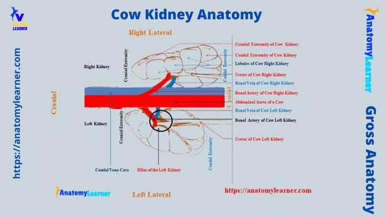
Now, I will show you the cow kidney anatomy labeled diagram that might help you to understand the details and features. Both from the right and left kidneys of a cow and ox, I tried to show the external and internal anatomical facts.
Let’s see the first cow kidney (bovine or ox) labeled diagram (more diagrams), where I tried to show you the location of both the right and left kidneys. Again, this labeled diagram also shows the external features of these 2 kidneys.
There are almost 15 – 19 lobules on the external surface of the cow kidneys. The cow kidney labeled diagram identifies all the lobules, fissures, and external renal fats.
The surfaces, borders, and extremities from both the right and left kidneys of the cow are also identified in the labeled diagram. In addition, the hilus from both the right and left kidneys of the cow are shown on the labeled diagram.
Now, let’s see the internal anatomical features of the cow kidney structure. Here, I tried to show all the important features in the cow kidney’s internal structure labeled diagram.
This diagram shows the cortex, medulla, calyx (minor and major), renal sinus, and renal fat from the internal structure of a cow kidney.
Finally, from the hilus of both the right and left kidneys of the cow, I tried to show you the renal vein, artery, and ureter separately. I hope these diagrams on the cow kidney structure help you practically.
Blood and nerve supply to cow kidney
The renal arteries provide the main arterial supply (right and left renal arteries). These 2 renal arteries of the cow kidneys arise directly from the abdominal aorta.
The left renal artery is larger compared to the right renal artery of the cow. Each renal artery of the cow reaches to the hilus of the kidney as a single artery.
At the hilus of the cow kidney, each of the renal arteries divides into 4 – 5 segments (segmental arteries). These segmental arteries again divide into the 4 – 5 second interlobar arteries.
These interlobar arteries of the cow kidney structure course straight up to the corticomedullary junction. Then, they (interlobar arteries) divide into 3 – 5 arcuate arteries.
The afferent glomerular arteries originate from the cow kidney’s interlobar arteries. Again, from the arcuate arteries, some straight vessels radiate toward the surface of the kidney parenchyma.
The renal plexus innervate the cow kidneys’ structure.
Kidney of sheep and goat
You will find a great variation in the external and internal anatomy of the sheep and goat kidneys compared to the cow. First, let’s discuss the external features of the sheep and goat kidneys that differ from the cow.
The unique external anatomical features of the sheep and goat kidneys –
- Sheep and goats posses the typical bean-shaped kidneys (right and left),
- The surfaces of both the right and left kidneys of the sheep and goat are smooth,
- There is no superficial lobulation found in the sheep and goat kidneys, and
- The hilus locates in the middle of the medial border for both the right and left kidneys of the sheep and goat,
So, you see, the external appearance of the sheep and goat kidneys possess distinguishing features from the cow. You will also find the bean-shaped and smooth surface kidneys in the pig and dog.
The dorsal and ventral surfaces of the sheep and goat kidneys are convex like the cow (but no lobulation). Again, the cranial and caudal extremities of the right and left kidneys of the sheep and goat are rounded.
The average length of the sheep kidney is about 7 cm. Again, you will find average 5 cm width in the sheep or goat kidney. The thickness of the sheep or goat kidneys is 3 cm (average).
The location of the sheep and goat kidneys is almost similar to the cow or ox. But, the right kidney of a sheep locates further caudal to the cow’s right kidney.
The right kidney of the sheep lies ventral to the first 3 lumbar transverse processes. Again, the left kidney of the sheep and goat vary in their position (generally under the bodies of 4 – 5 lumbar vertebrae to pelvic inlet).
Internal anatomy of sheep and goat kidney
Now, let’s see the internal anatomical features of the sheep and goat kidneys. Here, the internal surface of the sheep and goat kidneys also possesses some unique anatomical features –
- The renal pyramid is not distinctly visible in the internal structure of the sheep or goat kidney,
- 12 – 16 pyramid fuses to form the renal crest in a sheep kidney, whereas 10 pyramid (average) fuses to form the renal crest in the goat kidney,
- There is a distinct renal pelvis present in the structure of the sheep and goat kidney structure,
The remaining spaces of the sheep and goat kidney parenchyma fill with adipose tissue. Again, the microscopic features of the sheep and goat kidneys are similar to the cow kidney.
Animal kidney anatomy compare to cow
Other different animals possess similar or dissimilar features in the anatomy of the kidney compared to the cow. Here, I will focus on the anatomical features of the horse, pig, and rabbit kidneys compared to the cow.
First, let’s know the unique anatomical features of the horse kidney anatomy.
Horse kidney anatomy
In the shape of the horse kidney, you will see the unique feature compared to the cow. You will find the below-mentioned unique anatomical features in the horse kidney –
- The shape of the horse’s right kidney is heart-shaped or equilateral triangle,
- This right kidney of the horse lies ventral to the dorsal part, last 2 or 3 ribs, and first lumbar transverse process,
- You will see an almost bean-shaped left kidney in the horse,
- This left kidney of the horse usually lies ventral to the last rib and first 2 or 3 lumbar transverse processes,
- The external surface of the horse’s right and left kidneys are smooth (no lobulation),
- The medial border of the right kidney is convex and rounded; it presents deep hilus on its middle aspect,
- You will also see the hilus in the horse’s left kidney on its medial border (middle position),
I hope those as mentioned above external anatomical features might help you to differentiate the horse’s kidney from the cow’s. Now, let’s know the unique features of the internal aspect of the horse kidney.
Internal features of horse kidney compare to cow
Internally you will find a similar structure in the horse kidney to the dog. You can easily identify the cortex and medulla from the horse kidney structure.
There are several renal pyramid present in the structure of the horse kidney. But, these renal pyramids are not so distinct from the pyramid of the cow kidney.
You will also find the renal crest in the structure of the horse kidney. The inner central part of the medulla forms a concave ridge that project into the renal pelvis of the horse kidney.
These small projections of this structure are the renal crest. So, this is an important anatomical feature in the internal aspect of the horse kidney compared to the cow kidney.
Again, there is a large dilated renal pelvis in the structure of the horse kidney. It is funnel-shaped but flattened dorsoventrally in the horse kidney.
The microscopic features of the horse kidney are similar to the other animal’s kidneys.
Unique features of dog kidney compare to cow
You will see the following important anatomical features in the dog kidney compared to the cow –
- Both the right and left kidneys of the dog are typically bean-shaped,
- The surfaces of both right and left kidneys are smooth (possess no lobulation),
- The hilus of both the right and left kidneys of the dog located on the middle of the medial border (like the horse kidney),
- You will see the dog’s right kidney below the bodies of the first three lumbar vertebrae,
- Again, the left kidney of the dog locates below the bodies of the third to fifth lumbar vertebrae,
- The internal features of the dog kidney are similar to the horse or sheep (already described),
Pig and rabbit kidney anatomy
The pig kidneys (right and left) also show a typical bean-shaped appearance. You will see the smooth surfaces in the pig kidney, but sometime some lobulation may see on their surface.
Both the right and left kidneys of the pig locate under the last 4 lumbar transverse processes. You will find the hilus in the pig kidney on their medial borders like the horse and dog.
The internal anatomical features of the pig kidneys are also similar to the dog or horse. You will find the distinct cortex and medulla in the internal structure of the pig kidney. The major calyx and minor calyx are distinctly visible in the structure of the pig kidney.
The kidneys of the rabbit are also bean-shaped structure. They also possess (rabbit kidneys) smooth surfaces. Again, the hilus of the rabbit kidney locates in the middle of the medial border.
Frequently asked questions on cow kidney anatomy.
In this section, I will try to solve the questions commonly asked by the learner of cow kidney anatomy. Here, you will find the concise answer to each query related to the cow kidney or other ruminant.
Let’s see the common inquiries on the cow kidney structure.
How many kidneys does a cow have?
The cow has 2 kidneys (right and left) in its urinary system. Here, the right kidney of a cow possesses an elongated elliptical outline. It is (the right kidney of a cow) also flattened dorsoventrally.
You will find the cow’s right kidney under the last ribs and the first two or three lumbar transverse processes. There are 2 surfaces, 2 borders, and 2 extremities in the structure of a cow’s right kidney.
The left kidney of a cow possesses an elongated or oval (bean) shaped outline. You will find this cow’s left kidney in a variable location. But, it extends from the bodies of the third, fourth, and fifth lumbar vertebrae.
This left kidney of the cow possesses 3 distinct surfaces, 3 easily identifiable borders, and 2 rounded extremities.
What is the size of a cow kidney?
The size of the cow kidney varies, and the weight is about 2% of its body weight. On average, you may find a 20 – 22 cm length in the right kidney of the cow. In contrast, the left kidney of a cow measured about 15 – 18 cm long.
There is a little difference in the width between the left and right kidneys of the cow. You will find average 10 – 12 cm width in the right kidney of the cow. In comparison, 8 – 10 cm width is found in the cow’s left kidney.
But, the thickness of the cow’s left kidney is more than the right kidney. You will see an average 5 – 6 cm thickness in the right kidney. In addition, the average thickness of the cow’s left kidney was measured as 7 – 8 cm.
Why is the bovine kidney lobulated?
You know the cow kidneys are the multilobulated organs in the urinary system. Due to the persistence of fetal lobulation, the cow kidney shows lobulation on its external surface.
But, in other animals’ kidney, the fetal lobulation disappears and become smooth on their surface. As the lobulation is visible over the surface of the cow kidney, it is known as the lobulated kidney.
Normally, the number of these lobulations may vary in cows. You may find 12 – 19 lobulations on the external surface of the cow’s kidneys (both right and left).
What animal has a shaped kidney?
The kidney of a cow is called bean-shaped, but they are elongated elliptical structure. You will find the typical bean-shaped kidneys in the dog’s urinary system.
Some other species, like sheep, horses, pigs, and rabbits, also possess typical bean-shaped kidneys. But, only the left kidney of the horse is bean-shaped.
Conclusion
So the cow right and left kidney anatomy show a little difference externally. You will find these differences in their surfaces, borders, and location of the hilus.
Externally, the location of the hilus for a cow’s right and left kidneys are important to differentiate them. The right kidney possesses the hilus on its cranial part of the ventral surface, whereas the left kidney posses on its dorsal surface.
All the external and internal features from the cow kidney anatomy with the labeled diagram are important for understanding the species variation. Now, you may practice all the anatomical components of the cow kidney with the actual samples.