The cow uterus anatomy consists of 2 horns, a body, and a neck. It is a hollow muscular organ that continues cranially with the uterine tube.
The cow uterus is chiefly in the abdominal cavity, but it extends into the pelvic cavity. This organ attaches to the sublumbar region and the lateral wall of the pelvic cavity by 2 folds of the peritoneum (known as broad ligaments).
This article might help you to get a basic idea of the different features of the cow uterus anatomy with the labeled diagram. I will also show you the basic difference between the other species of the uterus, like horses, dogs, pigs, and rabbits, compared to the cow.
So, if you want to get a basic idea of the anatomical features of the cow (ruminant) uterus and compare it with other animals, let’s continue this article till the end.
Cow uterus anatomy
I will show you its location, shape, and different parts in the cow uterus anatomy. Again, I will show you the 3 different layers (perimetrium, myometrium, and endometrium) of the cow’s uterus wall.
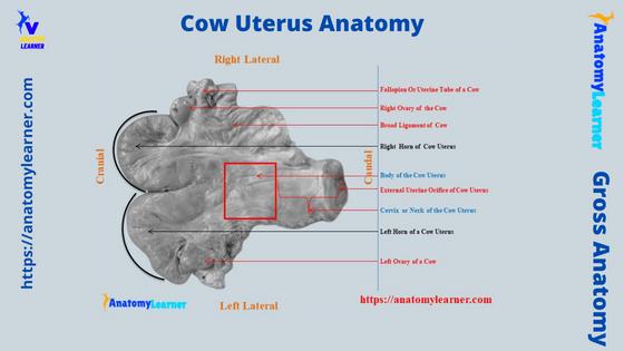
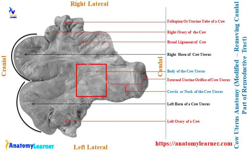
But first, see the different important anatomical features of the cow uterus. The below-mentioned 2 labeled diagrams show both the external and internal anatomical features of the cow uterus.
Okay, let’s try to identify the following anatomical features from the cow uterus structure –
- Horn of the cow uterus (just caudal to the uterine tube),
- Body of the cow uterus (2 horns join to form a single body),
- The neck of the cow uterus (cervix),
- Different parts of the uterine tube (not included under the structure of the cow uterus),
- Broad ligament of the cow uterus,
- Round ligament of the cow uterus, and
- Mesovarium and mesosalpinx of the cow uterus and uterine tube,
The neck is the short part; you may identify it from the surface approach. It is a very hard part of the cow’s uterus compared to the body and horns.
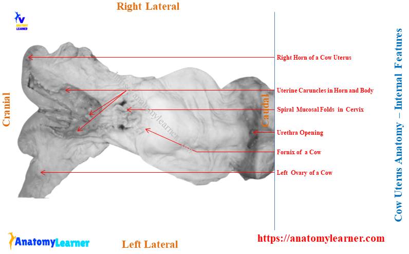
Now, let’s identify the internal features of the cow uterus structure –
- The folded mucous membrane in the horns of a cow’s uterus,
- Uterine caruncles or cotyledon in the mucous membrane of the uterus horn,
- Spiral mucosal fold in the neck of the cow’s uterus,
- Plug of mucus in the neck of cow’s uterus,
- Internal uterine orifice of the uterus,
- External uterine orifice of the uterus, and
- Cervical canal or birth canal of the cow’s uterus,
The cow uterus labelled diagrams show all these anatomical features (both external and internal). These features might provide the basic idea of the structure of a cow’s uterus.
Where is the uterus located in a cow?
The uterus is a hollow muscular organ of the cow’s reproductive system that locates almost in the abdominal cavity. Again, this organ partly locates in the cow’s pelvic cavity between the urinary bladder below and the large intestine above.
If you see the diagram, this uterus of the cow continues cranially with the last part of the uterine or fallopian tube. Again, you will find the connection with the fornix caudally.
So, you may tell, the cow uterus attaches and extend from the sublumbar region to the lateral wall of abdominal and pelvic cavities. But who attaches the cow uterus to these regions?
There is a double fold of the peritoneum, which is known as the broad ligament, that helps the cow uterus to attach to these sublumbar regions and the lateral wall of the cavities. In the structure of the broad ligament, you will find the peritoneum along with the connective tissue, muscle fibres, nerves and vessels.
The muscle fibres continue with the muscle of the uterus wall. Broad ligament from 2 lateral aspects gives a fold to the cow’s uterus.
This fold extends up to the internal inguinal ring and is known as the round ligament of the cow uterus. The same broad ligament of the cow uterus is named differently according to its position.
When the broad ligament supports and covers the ovary of the cow, then it is the mesovarium. Again, the part of the broad ligament covers the uterine tube and mesometrium; then, it is the mesosalpinx.
Unique features of cow uterus anatomy (bovine)
Now, you may point out some of the important anatomical features of the cow uterus. Let’s see what the important features of cow uterus that you should know are –
- There are 3 different parts in the cow uterus (horn, body, and neck or cervix),
- You will find 2 horns in the cow’s uterus that form a gentle curved (as shown in the diagram),
- The horns are comparatively larger than the body of the uterus,
- Internally, the horns possess mucosal fold and uterine caruncles (cotyledon),
- The body is small compared to the body of the horse’s uterus,
- You will find a spiral mucosal fold, mucosal plug, and cervical canal in the neck of the cow uterus (internally),
- The cervical canal of the cow uterus is spiral, whereas it is straight in the horse uterus,
- Again, the neck of the cow uterus presents the internal and external uterine orifices,
Now, you may learn the details anatomical facts of the cow uterus with the labeled diagram. Don’t miss the unique anatomical features of the other animals’ uterus (like a horse, pigs, dogs, and rabbits) from the last section of this article.
Now, I will discuss the different parts of the cow uterus with the labeled diagram. Let’s start with the anatomical features of the horns of the cow uterus.
Horns of cow uterus
You will see 2 horns in the cow uterus anatomy – right and left horns. The cow uterus’s horns are entirely located in the abdominal cavity and have an attachment with the sublumbar region.
Both the right and left horns of the cow’s uterus show a spiral appearance. Normally, they are the muscular spiral tube tapered cranially and joined with the uterine tube. Again, they become broad caudally and unite together to form the body of the cow’s uterus.
The horns of the cow uterus are usually more extensive than the horse and dog. You will find the average 35 to 40 cm length in the cow uterus.
As the horns of the cow uterus are hollow organs, you will find roughly 2 borders – dorsal and ventral. The dorsal border attaches to the sublumbar and abdominal cavity with the broad ligaments.
The ventral border of the horns is concave and free. Now, the free part of the uterine horn directs downward, forward, outward, backward, and finally upward.
Thus, the horns of the cow uterus present a spiral appearance like the horns of the ram. This part of the cow uterus is known as the horn.
Caruncles and cotyledon of cow uterus
The mucous membrane of the horns and body (of cow uterus) shows a unique feature – uterine caruncles. These are the oval to circular prominence that is irregularly scattered over the surface of the mucous membrane (endometrium) of the cow’s uterus (horn and body).
Sometimes, you may also find them arranged in rows in the mucous membrane of the uterine horn and body of the cow. Anatomically, they are the specialized thickening of the ruminant endometrium.
The number of uterine caruncles in the endometrium of the cow’s uterus may vary. A normal healthy uterus of a cow may possess 80 – 100 uterine caruncles in their endometrium.
The caruncles make up the maternal component of the placenta. Again, the fetal component is the cotyledon.
So, together the caruncles and cotyledons make up the placentome. You may see the enlarged placentome during the pregnancy of the cow. This placentome can be palpated to estimate the stage of the cow’s pregnancy.
But, in the horse and dog uterus, you will not find any cotyledons in their endometrium. You will find the full features of the horse and dog uterus anatomy in the last section of this article.
Body of the bovine uterus anatomy
The body is very small in cow uterus, but it appears to be longer. This is due to the presence of a common peritoneal covering on the caudal most segment of the horns before their union.
The body of the cow uterus is partly located in the abdominal cavity and partly in the pelvic cavity. This body is cylindrical but considerably flattened dorsoventrally. So, you will find the elliptical appearance in the cross-section of the body of the cow’s uterus.
The average length of the body of a cow uterus is 5 – 6 cm, and the diameter is about 7 – 8 cm. You will find thicker walls in the body of the cow uterus compared to the horns.
The 2 lateral aspect of the body of the cow’s uterus attaches with the broad ligaments. As the body of the cow uterus is dorsoventrally flattened, you will find 2 distinct surfaces – dorsal and ventral.
Here, the dorsal surface of the body relates to the different parts of the cow’s large intestine (shown in the diagram). Again, the ventral part of the body of the cow uterus has contact with the urinary bladder.
You will also find an inconstant relationship of the cow uterus with the different parts of the cow’s small and large intestines. The point of the body from where the horns separate is known as the fundus.
Finally, the caudal part of the body (of a cow’s uterus) continues with the cranial part of the neck or cervix.
The internal features of the body of a cow uterus are similar to the horns. But, the number of mucosal folds and uterine caruncles is less than the horns.
The uterine glands in the horns and body are long and branch in the cow uterus.
The cervix of cow uterus anatomy
The neck or cervix of the cow uterus anatomy is the most caudal part that joins with the fornix. It is (neck of the uterus) about 8 – 10 cm long in a healthy cow.
The wall of the cervix is very dense and may be about 3 cm in thickness. Thus, this structure is transformed into a narrow canal and remains filled by a spiral fold of mucous membranes.
The mucosal plugs present in the cervix make the lumen spiral. It isn’t very easy to dilate the lumen due to the spiral mucosal fold and mucosal plugs. You know, this spiral lumen (canal) of the cervix is the cervical canal.
So, you will find the tightly closed neck in the structure of the cow uterus. An external and internal cervical orifice are more distinct in the cow uterus.
The mucosal folds form rounded prominence arranged circularly at the external and internal uterine orifices. You will not find any glands (uterine) in the cervix or neck region of the cow uterus. But, the thick mucus is secreted by the goblet cells.
Structure of cow uterus wall
The wall of the cow uterus consists of 3 layers or coats –
- Outer serous coat or perimetrium of the cow uterus,
- Middle muscular coat or myometrium of the cow uterus, and
- Inner mucous coat or endometrium of the cow uterus,
the outer serous coat or perimetrium of the cow uterus covers the whole organ except the lateral border. Then, this perimetrium of the cow uterus is closely adherent to the myometrium layer.
Again, the perimetrium layer continues with the broad ligament of the cow’s uterus.
In the middle muscular coat or myometrium of the cow uterus is thick and composed of 3 layers of smooth muscles –
- Thin external longitudinal muscle fibres,
- A circular and oblique middle layer of smooth muscle fibers, and
- A thick internal layer of circular muscle fibers,
The circular muscle fibers are thicker in the neck or cervix part of the cow uterus. Here, you will find more circular muscles that act as the sphincter.
In the structure of the myometrium of the cow uterus wall, you will also find connective tissue and vessels. These uterine blood vessels remify in the muscular layers.
The oestrogen stimulates the growth of the uterus myometrium layer. In contrast, the length and number of muscle fibers increase during pregnancy.
So, you will find a great change in the wall of the cow uterus in normal and pregnant conditions. These features will well understand in the microscopic slide.
These are the microscopic features of the animal uterus (active and inactive conditions) that will provide the basic idea –
- Uterus histology slide with the labeled diagram,
Now, let’s discuss the inner endometrium layer from the cow uterus structure.
The endometrium of cow uterus
The endometrium of the cow uterus comprises the surface epithelium and lamina propria. You know, the endometrium or the mucous membrane of this segment, is the continuation of the mucous membrane of the uterine tube.
The lining epithelium of the cow uterus vary with the different segment –
- The Horn of the cow uterus – covers with the ciliated columnar epithelium,
- A body and neck of the cow uterus – cover with the columnar epithelium, and
- External uterine orifice – lines with the stratified epithelium,
These features are well described in the histology section of the animal uterus. So, you may read the details facts of these different parts under the microscope from another article of anatomy learner.
The lamina propria of the cow uterus (endometrium) possess stromal cells, tubular uterine glands, lymphatics, nerves, and blood vessels. You will find differently coiled and branched endometrial glands (uterine glands) in the endometrium of the cow uterus.
These glands open on the endometrial surface of the cow uterus. The nerve opens on the caruncular area of the cow’s uterus. In the endometrium of the body and horn of the cow uterus, you will see a pitted button-like structure that is scattered and distributed.
These are the uterine caruncles on the cow uterus (endometrium). Uterine vessels enter the caruncle through the attached surface with the uterine wall.
Attachment of cow uterus anatomy
I have already described the attachment of the cow uterus anatomy earlier in this article. Now, I will show the details of the attachment of the cow uterus with the sublumbar region, abdominal, and pelvic cavity.
In this section, you will also find the different organs, parts, and structures that have direct and indirect attachment or contact with the cow uterus. Let’s see the attachment and connection of the various organs or parts of the cow uterus.
The cow uterus’s body and horns (2) attach to the abdominal and pelvic wall with the 2 extensive peritoneal folds (double layered). This is the broad ligament of the cow uterus, also known as the ligament lata uteri.
These 2 broad ligaments extend on either side from the sublumbar region. It also extends from the lateral wall of the abdominal and pelvic wall to the dorsal and lateral wall of the cow uterus.
That means the dorsal border of the uterus and lateral margin of the body of the uterus attaches with the broad ligament to these regions. In the structure of the broad ligament, you will find the uterine vessels and nerves.
There are also large amounts of smooth muscle fibers and connective tissue present in the structure of the broad ligament. The muscle layers of the broad ligament continue with the muscular layer of the uterine wall.
Now, the lateral wall of the broad ligament provides a fold on both sides. And thus form the round ligament in the cow uterus. It bends with the parietal peritoneum over the deep inguinal ring of the cow.
Organs or parts attach to cow uterus
The cranial part of the horn of the cow uterus attaches to the caudal part of the fallopian tube (isthmus). Again, the caudal part of the uterus (neck) attaches to the fornix of the cow.
The 2 lateral aspect of the cow uterus (tube) attaches to the broad ligament, which again attaches them with the lateral wall of the abdominal and pelvic wall. The ventral surface of the cow uterus (horn, and body) are free and has a curve appearance (especially horn).
You will see indirect contact with the sublumbar muscle and direct contact with the last part of the large intestine on the dorsal surface of the cow uterus. Again, you will find direct or indirect contact with the urinary bladder and different parts of the intestine ventrally.
Vessels and nerves to cow uterus
2 main arteries supply the cow uterus – the uterine artery (2) and the uterine branch (1) of the ovarian artery. The uterine artery is the chief structure that supplies the cow uterus.
The uterine branch of the ovarian artery has a flexuous course in the broad ligament of the cow uterus. You will also find a branch from the internal pudendal artery that supplies to the cow’s uterus.
The caudal branch of the uterine artery supplies the caudal part of the horn and the adjacent part of the body of the cow uterus. You will find some anastomoses with the uterine artery branches in the cow uterus.
You will see numerous lymph vessels that go to the internal iliac lumbar lymph nodes. The uterine and pelvic plexus nerves innervate the cow uterus.
Cow uterus anatomy labeled diagram
Now, let’s see some of the cow uterus anatomy labeled diagrams that might help you get the full idea of it. Here, I will show you the different external and internal features of the cow uterus structure.
First, let’s see the full female cow reproductive system diagram, where I tried to identify the different segments of the fallopian tube (along with the ovary), uterus, and ligaments. The 2 horns (right and left), single body, and neck of the uterus also identify separately in the cow uterus labeled diagram.
The cow uterus labeled diagram shows the internal mucosal folds in the horn and body parts. Again, it shows the uterine caruncles in the mucous membrane of the horn and the body of the cow uterus.
The number of mucosal folds is more in the horns area compared to the body of the uterus. Again, the mucosal folds spiral in the cow uterus’s neck region (shown in the diagram).
The number of uterine caruncles decreases in the body compared to the horns of the cow uterus (shown in the diagram). No such uterine caruncles are found in the uterus neck diagram.
The cow uterus labeled diagram also shows the mucosal plugs in the neck region. Here, the diagram shows the spiral cervical canal in the cow uterus.
The external and internal uterine orifices are also identified in the cow uterus labeled diagram. You may get more labeled diagrams on the cow uterus structure on social media of anatomy learners.
Again, there is a video on the cow uterus where I explain every organ structure. Let’s check that video and better understand the cow uterus.
Uterus in horse and dog compare to cow
The anatomical facts of the horse and dog uterus are somewhat different compared to the cow. You will find a great variation (difference) in the shape and structure of the dog and pig uterus compared to the cows.
Here, I will enlist some (unique) of the important anatomical facts of the horse, dog, and pig’s uterus with the labeled diagram. The labeled diagrams on the horse, dog, and pig uterus might help you to understand the unique features.
Okay, let’s start with the unique features of the horse uterus anatomy.
Horse uterus anatomy
In the horse uterus anatomy, you will find the below-mentioned unique features –
- The horse uterus divides into 3 different parts – horns, body, and neck (like the ruminant),
- You will see the short horns (right and left) compared to the cow,
- The cranial extremity of the horns (uterus) is blunt and continues with the uterine tube,
- You will find a more extensive and longer body in the horse’s uterus in comparison to the cow,
- The neck is very short and possesses all the anatomical features that you have found in the cow uterus,
- Here, the cervical canal is straight, whereas it is spiral in the cow,
- You will not find any uterine caruncles or cotyledon in the endometrium of the horse uterus,
These are concise information on the horse’s uterus anatomy. But, you may know more about the horse uterus structure from another article of anatomy learner.
Now. Let’s see the unique (basic) anatomical features of the dog uterus anatomy with the labeled diagram.
Dog uterus anatomy
The shape and the location of the dog’s uterus are somewhat unique compared to the cows. Here, I will only enlist the unique anatomical features of the dog’s uterus with the labeled diagram.
But, you are suggested to read the full guide on the dog uterus anatomy from another article of anatomy learners.
Okay, let’s see the unique features of the dog’s uterus anatomy –
- The whole structure of the dog uterus appears as V-shaped, whereas the horn of a cow becomes S-shaped,
- You will find a longer body in the dog uterus anatomy and a very short body,
- The horns (right and left) are not tapered cranially like the cow’s uterus (horns),
- You will not find any cotyledons in the endometrium of the dog uterus structure,
- The neck region of the dog uterus possesses similar structures, but the cervical canal is straight in the dog,
Now, let’s know very little about the unique anatomical features of the pig and rabbit uterus. There is a great variation (difference) in the shape and structure of the horns of a pig uterus.
The horns of the pig uterus are very long, flexuous, and coiled structure compared to the other animals. You will see a comparatively small body in the pig’s uterus.
In the rabbit uterus, you will also find a unique feature. The horns of the rabbit uterus are curved, narrow, slightly flexuous tubes.
The thickness of the uterus wall is comparatively more in rabbits. There are 3 – 4 longitudinal mucosal folds in the internal aspect of the uterine horn of the rabbit.
The neck of the rabbit uterus shows a double cervical opening which is very exceptional compared to the cow.
Common inquiries on cow uterus anatomy
In this section, I will provide a short answer to the common inquiries on the cow uterus anatomy that the learner asks. I recommend reading the full guide on the cow uterus structure (bovine uterus) from start to end.
Let’s see what the common inquiries on the cow uterus from the learners are.
Does a cow have a uterus?
Yes, cows have a uterus that extends from the sublumbar region to the abdominal and pelvic cavities. There are 2 horns (right and left) in the structure of the cow uterus, which is comparatively longer than other parts of the cow uterus.
Here, the right and left horns join to the body of the cow’s uterus caudally. You may easily identify the 3 different parts of the cow uterus (horns, body, and neck) by external and internal anatomical facts.
In this article, I have already described all these features that help you to identify the different parts of the cow uterus.
How much does a cow’s uterus weigh?
The weight of the cow uterus varies with the animals. A healthy uterus may weigh 1.5 – 2.25 kg (not constant).
If you want to weigh the cow’s uterus, you might separate this part from the reproductive tract.
What does a cow’s uterus look like?
The horns of the cow’s uterus look S-shaped. Again, the body and neck of the cow’s uterus look like a straight muscular tube.
The arrangement of the horns of the cow’s uterus is already described in this article. Its cranial extremity is narrow and continues with the caudal part of the uterine tube.
Practically, the body of a cow uterus is very short and easily identified with its longitudinal folds (less than the horn). The neck of the cow uterus is tough to touch from the surface because of the spiral mucosal folds and plugs.
Where is a cows uterus?
The cow uterus is in the sublumbar region and abdominal cavity, and a small portion remains in the pelvic cavity. A broad ligament (modification of the peritoneum) connects the uterus with the sublumbar, abdominal, and pelvic cavities.
There are some organs and structures that have direct or indirect contact with the different parts of the cow uterus. In this article, I have already described the attachment of the different organs to the cow uterus.
What type of uterus do cows have?
The cows have a bipartite type of uterus. You will also find the same type of uterus in the horse, sheep, and goats also.
The cow’s uterus possesses 3 distinct segments – horns, body, and neck. Two horns arrange in two lateral sides of the abdominal and pelvic cavities.
Each of these horns contains an oviduct or uterine tube. The first segment of the uterine tube contains the ovary.
How many uterus do cows have?
There is only one uterus present in the female cow reproductive tract. But, a cow’s uterus possesses 3 portions or parts – a long horn, a short body, and a short neck.
The horn is longer than any other part of the cow uterus. Anatomically, the horns (both right and left) are curved and form an S-shaped appearance.
Conclusion
So, the cow uterus anatomy consists of 3 main parts – 2 horns (right and left), 1 cylindrical body, and a dorsoventrally flattened neck. The internal structure of the cow uterus possesses unique uterine caruncles and cotyledons.
In most species, you will find the fundamental difference in the horns and body parts of their uterus. Again, a distinct structural difference is also present in the cervical canal of the animal uterus.
All the labeled diagrams on the cow uterus anatomy might help you ideally identify all the actual sample features perfectly.