The cow leg anatomy consists of bones, muscles, nerves, and vessels. Bones are the hardest and main component of the cow leg structure.
Again, the muscles are also essential as most vessels and nerves pass along or within them. Here, I will share details information on the cow front and hind leg anatomy with the labeled diagram.
First, you will find all the bones from both legs of the cow with their main identifying features. As many muscles are in the structure of a cow leg, I will only share the most important ones from them.
Again, I will focus on the main branches of the nerves and vessels that run along both the front and back legs of a cow. So, if you want to get a basic concept of the anatomical facts from the cow’s front and hind limbs (leg), let’s continue this article till the end.
Cow leg anatomy
The term cow leg anatomy means only the anatomical facts of the tibia fibula region. But, here, I will discuss the anatomical points from different areas of the front and hind legs of the cow.
First, make sure you have a good piece of knowledge on the different regions from both the front and the hind leg of the cow –
- The front or thoracic limb of a cow divides into below-mentioned regions –
- A thoracic girdle or shoulder girdle – includes the scapula bone of the cow,
- Arm or brachium region – includes the humerus bone,
- Forearm or antebrachium region – consists of the radius and ulna bones of the cow,
- Manus region – consists of the carpals, metacarpals, and phalanges,
Again, the pelvic limb (leg) of the cow divides into the following different areas –
- The pelvic girdle of the cow – includes the os coxae and sacrum,
- A thigh region of the cow – consists of the femur bone,
- Leg region of the cow – includes the tibia and fibula bones, and
- Pes region – consists of the tarsals, metatarsals, and phalanges of the cow’s hind limb,
All these regions from both the front and hind legs of the cow are identified with their specific bones in the labeled diagram.
Again, you might identify the muscles, vessels, and nerves from the cow’s front and back legs. The major muscles from the front leg of a cow are –
- Supraspinatus, infraspinatus, and subscapularis from the shoulder girdle,
- Triceps brachi and brachialis from the arm region,
- Extensor and flexor muscles from the forearm and manus,
Again, the hind leg of a cow possesses the below-mentioned important muscles –
- Biceps femoris, quadriceps femoris, semitendinosus, and semimembranosus from pelvic girdle and thigh,
- Gastrocnemius, and other extensor and flexor muscles from the leg and pes region,
But you will learn more muscles from the cow leg in the next section.
Major vessels and nerves in cow leg
While studying the anatomical facts of the cow leg (both front and back or hind legs), you might also have a good knowledge of the major vessels and nerves. Here, the front leg of cow posses following major vessels –
- Left and right axillary arteries,
- Brachial arteries (right and left – continuation of the axillary arteries), and
- Branches of the brachial arteries – radial, ulnar, and median that supply up to the digits of the cow,
You will also find the cephalic, radial, and median veins in the structure of the cow’s front leg anatomy. Again, from the hind or back leg of a cow, you will find the below-mentioned vessels –
- External iliac and deep femoral arteries,
- Saphenous artery and cranial tibia artery,
- Medial and lateral plantar arteries of the cow (continuation of the saphenous artery),
- Dorsal pedal, metatarsal, and dorsal common digital arteries (continuation of the cranial tibia artery),
The major veins from the cow’s back or hind leg structure are femoral and saphenous. You may learn the short course of these veins from another article by an anatomy learner.
Two major nerve plexuses in a cow’s body – brachial and lumbosacral plexus innervate the legs. Here, the brachial plexus of the cow innervate the different aspect and digits of the front leg. Again, the nerves (branches of an ischiatic nerve) from the lumbosacral plexus innervate the different regions and digits of the cow’s hind leg.
There are 12 nerves in the brachial plexus of a cow, but the following 3 major nerves innervate up to the digits –
- Radial, median, and ulnar nerves of the cow,
You may learn details anatomical facts (courses and supply) of these 3 major nerves from the below-mentioned article –
- Formation of brachial plexus in ox – nerves and their course with innervation,
Unique features of cow leg anatomy
So, in the cow leg anatomy, you will find the following unique features –
- The bones from both the front and the hind leg of a cow possess typical osteological features compared to the other animal,
- Major important muscles from the cow front legs are – supraspinatus, infraspinatus, subscapularis, deltoideus, triceps brachii, biceps brachii, brachialis, and extensor and flexor group of muscles,
- The essential muscles from the hind leg of a cow are – the gluteiobiceps, quadriceps, semitendinosus, semimembranosus, gastrocnemius, and other extensor and flexor groups of muscles,
- The axillary artery and its major branches supply the different segments of a cow’s front leg,
- Different branches of the external iliac artery provide the different parts of the cow’s hind leg,
- Nerves from the brachial plexus are another main component of the cow’s front leg structure,
- The ischiatic nerve and its 2 major branches (tibia and fibula) innervate the different segments of the cow’s hind leg,
You will find some other organs or parts or features like the presence of lymph nodes, lymph vessels, joints, ligaments, and tendons in the structure of both the hind and front legs of the cow. If you want to know (learn) more about the anatomical facts of the cow leg, let’s continue this article.
Okay, first, start with the bones and joints of the cow’s front and back legs.
Cow leg bone anatomy
You have already got the idea of the different bones both from the cow’s front and hind legs. The front and hind legs of the cow possess the following bones –
- The front leg of a cow – possesses scapula, humerus, radius, ulna, and manus (carpal, metacarpal, and phalanges), and
- Hind leg of a cow – posses os coxae (ilium, ischium, and pubis), femur, tibia, fibula, and pes (includes tarsal, metatarsal, and phalanges),
So, in the front and hind leg of a cow, you will find different types of bone-like –
- Flat bones of cow leg – scapula, ilium, ischium, and pubis,
- Long bones of cow leg – humerus, radius, femur, tibia, metacarpal, and metatarsal,
- Modified long bones in a cow leg (miniature long bone) – ulna and fibula,
- Short bones in a cow leg – carpal, tarsal, and phalanges,
- Sesamoid bones in a cow leg – patella,
You may know more about these different types of bone from the cow or other animal bodies from the below-mentioned article –
- Types of bone in animals with the labeled diagram,
Now, you may easily calculate the total number of bones from both the front and hind leg of the cow. Let’s see the number of bones from both 4 legs (2 front and 2 hinds) of the cow skeleton –
| Cow front leg bones | Cow hind leg bones |
| Scapula – 1 | Os coxae – 3 |
| Humerus – 1 | Femur – 1 |
| Radius and ulna – 2 | Tibia fibula – 2 |
| Carpal – 6 | Patella – 1 |
| Metacarpal – 2 + 1 = 3 | Tarsal – 5 |
| Phalanges – 3 x 2 = 6 | Metatarsal – 2+1 = 3 |
| Sesamoid bone – 4+2 = 6 | Phalanges – 3 x 2 = 6 |
| Sesamoid bone – 4+2 = 6 | |
| Total = 25 | Total = 27 |
| 25 bones in each front leg | 27 bones in each hind leg |
Table 1 shows the name of bones with their number for both 2 front and 2 hind legs of a cow. Each of the front leg of a cow posses in a total of 25 bones. Whereas the hind leg of a cow possesses 27 bones on each right and left.
But, you might also have a good piece of knowledge on the total number of bones from the cow skeleton.
How many bones are in the cow skeleton?
If you consider the main bones, you will find almost 208 bones in the cow skeleton. When you consider the small metacarpal, small metatarsal, and all sesamoid bones from both the front and hind leg of the cow, you will find 217 bones in a cow.
Let’s see how there are 217 bones in the cow skeleton. I will show you the summary of the bones from the cow skeleton in Table 2 –
| Bones of cow | Numbers |
| Bones in skull | 32 |
| Bones in vertebrae | 52 |
| Sternum, and ribs | 27 |
| Bones in front leg | 50 |
| Bones in hind leg | 54 |
| Visceral bones | 2 |
| Total Bones | 217 |
Again, you may know the details of the cow bones from another article by anatomy learners.
Cow leg bone structure
As most of the prominent bones of the cow leg are long bones, so, you might have an idea of the structure of the long bone. I have already described all the features of an animal’s long bone here on anatomy learner previously.
You will get the full guide on the long bone from the below-mentioned article –
- Long bone structure with the labeled diagram,
So, in a cow leg bone like the humerus, femur, radius, and tibia, you will find the following structures in common –
- Epiphysis of the cow leg bone,
- The diaphysis of the leg bone,
- An epiphyseal plate of the cow leg bone,
- Hyaline or atricualr cartilage,
- Parts of cancellous and compact bones,
- Marrow cavity of the cow leg bone, and
- Periosteum of the cow leg bone,
The labelled diagram shows all these features from the cow leg bones.
Now, let’s know the essential osteological features of the cow’s front and hind legs bones.
Cow front leg bones anatomy
Here, I will show you the important osteological features from every single bone of cow front leg anatomy. First, let’s start with the cow scapula bone that forms the thoracic or pectoral girdle.
Cow scapula features
It is a flat triangular bone in the front leg of a cow that locates in the cranial part of the lateral wall of the thorax. The long axis of the cow scapula extends obliquely from the 4th (fourth) thoracic spine to the ventral end of the 1st (first) rib.
You will find the following important (main) osteological features in the cow scapula –
- The cow scapula possesses 2 surfaces, 3 borders, and 3 angles (shown in the diagram),
- The lateral surface of the cow scapula possesses a spine that divides the bone into infraspinatus and supraspinous fossa,
- An acromion process is well developed in the cow scapula,
- You will see a hollow medial or coastal surface in the scapula that possesses subscapular fossa and facies serrate,
- The cranial border is rough, whereas the caudal border is slightly concave,
- You will find scapular cartilage in the dorsal border of the cow scapula,
- The ventral angle of the cow scapula possesses some essential structures like the glenoid cavity, supraglenoid tubercle, and small coracoid process,
If you want to know (learn) more about the details anatomical facts of the cow scapula with the labeled diagram and video, you may read the below-mentioned suggested article –
Now, let’s see the osteological features of the cow humerus from its front leg.
Cow humerus anatomical features
As the cow humerus is a long cylindrical bone, it possesses a body and 2 defined extremities (proximal and distal). In the anatomy of a cow humerus, you will see the following important osteological features –
- The femur of a cow is slightly twisted and possesses 4 defined surfaces – lateral, medial, cranial, and caudal,
- A musculo spiral groove and a small deltoid tuberosity are located at the lateral surface of the cow humerus,
- The medial surface of the cow humerus possesses the teres major tuberosity,
- The crest of the humerus locates on the cranial surface of the cow humerus,
- In the proximal extremity of the cow humerus, you will see the head, neck, 2 tuberosities (lateral and medial), and intertuberal groove,
- The distal extremity possesses medial and lateral condyles,
- You will see olecranon fossa (caudally) and radial fossa (cranially) on the distal extremity of the cow humerus bone,
You may also learn the details anatomical facts of the cow humerus bone from the below-mentioned article –
- The humerus of an ox or cow with the labeled diagram,
Features of cow radius and ulna bones
The cow radius bone is larger than the ulna bone and extends vertically. You will find the below-mentioned important osteological features in the cow radius and ulna bones –
- The cow radius possesses a body and 2 extremities (proximal and distal),
- There are 2 surfaces (cranial and caudal) and 2 borders (lateral and medial) in the structure of a cow radius bone,
- The caudolateral surface of the cow radius attaches with the cranial surface of the ulna bone (also forms the interosseous spaces between the radius and ulna),
- The proximal extremity of the cow radius possesses capitular fossa and radial tuberosity, whereas the distal extremity possesses the 3 facets for the carpal bones,
- The cow ulna is an ill-developed bone that includes 3 surfaces and 3 borders,
- A proximal extremity of the cow ulna bones possesses 2 important features – anconeal process and olecranon tuberosity,
- The distal extremity contributes to form the lateral facet for the ulnar carpal and also forms the styloid process,
You may also learn more about the cow radius and ulna bones from the below-mentioned article –
- Radius and ulna of ox – details anatomical facts with the labeled diagram,
Cow manus anatomy
The manus from the cow front leg anatomy comprises 6 carpals, 4 metacarpals (2 large fused to form a single one and other 2 small), and 2 developed digits. Each of the digits of a cow contains 3 phalanges – the first, second, and third phalanges.
Let’s see the unique osteological features of the cow manus anatomy –
There are 6 carpal bones in the cow that arranges into 2 rows. In the first or proximal row, you will find the radial, intermediate, ulnar, and accessory carpal bones in the cow. Again, the distal row shows the second and third fused carpal and fourth carpal bones.
There are 2 large metacarpal (III, IV) and 1 small metacarpal (V – lateral) bone in the structure of the cow front leg. These 2 large metacarpal bones fuse to form the single metacarpal in the cow.
The single metacarpal of the cow possesses a shaft and 2 extremities. In the anatomy of the cow metacarpal, you will also find the 2 surfaces (cranial and medial) and 2 borders (lateral and medial).
The proximal extremity of the cow metacarpal possesses 2 articular surfaces (lateral and medial) and metacarpal tuberosity at the dorsomedial surface. Again, the distal extremity of the cow metacarpal possesses the lateral and medial condyles with an intercondyloid cleft.
You will also find 4 depressions on the distal extremity (caudally) on the cow metacarpal bone for the sesamoid bones.
Digits III and IV are developed in the cow’s front leg, consisting of 3 phalanges – proximal, middle, and distal. You will also find 2 distal sesamoid bones in each digit of the cow.
Cow hind leg bones anatomy
Now, you will learn the unique osteological features of the cow hind leg bones. You have already identified all the bones like the hip bone, femur, tibia, fibula, tarsal, metatarsal, and phalanges from the cow hind leg structure.
Here, the hip bones mean the ilium, ischium, and pubis (flat bones in the cow skeleton). The right and left hip bones (oscoxae) of the cow skeleton fuse to form the os coxae. Again, the os coxae and sacrum bones form the pelvic girdle or bony pelvis in the cow.
Let’s see the unique osteological features of every single bone of the cow hind leg structure. First, start with the unique osteological features of the hip bone (os coxae).
Bones of cow pelvic girdle or hip
Ilium is the largest and triangular bone in the cow hip anatomy. You will find 2 surfaces, 3 borders, and 3 angles in the structure of the cow ilium bone.
The gluteal surface of the cow ilium is wide and possesses a gluteal line. Again, the sacropelvic part attaches to the articular surface of the cow sacrum.
There is a psoas tubercle in the medial part of the sacropelvic surface of the cow ilium bone. The medial border of the cow ilium bone forms the ischiatic notch and spine.
The lateral angle of the cow ilium bone form the coxal tuber, whereas the medial angle help to form the sacral tuber. Finally, the distal angle of the cow ilium bone joins with the other bones of the hip and forms a cotyloid cavity (acetabulum).
Cow ischium is the rough quadrilateral flat bone that forms the caudal and ventral walls of the bony pelvis. This bone of the cow hip possesses 2 surfaces (pelvic and ventral), 4 borders (cranial, caudal, lateral, and medial), and 4 angles.
The caudal border of the cow ischium bones possesses an ischiatic arch, and the lateral border forms the lesser ischiatic notch. Again, the caudolateral angle of the ischium bone forms the important structure – thick three-sided ischiatic tuberosity or tuber ischii in the cow.
The cow pubis is a small triangular bone that forms the craniomedial aspect of the pelvic floor. You will see a body, 2 surfaces (pelvic and ventral), and 3 borders (cranial, medial, and caudal) in the structure of the cow pubis bone.
The cranial border of the pubis bone possesses an ilopubic eminence and ventral tubercle. In contrast, the caudal border of the cow pubis forms the margin of the obturator foramen (the obturator nerve passes through this foramen).
Cow femur bone anatomy
The cow femur is the largest and more massive bone in the hind leg. This bone of the cow’s hind leg directs obliquely downward and forward.
As this is a long bone in the cow’s hind limb, you will find a cylindrical body and 2 extremities in the femur. The body of the cow femur bone is cylindrical but flattened caudally and possesses 4 different surfaces – cranial, caudal, medial, and lateral.
Here, the cranial, medial, and lateral surfaces are smooth and convex side by side, and thus they form a round structure in the cross-section. Again, the caudal surface of the cow femur is wide and possesses some nutrient foramina.
The lateral and medial borders of the cow femur possess some important osteological features. On its medial border, you will find the lesser trochanter at the proximal part of the cow femur bone. Again, the lateral border of the cow femur possesses supracondyloid fossa on its distal extremity.
The proximal extremity of the cow femur bone possesses a head, neck, and greater trochanter (laterally). You will find a deep fovea capitis femoris in the structure of the head of a cow femur bone. The caudal border of the greater trochanter posses a trochanteric crest and fossa.
Finally, the distal extremity of the cow femur bone possesses trochlea (cranially) and condyle (caudally). There are 2 condyles (lateral and medial) in the distal extremity of the cow femur bone that is separated by the intercondyloid fossa.
You may learn more about the cow femur bone from the following article of anatomy learner –
- Femur bone of an ox or cow with the labeled diagram,
Cow patella anatomy
Patella is the large triangular sesamoid bone in the cow hind leg anatomy. This sesamoid bone articulates with the trochlea of the cow femur bone and presents 2 surfaces, 2 borders, a base, and an apex.
The cranial or free surface of the cow patella is quadrilateral and convex. You will find a rough area on this surface to attach muscles and ligaments.
The articular surface of the cow patella is also quadrilateral and less extensive. You will see a verticle rounded ridge on the articular surface of the cow patella that corresponds to the groove on the trochlea of the femur bone.
The medial and lateral borders of the cow patella join distally and form the apex. There is a concave craniocaudal base in the cow patella that faces proximally and caudally.
Cow tibia and fibula anatomy
The cow tibia is another strong and massive bone from the hind limb. In the structure of the cow tibia, you will see a large, three-sided (proximally) body and 2 extremities.
But, the body of the cow tibia is also flattened and smaller at its distal extremity. It is also twisted and presents 3 surfaces (medial, lateral, and caudal) and 3 borders (cranial, medial, and lateral).
The lateral surface of the cow tibia is spiral and smooth. You will find the tibial crest at the cranial border of the cow tibia bone.
The proximal extremity of the cow tibia bone posses – articular eminence (lateral and medial condyles) and intercondyloid eminence or spine. In comparison, the distal extremity of the cow tibia possesses an articular surface adapted to the trochlea of the talus.
The cow fibula is the ill-developed or reduced bone that locates the lateral border of the tibia. You will not find such unique osteological features in the structure of the cow fibula bone. The distal extremity of the cow fibula bone fuses with the tibia and continues with the lateral malleolus.
Cow pes anatomy
There are 5 tarsals, 2 large metatarsals, 1 small metatarsal, and 2 well-developed digits (possess 3 phalanges in each) in the structure of cow pes anatomy. The tarsal of the cow pes arrange into 3 rows –
- The proximal row of the cow pes consists of the tibial tarsal (talus) and fibular tarsal (calcaneus),
- A middle row of the cow pes comprises the central and fourth fused tarsal, and
- The distal row of the cow pes contains the first tarsal and second and third fused tarsal bones,
There are also 2 large metatarsals (III, IV) in the cow that fuses to form a single bone. Again, there is a small metatarsal (II) structure of the cow pes.
You will find similar features in the cow’s hind leg digit as you found in the front leg. So, there are also 2 developed digits which contain 3 developed phalanges in each – proximal, middle, and distal.
Again, the number of the proximal and distal sesamoid bones in the cow hind leg is similar to the front leg.
I hope you got the basic idea of the osteological features of the cow leg bones with the labeled diagram. Now, let’s know the muscles anatomy of the cow legs (both front and back).
Cow front leg muscles anatomy
You might identify both superficial and deep muscles from the different regions of the cow front leg. There are many muscles in the cow front leg, so it is difficult to describe them in detail. Instead, it will be better to identify the most superficial muscles and describe some essential ones.
First, let’s see the cow front leg muscles labeled diagram and try to identify all from the actual sample. From the thoracic girdle of the cow’s front leg, you might see the following muscles –
- Trapezius muscle of a cow,
- Omotransversarius muscle,
- Cutaneous omobrachialis,
- Rhomboideus muscle,
- Lattisimus dorsi muscle of a cow,
- Part of the brachiocephalic muscle,
- Different parts of the pectoralis muscle, and
- Serratus ventralis thoracic and cervicis,
Again, from the shoulder region of the cow, you might identify the below-mentioned muscles –
- Deltoideus muscle,
- Teres minor muscle of a cow,
- Infraspinatus muscle,
- Supraspinatus muscle,
- Subscapularis muscle,
- Teres major muscle, and
- Coracobrachialis muscle,
Now, let’s see the extensor and flexor muscles from the arm region of the cow front leg –
- Biceps brachii muscle,
- Brachialis muscle,
- Triceps brachii muscle of a cow,
- Pronator teres muscle,
- Tensor fascia antebrachii muscle,
- Anconeus muscle of a cow,
The labelled diagram identifies all these muscles from the cow’s front leg (shoulder girdle to arm). Now, let’s see the muscles from the antebracium (forearm) and manus of the cow front leg.
Muscles of the cow’s antebrachium and manus
There are extensor and flexor groups of muscles in the antebrachium and manus of a cow. These muscles are clinically important as most of the nerves and vessels pass along with these muscles.
From the extensor group muscles of the cow’s antebrachium, you will find the below-mentioned muscles –
- Extensor carpi radialis muscle of the cow,
- Extensor digitorum communis muscle,
- Lateral digitorum extensor muscle, and
- Abductor digit I longus muscle,
Again, the flexor muscles from the cow’s antebrachium and manus are listed below –
- Ulnaris lateralis muscle of the cow leg,
- Flexor carpi radialis muscle of the cow,
- Flexor carpi ulnaris muscle of a cow,
- The flexor digitorum profundus muscle of a cow,
- Flexor digitorum superficialis muscle of a cow, and
- Interosseous muscle of the cow front leg,
All these extensor and flexor groups of muscles are identified in the cow front leg muscle labeled diagram. Now, you will learn the details and anatomical facts of some of the major muscles from the cow front leg.
Lateral flexor muscle of cow shoulder
The lateral flexor muscles of the cow front leg anatomy consist of the deltoideus, infraspinatus, and teres minor. Here, the cow deltoideus muscle divides into an acromial and scapular part.
So, the cow deltoideus arises from the acromion process of the scapula and also from the caudal border of the scapula. And both part of the deltoideus inserts into the deltoid tuberosity of the humerus bone.
An infraspinatus is a powerful, heavy tendinous muscle in the cow front leg that fills the entire infraspinous fossa. This muscle arises from the infraspinatus fossa, the spine of the scapula, and scapular cartilage.
The tendon of the cow infraspinous muscle inserts into the greater and lesser tubercles of the humerus bone. This infraspinatus muscle serves as the lateral collateral ligament of the shoulder joint of the cow front leg.
On the other hand, teres minor muscle is a small muscle that lies caudolateral to the shoulder joint under the deltoid muscle. It arises from the distal half of the caudal border of the cow scapula.
The main action of the teres minor muscle is to flex the cow shoulder joint.
Medial flexor muscle of cow shoulder
The medial flexor muscles of the cow front leg include the subscapularis, teres major, and coracobrachialis. You will see 3 parts in the structure of the subscapularis muscle of the cow.
The subscapularis muscle of a cow is falt and follows the outline of the costal surface of the scapula. It arises from the scapular cartilage and the entire subscapular fossa of the cow scapula.
All the 3 parts of the cow subscapularis muscles insert into the lesser tubercle of the humerus bone.
The teres major is a flattened bone that extends from the caudal border of the scapula to the teres tuberosity of the humerus bone. So, this muscle of the cow front leg arises from the caudal angle of the scapula and inserts into the humerus’s tuberosity.
This teres major muscle of the cow also helps to flex the shoulder joint.
On the other hand, the coracobrachialis muscle of the cow is flat and lies on the medial border of the shoulder joint. This muscle arises from the coracoid process of the scapula and inserts into the teres tuberosity of the humerus bone.
The primary function of the cow coracobrachialis muscle – adduct the arm and flex the shoulder joint.
You will also find the supraspinatus muscle, the powerful extensor muscle in the cow front leg. It arises from the supraspinatus fossa, spine, and ventral part of the cartilage of the scapula. Finally, this muscle inserts into the proximal tubercle of the humerus.
Flexor muscles of cow elbow (arm)
The flexor muscles of the cow elbow consist of biceps brachii, brachialis, and pronator teres. Here, the biceps brachii of the cow is a strong muscle that lies (runs) on the cranial surface of the humerus bone.
It arises from the supraglenoid tubercle of the cow scapula. You will find both deep and superficial insertion of the biceps brachii muscle. Superficially, the biceps brachii muscle of the cow inserts into the radial tuberosity of the radius bone.
The cow brachialis muscle locates on the musculospiral groove of the humerus bone. It arises from the proximal third of the caudal surface of the humerus.
This brachialis muscle of a cow inserts on the medial border of the radius bone just distal to the radial tuberosity. These muscles of the cow front leg help to flex the elbow joint.
The cow pronator teres is a weak, narrow band-type muscle located on the medial surface of the elbow joint. It arises (origins) from the medial epicondyle of the humerus with the medial collateral ligament of the elbow joint.
This muscle inserts on the medial border of the radius bone and produces a slight flexion of the elbow joint.
Extensor muscle of cow elbow (arm)
The extensor muscles of the cow elbow or arm are well-developed and include tensor fascia antebrachii, triceps brachii, and anconeus. Here, the tensor fascia antebrachii of the cow front leg is a slender muscle that lies caudal to the triceps brachii muscle.
This tensor fascia antebrachii arises from the caudal border of the scapula and also from the lattisimmus dorsi. It terminates as the narrow tendon on the medial surface of the olecranon process of the cow ulna bone.
The triceps brachii is the largest muscle in the arm region of the cow front leg. You will find 3 main heads in the cow triceps brachii muscle structure – long, lateral, and medial. But, sometimes, you may also find an accessory head in the structure of the cow triceps brachii muscle.
The long head of the cow triceps brachii is the largest part that arises from the caudal border of the scapula. Again, the lateral head of the triceps arises from the lateral surface of the arm. Finally, the medial head of cow triceps arises from the medial surface of the body of the humerus bone.
The anconeus is a very small muscle on the cow front leg structure that occupies the olecranon fossa. It arises from the humerus’s caudal surface and the olecranon fossa.
This muscle is inserted on the cranial border and lateral surface of the olecranon process of the cow ulna bone. The primary function of the anconeus muscle is to extend the elbow joint and raise the joint capsule.
Extensor muscles of cow antebrachium
The extensor carpi radialis muscle of the cow arises from the lateral epicondylar crest of the humerus and radial fossa. This muscle is inserted into the metacarpal tuberosity as the broad and flat tendon.
The main function (action) of the extensor carpi radialis – is to extend and flex the carpal joint and flex the elbow joint,
The extensor digitorum communis muscle of a cow is more complex that comprises 2 parts – lateral and medial belly. Here, the lateral belly of the extensor digitorum communis muscle is slender and possesses a deep, superficial head.
The superficial head of the lateral belly arises from the radial fossa and lateral epicondyle of the humerus. Again, the deep head of the lateral belly arises from the proximal third of the caudolateral aspect of the radius bone.
The medial belly of the extensor digitorum communis muscle arises from the radial fossa and lateral epicondyle of the humerus. It helps to extend the carpal and digital joints of the cow front leg. Again, this muscle flexes the elbow joint of the cow.
The extensor digitorum lateralis muscle arises from the lateral epicondyle of the humerus and the lateral aspect of the radial head. This muscle extends the lateral digital and carpus. Again, it also flexes the elbow joint like the extensor digitorum communis muscle.
The abductor digit I longus muscle is a thin and flat muscle in the cow’s front leg that lies deep into the other muscles. It arises from the lateral surface (1) of the distal half of the radius and craniolateral aspect of the ulna bone.
Flexor muscles of cow front leg anatomy
The ulnaris lateralis of a cow front leg is a long, straplike muscle in the craniolateral aspect of the antebrachium. This ulnaris lateralis arises from the lateral epicondyle of the humerus bone.
Finally, it inserts on the lateral surface of the accessory carpal bone and a lateral surface of the large metacarpal bone. To flex the carpal and extend the elbow joint is the primary function of the ulnaris lateralis muscle of the cow.
The flexor carpi lateralis of the cow front leg lies along the caudomedial aspect. It arises (oirgins) from the medial epicondyle of the humerus bone and is inserted on the mediopalanter surface of the large metacarpal bone.
The flexor carpi ulnaris is a broad and flat muscle in the antebrachium of the cow. It arises from the caudal and medial surface of the olecranon process of the ulna. You will find a common insertion of this muscle with the ulnaris lateralis.
This muscle of the cow front leg also helps to flex the carpal joint and extend the elbow joint. In the structure of the flexor digitorum profundus muscle of a cow, you will see 3 heads – radial, humeral, and ulnar.
- Radial head – arises from the caudal surface of the proximal extremity of the radial head,
- Humeral head – arises from the medial epicondyle of the humerus bone, and
- Ulnar head – arises from the medial, caudal, and lateral surfaces of the ulna bone,
All these 3 heads of the flexor digitorum profundus muscle insert on the distal phalanges (3) of the third and fourth digits. It flexes the digits and carpus and extends the elbow joint of the cow.
The flexor digitorum superficial of a cow arises with the flexor carpi ulnaris and divides into 3 parts.
Cow back leg muscles anatomy
In the cow back leg anatomy, you will also find a lot of muscles in the different regions. The lateral and medial muscles of the hip and thigh are clinically significant in the cow.
Again, the muscles of the leg region of a cow are also important as you might learn the course of the vessels and nerves along with these muscles. First, I will show you different important muscles from the cow back leg with the labeled diagram; then, I will describe the anatomical facts.
Okay, let’s try to identify the below-mentioned muscles from the cow back or hind leg structure –
The lateral aspect of the cow hip and thigh consists of the following muscles –
- Tensor fascia latae of cow hip and thigh,
- Gluteus medius and profundus of a cow,
- Gluteobiceps or biceps gluteus muscle,
- Semitendinosus and semimembranosus muscle of a cow,
Again, at the cranial aspect of the cow hip and thigh, you will see the large quadriceps femoris muscle that possesses the following parts –
- Vastus lateralis muscle,
- Straight femoris muscle,
- Vastus medialis muscle of a cow, and
- Vastus intermediate muscle of the cow,
Finally, at the medial aspect of the cow hip and thigh, you will find the below-mentioned muscles –
- Sartorius muscle of the cow,
- Gracilis muscle of cow thigh,
- Pectineus muscle of a cow,
- Quadratus femoris muscle of a cow,
- Gemelli muscle of a cow, and
- Obturator externus and internus muscles,
In the leg and foot of a cow, you will find the dorsolateral and planter group of muscles. Let’sLet’s see what are the muscles under these 2 groups –
- Dorsolateral group – includes extensor digitorum longus, extensor digitorum lateralis, extensor digitorum brevis, fibularis longus, fibularis tertius, and tibialis cranialis, and
- Palnter group – includes gastrocnemius, soleus, flexor digitorum superficialis, flexor digitorum profundus, and popliteus muscles,
Lateral muscles of the cow hip and thigh
The tensor fascia latae of a cow hind leg arises from the tuber coxae and also from the gluteal fascia. The tensor fascia lata attaches to the patella and tibia of the cow hind leg.
The gluteus medius is the thick, fleshy muscle in the cow in between the tuber coxae and the greater trochanter. This muscle arises from the dorsal and lateral sacroiliac, broad sacrotuberous ligament, and the coxal tuber.
The gluteus profundus of a cow hip is a fan-shaped muscle that arises from the tuber coxae and inserts on the neck of a femur bone. You will find the large gluteobiceps muscle in the lateral aspect of the cow thigh, which arises from the sacral spine, and ischiatic tuber.
The tendon from the gluteobiceps femoris muscle inserts on the patella, cranial border of the tibia, and calcaneal tuber. This muscle helps to extend the hip and stifle joints as well as the hock joint of the cow.
You will find a long, fusiform, fleshy semitendinosus muscle in the caudolateral aspect of the cow thigh. This muscle arises from the caudoventral surface of the ischiatic tuber and inserts on the calcaneus tuberosity.
Finally, you will find the long, thick, and fleshy semimembranosus muscle at the caudal aspect of the cow’s thigh. It also arises from the ischiatic tuber and ventral surface of the caudal part of the ischium bone.
Quadriceps femoris muscle of the cow
The quadriceps femoris muscle is the strong extensor of the cow’s stifle joint. Let’s see the anatomical facts of the 4 parts of the cow’s quadriceps femoris muscle.
The vastus lateralis (1)of the quadriceps femoris muscle arises from the lateral surface of the greater trochanter and also from the caudolateral surface of the femur bone. It inserts on the lateral part of the patella and lateral patellar ligament.
This part of the quadriceps femoris muscle help to extend the cow’s stifle joint.
The straight part of the quadriceps femoris muscle arises from the os coxae and cranial part of the acetabulum. This muscle inserts on the base and cranial surface of the cow patella.
The vastus medialis of the cow arises from the neck of the femur and insert on the craniomedial surface of the patella. On the other hand, the vastus intermediate is the extensive part of the quadriceps femoris muscle that arises from the caudal aspect of the femur bone.
The tendon of the vastus intermediate attaches to the cranial surface of the patella and femoropatellar ligament. This muscle of the cow hind leg extends the stifle joint and raises the femoropatellar capsule.
Medial muscles of cow hip and thigh
A straplike Sartorius muscle occurs in the medial cranial surface of the cow thigh. This muscle of the cow thigh arises from the tendon of the psoas minor and caudal part of the body of the ilium bone.
The aponeurotic part of the Sartorius’s tendon inserts on the proximal part of the medial surface of the tibia and also the medial patellar ligament. It flexes the cow hip joint and adducts the hind limb.
There is a flat, broad gracilis muscle on the caudal part of the medial aspect of the cow thigh. This muscle adduct the hind limb, flex the stifle joint and extend the hock joint.
The pectineus is the large and fleshy muscle in the hind leg of a cow. It is somewhat triangular and arises from the prepubic tendon and cranial border of the pubis bone.
The pectineus muscle tendon inserts on the femur’s medial epicondyle. This muscle of the cow’s hind leg adducts the limb and flexes the hip joint.
You will find another thick and fleshy adductor muscle in the medial aspect of the cow thigh. It arises (origins) from the ventral surface of the ischium and pubis and inserts (insertion)on the caudal surface of the femur bone.
The quadratus femoris is a very small muscle in the medial aspect of the cow thigh. This small muscle of the cow thigh arises from the ventrolateral angle of the ischium and insert on the intertrochanteric crest of the femur.
A triangular Gemelli muscle in the medial aspect of the cow thigh extends from the ventrolateral aspect of the ischium to the trochanteric fossa. The obturator externus and internus are the fan-shaped muscles in the medial aspect of the cow thigh.
Muscles of cow leg and foot
You will find a complex extensor digitorum longus muscle in the dorsolateral aspect of the cow leg. This muscle arises from the extensor fossa of the femur in common with the fibularis tertius muscle.
It extends the digits of the cow’s hind leg and flexes the hock joint. The extensor digitorum lateralis of a cow lies on the lateral aspect of the leg and arises from the lateral condyle of the tibia.
This muscle (extensor digitorum lateralis) extends the fourth digit of the cow’s hind leg.
You will see a very small extensor digitorum Brevis muscle in the hind leg of the cow. In contrast, the fibularis longus is a long and triangular-shaped muscle on the lateral surface of the cow leg.
This fibularis longus of the cow leg flex the hock joint as well as rotates it medially.
The fibularis tertius is a superficial and fusiform muscle in the dorsolateral aspect of the cow leg. It arises from the extensor fossa of the femur bone and inserts on the metatarsal tuberosity.
The main function of the fibularis tertius muscle – flex the hock joint of the cow,
The tibialis cranialis is the deepest muscle in the dorsolateral group of the cow leg. This muscle of the cow leg arises from the lateral surface of the tibial tuberosity and cranial border of the tibia bone.
The tendon from the tibialis cranialis muscle inserts on the first tarsal, second and third fused tarsal, as well as the metatarsal bones.
Planter muscle of cow leg
The gastrocnemius is the large and fleshy muscle in the structure of the cow’s hind or back leg. It is a significant muscle in the cow leg and comprises 2 heads – lateral and medial.
This gastrocnemius muscle flexes the stifle joint of the cow and extends the hock joint as well.
There is a fusiform, well-developed, fleshy flexor digitorum superficialis muscle in the structure of the cow back leg. It extends the cow hock and flexes the digits of the hind leg.
On the other hand, the flexor digitorum profundus of the cow leg is a very complex muscle with 3 heads. This muscle also flex the digits and extends the cow hock joint.
The soleus is a thin, ribbon-like muscle along the cranial aspect of the flexor digitorum I longus muscle. It arises (locate) from the lateral surface of the head of the cow fibula bone. This muscle assists the gastrocnemius muscle in extending the cow hock joint.
You will find a fleshy and triangular popliteus muscle at the distal part of the caudal aspect of the cow stifle joint. It arises from the lateral epicondyle of the femur and inserts on the medial aspect of the proximal extremity of the cow tibia bone.
Bovine leg joints anatomy
While reading the bovine or cow leg anatomy, you already got the different joints. Here, I will show you the different joints from both the front and back legs of the cow.
But, you may learn the details anatomy of the cow leg joint from another article by an anatomy learner. You may read the below-mentioned article where I show the different joints of the animal body with their bone involvement –
But, if you want to get a basic idea of the different joints of the cow leg, you may continue this part of the article. Let’s see the different joints and their bone involvement from both the front and back legs of the cow.
Cow front leg joints
The joints of the cow front leg consist of –
Shoulder or humeral joint of cow – in between the glenoid cavity (1) of the scapula and head of the humerus,
The Elbow joint of a cow – divides into humero radial, humero ulnar, and radioulnar joints,
The carpal or knee joint of the cow – divides into ante brachiocarpal, intercarpal, and carpometacarpal joints,
An intermetacarpal joint of cow leg – in between the large and small metacarpals,
Metacarpophalangeal or fetlock joint of cow – joint among the proximal end of the first phalanx, distal end of the large metacarpal, and proximal sesamoid bone,
Proximal interphalangeal or pastern joint of cow – in between the convex lower end of the proximal phalanx and a concave upper end of the middle phalanx, and
Distal interphalangeal or coffin joint in a cow – joint forms among the distal end of the middle phalanx, proximal concave end of the distal phalanx, and distal sesamoid bone,
All the joints from the cow front leg are identified in the labeled diagram with their bone involvement. But, the ligament and other binding material of these joints are not shown in the diagram. So, it is highly recommended to read the full guide on the animal joint (as I previously mentioned in that article).
Now, let’s see the joints from the pelvic or back leg of the cow.
Bovine back leg joints anatomy
The bovine back leg consists of the following joints –
Sacro-iliac joint – between the articular surface of the sacrum and the articular surface of the ilium bone,
Pelvic symphysis of a cow – between os coxae of two sides (cartilaginous joint),
The coxal or hip joint of a cow – in between the proximal end of the femur (head) and acetabulum,
A genual or stifle joint of a cow – divides into femoropatellar and femorotibial joints,
Tibia fibular joint – in between head of the fibula and the outer margin of the lateral condyle of tibia bone,
The pedal or tarsal or hock joint in a cow – divides into tarsocrural and intertarsal joints (proximal and distal intertarsal joints), and tarsometatarsal joint,
Metatarsophalangeal or fetlock joint in cow–joint form among the distal end metatarsal, a proximal end of the first phalanx, and proximal sesamoid bone,
Proximal interphalangeal or pastern joint in the hind leg – in between the distal end of the proximal phalanx (1) and a proximal end of middle or second phalanx, and
Distal interphalangeal or coffin join in cow’s hind leg – joint form among distal end of the middle phalanx, proximal concave end of middle phalanx, and distal sesamoid bone,
Again, all the joints mentioned above from the cow hind or back leg are also identified in the labeled diagram. But, you might know the bone involvement and the different binding materials from these cow’s hind leg joints.
Cow front leg nerves
Nerves are another important structure in the cow front and back legs. Here, I will only show you the list of nerves from the cow’s front leg and their innervation.
But, you may learn the details course of these nerves that innervate into the front leg of a cow. You may read the below-mentioned article to get the full idea on the courses of the cow front leg nerves –
- Brachial plexus and their nerves – formation, courses, and distributions,
Let’s see the main nerves from the cow brachial plexus that innervate the different areas of the forelimb or front leg –
- Suprascapular nerve – innervate suprascapular, supraspinatus, and infraspinatus muscles of the cow,
- Subscapularis nerve in cow – innervate subscapularis muscle (2 in number),
- A pectoral nerve in a cow – innervate to pectoral ascends muscle,
- A musculocutaneous nerve in a cow – innervates to the coracobrachialis and biceps brachii muscles,
- An axillary nerve in a cow – innervate to deltoideus, and teres major muscles,
- Radial, median, and ulnar – innervate different aspects of the digits (described in detail in the suggested article),
- Thoracodorsal nerve in a cow – innervates to latissimus dorsi muscle,
- A long thoracic nerve in a cow – innervates to the serratus ventralis muscle, and
- The lateral thoracic muscle of a cow – innervates to the skin, and the lateral thoracic wall,
All the brachial plexus nerves from the cow front leg are shown in the labeled diagram. Now, let’s see the nerves from the lumbosacral plexus that innervate the different areas of the cow hind leg.
Cow back or hind leg nerves structure
The back or hind leg of a cow is innervated by the different branches of the lumbosacral plexus nerve. In the lumbosacral plexus anatomy, you will find the below-mentioned nerves –
In the lumbar plexus of a cow, you will find the followings –
- A lateral cutaneous femoral nerve – extends along the deep face of the external iliac artery,
- The femoral nerve – the strongest nerve that gives branches into the skin and fascia of the medial aspect of the thigh,
- Obturator nerve of the cow – innervates to the adductor, pectineus, and gracilis muscles of the cow,
Again, in the anatomy of the cow sacral plexus, you will see the below-mentioned major nerves –
- Cranial gluteal nerve of a cow – innervate gluteus medius, profundus, and tensor fascia latae,
- A caudal gluteal nerve of the cow – innervates gluteus medius and gluteiobiceps,
- The ischiatic nerve of the cow – the largest nerve in the sacral plexus and gives different branches like – thin muscular, small muscular, thick muscular, caudal cutaneous, peroneal or fibular nerve, and tibial nerve,
Here, the thin muscular branch of the cow ischiatic nerve innervates to the obturator internus muscle. Whereas the small muscular branch innervates to the gamellus and quadratus femoris muscle.
The thick muscular branch of the cow ischiatic nerve innervates to the gluteobiceps, semitendinosus, and semimembranosus muscles. Again, the branches of the tibia and fibular nerves innervate different regions of the cow leg and also up to the digits.
You will get the complete guide on the lumbosacral plexus of a cow from the following article –
Lumbosacral plexus formation in a cow or ox – nerves and their distribution with the labeled diagram,
Cow leg vessels anatomy
The vessels are another important component in the cow leg anatomy. From the cow front leg structure, I will show you the short course of the axillary artery. Again, from the hind leg of a cow, I will show you the course of the external iliac artery.
But, you might also know the different important veins from both the front and back legs of the cow. Okay, let’s see the short course of these following arteries from the cow legs –
- A course of the axillary artery of a cow, and
- Branches and courses of the external iliac artery of the cow,
Let’s start with the branches and course of the cow axillary artery with the labeled diagram.
Axillary artery in the cow front leg
First, let’s see the diagram of the course of the axillary artery of the cow. This diagram shows most of the branches of the cow axillary arteries that supply different regions of the front leg.
So, here, you will find the below-mentioned arteries in the front leg of the cow –
- Axillary artery of the cow,
- External thoracic artery of the cow,
- Suprascapular artery,
- Circumflex scapular artery,
- Thoracodorsal artery,
- Caudal circumflex humeral artery,
- Brachial artery of the cow,
- Cranial circumflex humeral artery,
- The deep brachial artery,
- Collateral ulnar artery,
- Transverse cubital artery,
- Common interosseous artery (cranial and caudal),
- Radial artery of the cow,
- Deep palmar artery,
- Palmar metacarpal artery, and
- Palmar proper digital arteries of the cow front leg,
The axillary artery of the cow arises from the brachiocephalic trunk and continues as the brachial artery. This brachial artery gives branches in the different areas of the cow’s front leg and is termed a different artery.
It would help if you focused (know) on the following to understand the course of the axillary artery of the cow –
Origin of an artery – from the brachiocephalic trunk as an axillary artery,
Brachial artery – distal to the elbow joint (caudally),
Median artery – continuation of the brachial artery after common interosseous artery,
Radial artery – arises at the middle of the forearm,
Palmar metacarpal artery – at the distal part of the dorsal metacarpal, and
Palmar proper digital arteries – on the specific digits (III and IV),
Now, let’s see the course of the cow axillary artery in the below-mentioned labeled diagram.
You will get the details courses of the axillary and external iliac artery in the below-mentioned article of anatomy learner –
- Cow artery anatomy – courses of axillary, abdominal, and external arteries with the labeled diagram,
External iliac artery of cow leg
The external iliac artery arises from the abdominal aorta of a cow and courses caudoventrally. It continues as the femoral artery in the cow on the medial aspect of the thigh.
You will find the following branches of the cow external iliac artery that different supply areas of the cow back leg structure –
- External iliac artery (main),
- The femoral artery at the medial aspect of the thigh,
- A deep circumflex femoral artery,
- The deep femoral artery,
- A popliteal artery at the caudal aspect of the popliteal region,
- Cranial tibia artery,
- Dorsal pedal artery (dorsal aspect of the tarsal bone),
- Dorsal metatarsal artery,
- A common digital artery III,
- Dorsal proper digital arteries III and IV,
Again, the popliteal artery also gives off the saphenous, descending genicular, caudal femoral, and lateral circumflex arteries. Now, the saphenous artery of the cow hind leg divides into the followings –
- Medial and lateral plantar arteries of the cow leg,
- Planter metatarsal arteries II, III, and IV,
- Common plantar digital arteries II, III, and IV, and
- Planter proper digital arteries II, III, and IV,
You will find all these arteries from the cow hind leg in the below-mentioned flow chart or diagram with their course.
But, the details description of the courses of these arteries is explained in the previously suggested article. If you want to know (read) the details of the external iliac artery from an animal (cow), you might go through that article.
Cow leg anatomy labeled diagram
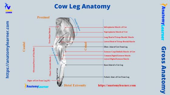
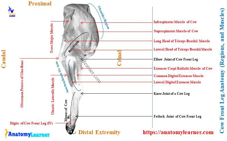
Here, I will provide the labeled diagram where you will find every component of the cow leg structure. The first diagram shows the bones from the cow front leg along with the joints, nerves, and vessels.
Again, the second diagram shows the muscles from the different regions of the cow’s front leg. But, for a better view of the cow front leg muscles, you may find more diagrams at here (social media of anatomy learner).
In the third diagram, I tried to show you the different bones from the cow hind leg structure. This diagram also shows the different important nerves and vessels from the cow back leg.
Finally, in the fourth diagram, I tried to show you the different important muscles from the cow back or hind leg.
Frequently asked questions on cow legs
Now, I will provide concise answers to some of the questions on the cow leg structure that the learners commonly ask. But, it is highly suggested to read the whole article to get the basic idea of the cow’s front and back legs features.
Okay, let’s see what other learners want to know about the cow legs.
What is the leg part of a cow called?
Actually, the leg part of a cow means the tibia and fibular region (back limb). The different segments of the cow front leg are familiar as the followings –
Scapula of a cow – shoulder region,
The humerus of a cow – arm or brachium region,
Radius and ulna of a cow – forearm or antebrachium region,
Carpal, metacarpal, and digits of the cow – known as the manus region,
Again, the different segments of the cow hind leg are known as the followings –
Hip bones of a cow – pelvic region,
The femur of the cow – known as the thigh,
Tibia and fibula of the cow – known as the leg region, and
Tarsal, metatarsal, and digits of the cow’s back leg – known as the pes region,
How do cows legs bend?
The cow’s hind and back legs possess different synovial joint types. Most of the cow leg’s joints can extend and flexion.
These extensions and flexion of the leg’s joint help to bend the cow’s leg. To understand the bending capabilities of the cow’s legs, you might have a good piece of knowledge on the joint anatomy as well as the different types of joint movement.
What is the hip of a cow called?
The hip of a cow is called the pelvic girdle. In the formation of the cow pelvic girdle, you will find the hip bones (ilium, ischium, and pubis) from both 2 lateral aspects along with the sacrum.
Again, you will also find one or two caudal vertebrae in the structure of the pelvic girdle of a cow. You know the 2 sides hip bone to fuse with the cartilaginous joint and is also termed the os coxarum.
You may read the article on the animal hip bone and joint anatomy for information on the cow hip.
What are the bones in the feet of cows called?
The feet of the cow’s front leg comprise 2 digits or claws. In each digit of a cow, there are 3 developed phalanges.
The phalanges of the cow digits are the proximal, middle, and distal phalanx. The Carpal, metacarpal along with feet of the cow are known as the manus.
Again, the feet of the cow’s hind leg consist of 2 digits (III, and IV). You will also find similar features in the structure of each digit of the cow’s hind leg.
The cow’s digits, along with the tarsal and metatarsal, are known as the pes of the hind leg.
What is the leg of a cow called?
The leg of a cow consists of the tibia and fibula bones. You know cow tibia is a long bone with a body and 2 distinct extremities.
In the structure of the 2 extremities of the cow tibia, you will find the condyles and spine at its proximal extremity. Again, the distal extremity of the cow tibia possesses the articular facets for the tarsal bones.
Again, the cow fibula is the modified or reduced long bone which doesn’t posses any distinct unique features.
Conclusion
So, the main components of the cow leg anatomy are the bones, joints, muscles, nerves, and vessels. I hope you got the basic idea of these components, both from the cow’s front and back legs.
The nerves of the brachial plexus and branches of the axillary artery are the principal component in the cow front leg. Again, the nerves of the lumbosacral plexus and branches of the external artery are significant structures in the cow hind leg.
All the labeled diagrams on the cow’s front and back leg anatomy might be helpful for you to understand all the features perfectly. You might try to identify all these features and components from the cow’s front and back legs with the help of a provided diagram.