The cow stifle joint is the compound joint between the hip and hock joints of the hindlimb. Here, I will describe the anatomical features of the cattle stifle joint with a diagram.
Quick overview: The cow stifle is the complex joint formed among the femur, patella, tibia, and fibula. It is a condylar articulation that acts as a hinge joint with a little rotation.
You will learn the involvement of the bones and ligaments from the cow’s stifle joint. I will also provide the type and movement of this stifle articulation from the hindlimb of a cow.
What is the stifle joint in a cow?
The stifle joint (genual) is a complex joint in a cow that comprises –
- Femoropatellar articulation – it is the articulation between the patella and trochlea of the femur bone. You will find a spacious joint capsule in the femoropatellar articulation of a cow.
- Femorotibial articulation – it is the joint among the condyle of the femur, the proximal end of the tibia, and interposed articular menisci (semilunar cartilage).
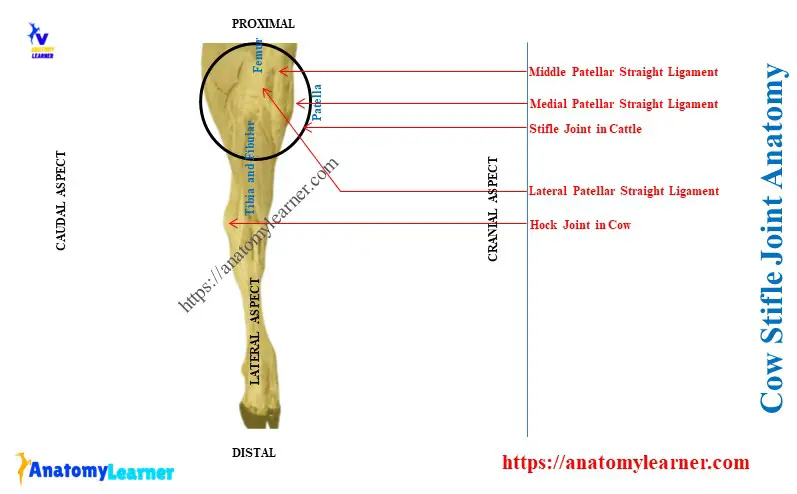
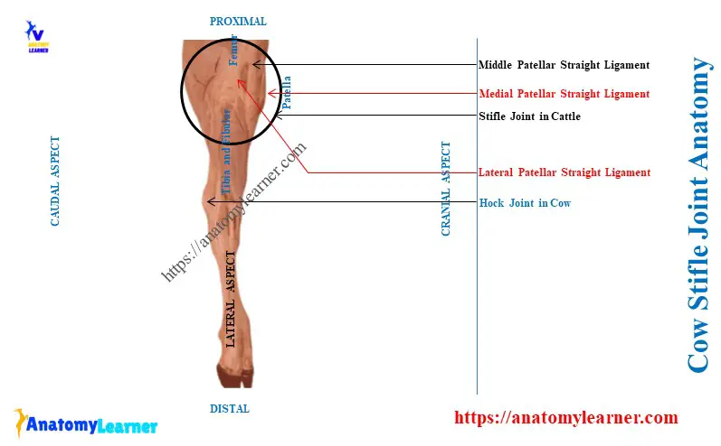
Extension, flexion, and little rotatory movements are the main movements of the cow stifle. Here, the diagram shows the different structures of the cattle stifle joint.
Again, Table 1 shows the summary of the cow’s stifle joint –
| Cow stifle | Femoropatellar joint | Femorotibial joint |
| Bones involvement | Trochlea of femur bone Articular surface of patella | Condyle of femur bone Proximal end of tibia Interposed articular menisci |
| Ligaments | Joint capsule (spacious) Lateral collateral ligament Medial collateral ligament Patellar straight ligaments – Lateral patella ligament Middle patellar ligament, and Medial patella ligament | Joint capsule (less spacious) Lateral collateral ligament Cruciate ligaments (two) – Cranial and caudal cruciates |
| Special structures | Absent | Lateral and medial menisci |
| Type of joint | Gliding type joint | Ginglymus type joint |
| Movement of joint | Translation joint movement | Extension and flexion, Little rotatory movement |
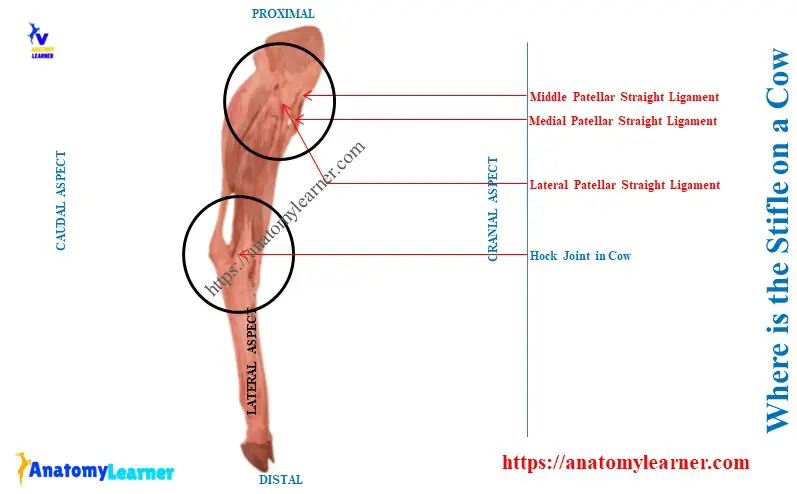
Where is the stifle on a cow?
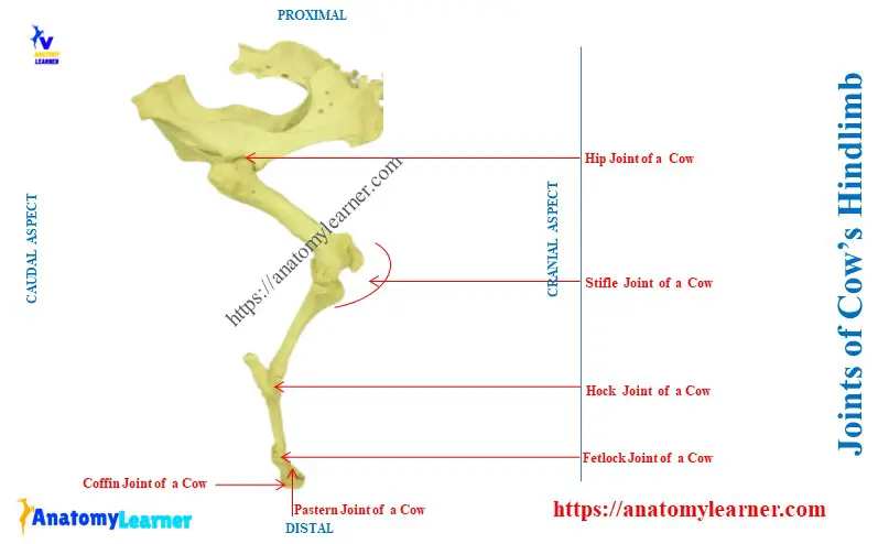
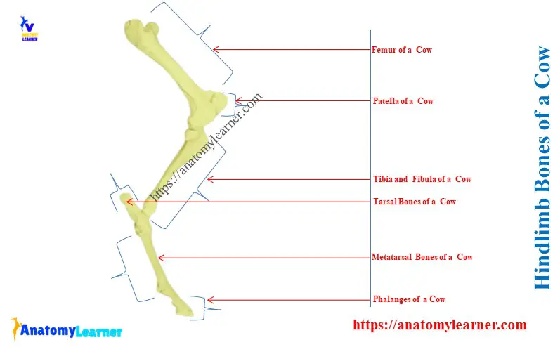
The stifle is located between the hip and hock joints on a cow’s hindlimb. Again, you may say this stifle is located between the distal end of the cow’s femur and the proximal end of the tibia bones.
Here, the patella and fibula bones also form this stifle joint in cattle. The diagram above shows the exact location of the stifle articulation of the cows.
So, to understand the stifle articulation from cattle, you might have the following knowledge –
- Bones of the cow’s hindlimb with their osteological features and
- Different joints of the cow’s hindlimb,
You might also have an idea of the typical features of a synovial joint of an animal. The below-mentioned article might help you to get an idea of bones and joints from a ruminant –
- Goat skeleton anatomy – skull, hindlimb, and forelimb bones,
- Animal joint anatomy – Name of the articulations with their bony involvement,
You may also know the osteological features of the femur and tibia-fibula bones directly related to stifle common formation.
“Another name of cow’s stifle joint is genual articulation.”
Cow stifle joint anatomy
In cow stifle joint anatomy, you might describe the following –
Articular surfaces of the bones that form the femoropatellar and femorotibial joints and
Ligaments that are involved in the formation of the femoropatellar and femorotibial joints,
Key features of the stifle joint anatomy –
- It divides into two parts – femoropatellar and femorotibial articulations,
- Articular menisci is present in femorotibial articulation, but femoropatellar don’t possess such structure,
- The joint capsule is thin and capacious in the femoropatellar joint, but it is less capacious in the femorotibial joint,
- Collateral ligaments are common in both femoropatellar and femorotibial articulations,
- There are three patellar straight ligaments in femoropatellar joint, where the medial one is clinically important, and
- The femorotibial joint possesses cranial and caudal cruciate ligaments that cross each other,
Now, let’s know the details of these two parts of the stifle joint from a cattle diagram.
The femoropatellar joint of a cow
The femoropatellar joint of a cow is a gliding type joint and provides translation movement in the hindlimb. The trochlea of the femur and the articular surface of the patella bones are involved in the formation of the femorpatellar joint.
Here, the diagram shows the trochlea from the distal end of the cow’s femur bone. Again, it also shows the articular surface of the cow’s patella.
You will find the below-mentioned ligaments in the structure of the femoropatellar ligament –
Joint capsule of femoropatellar joint
The joint capsule (capsular ligament) of the femorpatellar joint is thin and very capacious. You will see the following attachment of the joint capsule of a cow’s femoropatellar joint –
- On the patella – it attaches around the margin of the articular surface of the cow’s patella and
- On the femur – it attaches at a varying distance from the trochlear articular surface of the femur bone,
Collateral ligaments of the cow’s femoropatellar joint
The lateral collateral ligament of the femoropatellar joint attaches as follows –
It connects to the lateral condyle of the femur to the lateral border of the patella bone,
Again, the medial collateral ligament of the femoropatellar joint is thinner than the lateral ligament. It attaches to the medial epicondyle of the cow’s femur to the medial border of the patella bone.
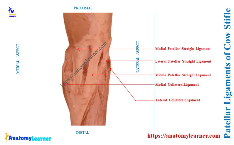
What are the patellar straight ligaments of a cow’s stifle joint?
The patellar straight ligaments are the strong bands that attach the patella to the tibial tuberosity of a cow. They are three in number in the cow’s stifle –
- Lateral patellar ligament – extensive and strong,
- Middle or anterior patellar ligament – cranially located and strong and
- Medial patellar ligament – narrow and weak ligament,
Lateral patellar ligament: it attaches to the lateral part of the cranial surface of the cow’s patella to the lateral part of the tibia tuberosity. It is extensive among the three ligaments and faces laterally.
This lateral patellar ligament receives the strongest tendons from the biceps femoris and fascia lata muscles. So, it is also important to know the hindlimb muscles of cows or ruminants.
Middle patellar ligament: it is the strongest ligament among the three of the cow stifle articulation. It extends from the cranial part of the apex of the cow’s patella to the distal portion of the groove of the tibial tuberosity.
Medial patellar ligament: it is the narrowest and weakest ligament of the cow stifle joint. The attachment of the medial patellar ligament of the cow stifle is as follows –
Starts from the proximal to the parapatellar fibrocartilage, and
Ends on the tuberosity of the tibia at the medial aspect of the groove,
This medial patellar ligament sometimes becomes thick and fixes the cow’s stifle joint. This condition is known as the patellar luxation in a cow.
“Other name of the patellar luxation – patellar lock.”
You will find two burase in the structure of the femoropatellar joint of a cow. One of the bursae is located between the middle patellar ligament and the upper part of the trochlea. Again, another bursa is located in the lateral patellar ligament and lateral condyle of the femur.
Femorotibial joint of a cow
The femorotibial joint is the ginglymus type of joint that provides extension, flexion, and little rotatory movements. The condyle of the cow’s femur and the proximal end of the tibia bones are formed this joint. You will also find the menisci between the articular surfaces of the femur and tibia.
You will find the below-mentioned ligaments in the structure of the cow’s femorotibial joint –
Capsular ligament of femorotibial joint
The menisci divide the femorpatellar joint cavity into two compartments. Thus, you will see two synovial membranes in the structure of the femorpatella joint in a cow.
The joint capsule or capsular ligament of the femoropatellar articulation is also capacious.
Collateral ligaments of the cow’s femorotibial joint
You will also find both the lateral and medial collateral ligaments in the structure of the femorotibial joint. Let’s see the attachment of the medial collateral ligament of the cow’s femorotibial articulation –
- Starts from the proximal to the prominent medial epicondyle of the femur bone and
- Ends on distal to the rough area of the medial epicondyle’s margin of the tibia bone,
The lateral collateral ligament is thicker than the medial collateral ligament. This ligament arises from the proximal depression of the lateral epicondyle and finishes on the head of the fibula.
What is the cruciate ligament in the cow’s stifle joint?
The cruciate ligaments are the two strong round bands in the structure of a cow stifle joint. They are mainly located in the intercondyloid fossa of the femur between two synovial sacs.
So, there are two cruciate ligaments named –
- Cranial cruciate ligament: it arises from the central fossa on the tibial spine. Again, it ends on the lateral wall of the intercondyloid fossa of the femur bone.
- Caudal cruciate ligament: it arises from the eminence at the popliteal notch of the cow’s tibia bone. Finally, it ends on the cranial part of the intercondyloid fossa of the cow’s femur bone.
What are the menisci in the cow stifle joint?
Answer: these are the lateral and medial crescentic plates of fibrocartilage structures in the cow’s stifle. They produce congruence in the articular surfaces of the femorotibial articulation of a cow.
Each of the meniscus has a proximal concave and distal convex surface. Here, the proximal surface of the menisci is adapted to the condyle of the femur bone.
Again, the distal surface of the menisci fits the corresponding condyle of the tibia bone. Let’s see the other key features of the menisci of the femorotibial articulation of the cow –
- The lateral meniscus doesn’t cover the lateral and caudal parts of the tibial condyle,
- Peripheral border of the menisci is thick and convex,
- The central part of the menisci are very thin and concave,
- Ligaments are attached to the tibial cranial and caudal to the spine and
- The lateral meniscus has a third attachment utilizing an oblique band,
Here, the lateral meniscus passes from the caudal end to the tail part of the intercondyloid fossa of the femur.
Species differences: the dog, pig, and a few small ruminants have only one patellar straight ligament.
Conclusion
The cow stifle joint consists of femoropatellar and femorotibial articulations. It is one of the complex joints of the cow’s hindlimb that provides the extension and flexion movements.
However, little rotatory movement is provided by the femorotibial part of the cow’s stifle joint. The patellar straight ligaments are the essential ligaments of the cow’s stifle joint.