Skeleton is the framework of rigid structures that support and protect the soft organs of the animal’s body. Goat skeleton anatomy is primarily divided into the axial and appendicular subdivision. In this article, I will show you the different peculiar osteological features of goat skeleton.
Hi, do you love to learn goat skeletal system anatomy? There is a tremendous osteological variation of goat bones in comparison to other ruminants like cattle or sheep.
Here, I will try to cover all the peculiarities of goat skeleton anatomy with a labeled diagram. But make sure you read my previous articles on animal’s osteology. If you don’t read, please go to the osteology section and check them.
Okay, let’s start to learn – how many bones in a goat skeleton, the unique features of goat bones, and more.
Goat skeleton anatomy
The skeleton of the higher animal consists mainly of bones, cartilage, and ligaments. In the early development of an animal, the skeleton consists of cartilages. You will find the following bones in the axial skeleton of goat anatomy.
- #1. Skull bones of a goat (cranial bones and facial bones)
- #2. Vertebrae bones (cervical, thoracic, lumbar, sacrum, and coccygeal bones)
- #3. Ribs of goat (sternal ribs, asternal ribs, and floating ribs) and
- #4. Sternum (different sternebrae, manubrium, xiphoid cartilage)
And, in the appendicular skeleton of a goat, you will find the following bones in other parts of limbs. You might know the identifying features of other bones from the goat skeleton.
- #1. Bones of thoracic or shoulder girdle of goat (scapula, clavicle, and coracoid bones)
- #2. Arm (humerus) and forearm bone (radius and ulna bones of a goat)
- #3. Knee bones (carpal bones of a goat)
- #4. Canon and splint bones of goat (metacarpal bones)
- #5. Phalanges and sesamoid bones of goat (proximal, middle, and distal phalanges)
- #6. A bone of pelvic girdle of goat (sacrum, ox coxae bones, and coccygeal bones)
- #7. A bone of the thigh region (femur)
- #8. Bones of leg region (tibia and fibula bones of the goat)
- #9. Tarsal (hock region), metatarsal bones (cannon and splint bones), phalanges bones
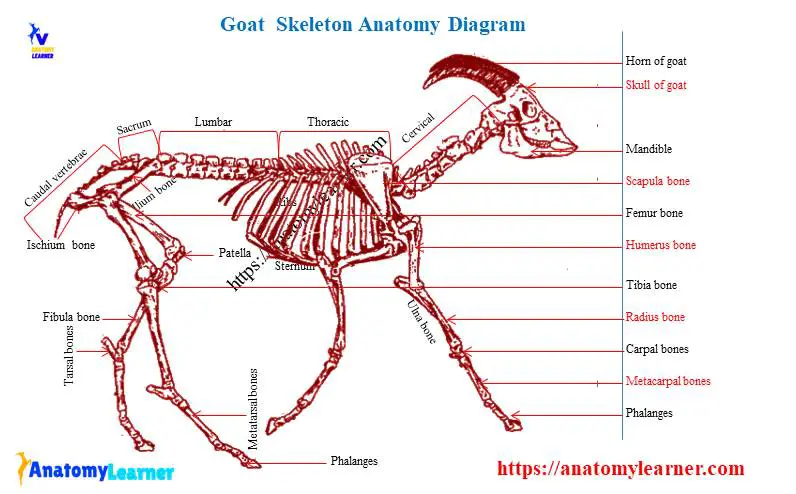
Now, I will share a goat skeleton labeled diagram where you will find all the bones. I hope that a labeled diagram of the skeletal goat system might help you know all the bones’ names. If I missed any bones, please let me know. I will try to identify those parts or bones in the next update.
The axial skeleton of goat anatomy
Let’s start with the axial skeleton of goat anatomy. You know, the axial skeleton consists of the skull, vertebrae, ribs, and sternum. I will show you all the essential and peculiar features of an axial skeleton from the caprian anatomy.
For description purposes, the goat skull is divided into two main parts – the cranium and the face. The cranium bones consist of those bones that immediately surround the brain, incorporate into the floor and vault of the brain cavity.
In the cranium bones of goat skull anatomy, you will find the following vital bones. Here, I am not going through the detailed anatomy of these goat skull bones. If you are interested in learning the details anatomy of skull bones, you may read my other article from anatomy learner. You will find the details guide on every single bone from the goat skull.
#1. Bones of cranium part of goat skull – (single bone – occipital, sphenoid and ethmoid; paired bones – frontal, temporal, parietal, and interparietal bones)
#2. Bones of a face from goat skull – (single bones – vomer and hyoid; paired bones – maxilla, premaxilla, palatine, pterygoid, nasal, lacrimal, malar, turbinate, and mandible bones)
You might check all these skull bones from the goat skull labeled diagram. If you found any mistakes in bone identification, please let me know.
Goat vertebrae column
The vertebrae column is the fundamental part of the goat skeleton. You will find the cervical, thoracic, lumbar, sacral, and coccygeal vertebrae in the skeletal goat system. The number of these vertebrae may vary with other ruminant animals. You will find the following numbers of vertebrae bones in the goat.
- #1. Cervical vertebrae – 7 (seven)
- #2. Thoracic vertebrae of goat – 13 (thirteen)
- #3. Lumbar vertebrae of goat – 6 to 7 (six to seven)
- #4. Sacral vertebrae – 4 (four) and
- #5. Coccygeal vertebrae of goat – 15 – 19 (fifteen to nineteen)
If you want to identify the different types of vertebrae from goat, you might know the structure of typical vertebrae and the unique features of a specific kind of vertebrae.
Cervical vertebrae of goat
The first (atlas) and second (axis) cervical vertebrae of the goat are highly modified in conformity with the head’s unique functions of support and movement. The goat’s sixth and seventh cervical vertebrae have unique characteristics (quadrangular, massive, and longer).
The goat atlas is atypical in form and structure. You will not find the body and spinous process in the goat atlas. The transverse process of the goat atlas is modified and curved to form a plate-like (wing) structure that projects laterally.
You will find the alar foramen, intervertebral foramen in the wing of the goat atlas. The dorsal arch of the atlas presents a median dorsal tubercle. You will find a narrow, curved, and thick ventral arch in the goat atlas. There are special features on the caudal end of the atlas, fovea dentist (transverse concave articular surface).
The cranial extremity of the goat’s second cervical vertebrae possesses odontoid processes that articulate with the fovea dentis of an atlas. Transverse processes of second cervical vertebrae are small, single, and projected caudally. The spinous process projects cranially and increases in height and thickness caudally.
You will find the typical vertebrae features in the goat’s third, fourth, and fifth cervical vertebrae. The body of these three vertebrae is long as compared to the other vertebrae of the goat. Cranial extremities of these vertebrae have a quadrangular convex articular surface, and the caudal extremity has a circular concave cavity. The articular surface of these vertebrae is flat and extensive in the goat. Again, the transverse process of these three vertebrae of a goat is a comprehensive and plate-like structure.
Sixth and seventh cervical vertebrae of the goat are short but broader than others. Transverse process is undivided and usually has no transverse foramen in the seventh cervical vertebrae of the goat.
Thoracic vertebrae of goat skeleton
You will find thirteen numbers of thoracic vertebrae in the skeletal goat system. The body of the thoracic vertebrae is short, but the ends are expanded that possess articular surfaces. There are small arches and articular processes in goat thoracic vertebrae.
The transverse processes are thick, short, and tuberous at the free ends. There are dorsally caudally directed large and narrow spinous processes in the thoracic vertebrae of the goat. The body of the first thoracic vertebrae of a goat is wider and dorso-ventrally flattened. There is an ahead-like structure found in the cranial extremity of the first thoracic vertebrae. The caudal extremity of the first thoracic vertebrae possesses a deeper concave cavity than any other thoracic vertebrae of the goat. The articular processes of the first thoracic are much larger than those of the other thoracic vertebrae of the goat skeleton.
The transverse process of the first thoracic vertebrae is short and has a sizeable concave facet on its ventral aspect for the articulation with the tubercle of ribs. But in the last thoracic vertebrae, caudal pari of costal facets are absent.
The bodies of all thoracic vertebrae of the goat gradually diminish in length and width to the middle of the region and then increase. The transverse process of thoracic vertebrae reduced in size and was placed more ventrally.
Lumbar vertebrae of goat
Usually, you will get some variation in the number of lumbar vertebrae of the goat. Sometimes you will find six numbers of lumbar vertebrae in the goat vertebral column. In a domestic goat, you may find five lumbar vertebrae.
The lumbar vertebrae are characterized by the semi-elliptical body and long transverse process. In the first three lumbar vertebrae of the goat, you will find a distinct ventral crest. The arches of the last three lumbar vertebrae of the goat increase in breadth and height.
A caudal notch on the vertebral pedicle is more profound than the cranial notch. Cranial articular process fused with the mamillary processes. The transverse processes are an elongated plate-like structure that flattened dorsoventrally.
You will find relatively low and wide spinous processes in the lumbar vertebrae of the goat. The spinous processes of the last lumbar vertebrae of a goat is smaller than others.
Please check the article from the general animal anatomy section to know the detailed anatomical features of lumbar vertebrae of animals with a labeled diagram and video.
Sacral vertebrae of goat anatomy
The sacrum of a goat is usually formed with the fusion of four sacral vertebrae and known as a single bone. It is triangular, and the long axis of this bone is genteelly curved and slightly oblique.
You will find two surfaces, two borders, a base, and an apex in the sacrum of the goat. In a young goat, you will find the four sacral spines that are directed dorsally and caudally. But in older goats, the dorsal sacral spine fused to form a single median sacral crest.
You will also find three dorsal sacral foramina in the dorsal surface of the goat sacrum bone. The pelvic surface of the goat sacrum is concave and wide cranially, narrow caudally. It is marked by three or more faint transverse lines that demark the bodies of the sacral vertebrae.
There are two lateral borders found in the sacrum of the goat. The border of the goat sacrum is thin and rough.
There is a long sacral canal in the goat sacrum.
The lateral part of the base of the sacrum is compressed and strongly curved masses. Apex of the sacrum is the small caudal aspect of the last sacral vertebrae.
If you want to know more about the sacral vertebrae of animals, I would like to suggest you check the article on sacrum anatomy.
Coccygeal vertebrae of goat
There is also a variation in the number of coccygeal or caudal vertebrae of the goat. But, you may find fifteen to nineteen caudal vertebrae in goat skeleton anatomy. From the first to last caudal vertebrae of the goat, gradually becomes reduced in size. First, the caudal vertebrae have their bodies, but in other caudal vertebrae, it is difficult to identify their bodies.
The first few caudal vertebrae body are flattened dorso-ventrally, constricted at the middle, and have both convex ends. The ventral surface of the caudal vertebrae possesses a ventral groove for the median coccygeal artery.
Cranial notches, articular processes are not distinct in the caudal vertebrae of the goat. The transverse processes of caudal vertebrae gradually fade out, and the vertebrae become cylindrical rods in shape.
Ribs and sternum of goat anatomy
Ribs are the modified long bones located in between the vertebrae and the sternum of animals. These are the paired long, curved bone that also forms the lateral thoracic wall. They arranged serially in pair which correspond in number to the thoracic vertebrae of any animals.
You know there are thirteen pairs of ribs in the goat, where eight pairs are sternal, five pairs are asternal, and the rest seven pairs are floating ribs. In a rib, you will typically find a body and two different extremities.
The first rib of goat may quickly identify with their short shaft and other features –
- #1. Present a giant head and two unequal facets.
- #2. Shaft windes extensively towards the ventral ends
- #3. There is no costal groove
- #4. Presence of very short and thick neck
- #5. Presence of more prominent tubercle with an extensive articular surface.
The last pair of ribs of a goat is more slender and regular curved. You will find the following characteristics in the goat ribs serially.
The length increase from the first to the fifth ribs, and eight ribs then diminishes. From first to seventh ribs, width increases then reduce. The groove of the lateral surface is more distinct from the fourth rib to the tenth rib. Head of the tubercle diminishes in size from first to last ribs.
The goat sternum is composed of six unpair segments or sternebrae. In the first sternebrae of the goat (known as manubrium), there is a club-like enlargement. The last sternebrae (xiphoid cartilage) of a goat is dorso-ventrally flattened. A thin plate of cartilage continues the caudal end of this structure.
The appendicular skeleton of a goat
Now, I will show you the bones from the appendicular skeleton of a goat. You already got an idea about all bones from the forelimb and hind limb of the goat. But here, I will try to cover the unique features of the different bones from goat’s limbs.
#1. Forelimb bones of goat and
#2. Hindlimb bones of goat
Forelimb bones of goat
First, go with the forelimb bones of the goat. You will find the following bones in the forelimb of a goat.
- #1. Scapula of goat (thoracic girdle)
- #2. Humerus (arm region)
- #3. Radius and ulna bones of goat (forearm region)
- #4. Carpal bones of goat (knee region)
- #5. Metacarpal bones of goat (splint bones) and
- #6. Phalanges or digits of goat
The maneus of goat consists of the following three types of bones – carpal bones, metacarpal bones, and phalanges. Let’s start to know the anatomical features of the forelimb bones of the goat.
Scapula of goat
The scapula of a goat is a flat bone that covers the cranial part and lateral part of the thoracic wall. It is roughly triangular in outline and posses two ends. The dorsal end is broader, and the ventral end is narrower. You will find two surfaces, three borders, and three angles in the goat scapula (like other scapula bones of animals).
You will get a detailed guide on animal scapula anatomy here in anatomy learner. If you want to know the detailed anatomical features of scapula bone, I suggest you check that article.
Okay, what are the unique features of goat scapula bone? The spine of the goat scapula increases gradually in height dorso-ventrally and inclined cranially. The supraspinous fossa is located cranially and is narrower than another ruminant. Again, the infraspinous fossa is found caudally and is vast.
The acromion process of goat scapula is short and blunt and is opposite to the neck. You will find the nutrient foramen in the ventral third of the caudal surface of the scapula.
The coastal surface is hollowed in its length by the subscapular fossa. You will find a triangular area (known as facies serrate) but not as prominent as found in cattle scapula.
The neck of the goat scapula is well defined and narrow. The glenoid cavity is continued with the ventral surface of the supraglenoid tubercles. You will find a small coracoid process at the medial surface of the supraglenoid tubercle in the goat.
Humerus bone of goat skeleton anatomy
This is one of the long bones of the goat skeleton and is located between the scapula and radius and ulna bone. It is an irregularly cylindrical and twisted bone in the goat (but less twisted than cattle humerus bone). You will find four surfaces and two different extremities in the humerus of the goat.
The osteological features of these four surfaces are similar to the cattle humerus bone. But the structures of cattle humerus is more prominent than the goat humerus bone.
You will find a musculospiral groove in the lateral surface of the goat humerus (less spiral than cattle). Proximal to the middle of goat humerus, there is a teres major tuberosity. You will also find the crest of the humerus and deltoid tuberosity in the body or shaft of the goat humerus.
If you want to know about these structures (deltoid tuberosity, crest of humerus) like their location, appearance, please check the details guide on humerus bone.
In the proximal extremity of the goat humerus bone, there are head, neck, two tuberosities, and inter-tuberal groove. The head of a goat humerus posses a circular convex articular surface that articulates with the glenoid cavity of the scapula.
The distal extremity of the goat humerus consists of lateral, medial epicondyles, olecranon process, and radial fossa. The distal groove and ridge are very well marked in goat humerus. The trochlea is comparatively larger in goat humerus bone.
There is an olecranon fossa in between two lateral epicondyles, which is quite deep in goat humerus bone. A radial fossa is also comparatively deep in goat humerus.
Radius and ulna bone from skeletal goat system
The radius of the goat lies between the humerus and carpus and is fused with the ulna bones with two interosseous spaces. This bone extends in an oblique direction from the elbow joint. You will find the same osteological features in the goat radius as you find in another ruminant.
For description purposes, the goat radius bone has a shaft and two different extremities. The shaft of the goat radius bone has four different surfaces – lateral, medial, cranial, and caudal surfaces. I have a detailed guide on the radius bone of an animal with a labeled diagram and video. If you wish to read that article and watch that video, please go and check now.
The proximal extremity of the goat radius bone is flattened and wide transversely. The medial tuberosity is continuing with the radial tuberosity and gove attachment to the elbow ligament. Again, the distal extremity of the goat radius bone is compressed craniocaudal. Carpal articular facets of radius are oblique, where the medial one is larger. That medial carpal facet articulates with the radial carpal bone of the goat.
Goat ulna bone is attached to the caudolateral surface of the radius of the forearm. The ulna of a goat is a three-sided bone in proximal third, compressed craniocaudal in the middle third. A distal end of the goat ulna bone is expanded.
Carpal and metacarpal bones of goat
There are two rows of carpal bones found in the goat skeleton. The proximal carpal bones of the goat consist of radial, intermediate, ulnar, and accessory carpals. Again in distal row carpals, you will find fourth and second, third fused carpal bones in goat.
The ulnar carpal is large and triangular in the goat. Accessory carpal of a goat is short, thick, and rounded. The second and third carpal in the distal row fused to form a large quadrilateral bone-in goat.
Goat metacarpal bones are located between the distal row of carpal bones and proximal phalanges. The large metacarpal bone of goat, results from the fusion of the third and fourth metacarpus bones. The fifth metacarpal bone of goat is a small metacarpal bone and known as splint bone.
The first and second metacarpal bones are absent in the goat skeleton. In a large metacarpal, you will find a shaft and two extremities. In the dorsal surface of the large goat metacarpal, there is a vascular groove – dorsal longitudinal groove. You will also find a ventral longitudinal groove at the ventral or palmar surface of the large metacarpal bones of the goat.
In a goat, there are present of four digits. But the third and fourth digits are well developed among these four digits. These two well-developed digits consist of three phalanges.
Hindlimb bones of goat skeleton
In goat hindlimb, you will find the following bones. I will show you the essential features of these bones from the goat skeleton.
- #1. Hip bone or ox coxae of goat (pelvic girdle)
- #2. Femur bone of goat (thigh region)
- #3. Tibia and fibula of goat (leg region)
- #4. Tarsal bones of goat
- #5. Metatarsal bones of goat and
- #6. Phalanges and digits of goat’s hindlimb
The pes of goat consists of tarsal bones, metatarsal bones, and phalanges bones. First, try to identify these hindlimb bones from the labeled diagram. If you want to learn the detailed anatomy of these hindlimb bones of an animal, please go and check the articles from the general anatomy section.
Hip bone or ox coxae of goat
The hip bone is the most extensive flat bone in goat skeletal anatomy. It consists of primarily three bones – ilium, ischium, and pubis bones.
Ilium is the modified long bone of goat skeletal anatomy and is the cranial component of the pelvic girdle. It is the most prominent bone among the three bones of the goat hip.
You will find some unique features in the goat ilium bones or hip bones. The gluteal surface of goat ilium bone is broad and concave. The curved gluteal line crosses a wide part of the bone. But the gluteal line of goat hip bone is not so prominent as you found in cattle hip bone.
The cranial border of the ilium bone is thick (know as an iliac crest) and rough at cranial ends and thin at the caudal end. Greater ischiatic notch is comparatively deep and continuous with the greater ischiatic spine. You will find the tuber coxae (lateral angle of ilium), which forms the basis of the point of the hip bone.
Goat ischium and pubis bones
Goat ischium is also a modified long bone that forms the caudal part of the ventral floor of the pelvis. It is irregularly quadrangular and posses two surfaces, four borders, and four angles. The caudal border of the ischium is thick and rough that forms the ischiatic arch. The medial border of goat ischium meets with the opposite part and forms the ischial symphysis.
The pubis bone of the goat locates the cranial to the ischium bone and forms the cranial part of the pelvic floor. It is the smallest of these three ox coxae bones and posses a body, two surfaces, and three borders.
In the ventral surface of the pubis bones (near symphysis), you will find a prominence known as the ventral tubercle. But the ventral tubercle is not so prominent in goat as cattle.
Femur of goat
The goat femur lies between the pelvic bone and tibia and fibula bones. It extends obliquely and posses a shaft and two extremities.
The shaft of the goat femur is cylindrical but flattened caudally. It presents four surfaces and two borders as found in the femur of cattle or another ruminant.
The cranial, lateral, and medial surfaces of the goat femur are smooth strongly convex from side to side. But the caudal surface is wide, flat, and smooth in the proximal end. Lesser tubercle found at the medial border of its proximal end of goat femur.
The proximal extremity of the goat femur consists of the head, neck, and greater trochanter. Head locates medially and posses a deep notch (fovea capitis femoris).
The distal extremity of the goat femur contains two condyles laterally and trochlea cranially. You will find an inter-condyloid fossa that separates the two lateral condyles. There are also lateral and medial epicondyles in the distal extremity of the goat femur. But the lateral epicondyle is less prominent in the goat.
Tibia and fibula bone from goat skeleton
The goat tibia bone articulates with the femur proximally and tarsus bones distally. It also posses a shaft and two extremities – proximal and distal.
The shaft of the goat tibia bone is large and three-sided (prismatic). It is distinctly curved so that the medial side is convex in proximal and concave in the distal part.
Medial surface of the tibia broad proximally and posses rough prominence for muscular attachments. The cranial surface is very prominent and bears a tibial crest.
Head of the goat tibia is large and three-sided. You will find the following osteological features in the proximal extremity of the goat tibia bone.
- #1. Medial and lateral epicondyles
- #2. Inter-condyloid eminence or spine of goat tibia
- #3. Inter-condyloid fossa of goat tibia
- #4. Semicircular smooth notch
- #5. Tibial tuberosity of goat
The distal extremity of the goat tibia is smaller than the proximal extremity, and it is quadrangular in form. The fibula bone of the goat fused with the lateral condyle of the tibia bone and continues with a small bone distally.
Tarsal and metatarsal bones of goat
You will find six pieces of tarsal bones in the goat skeleton. The tarsal bones of goat skeletal anatomy are –
#1. Tibial tarsal or talas of goat
#2. Fibular tarsal of goat
#3. Central and fourth tarsal of goat
#4. Second and third tarsal of goat
The fibular tarsal of a goat is a slender and long bone. Central and fourth tarsal fused to form Centro-quartal bone in a goat. Again the second and third tarsal bones combined to form a rhomboid bone in the goat.
You will find the large and small metatarsal bones in goat forelimb bones. From the metacarpal of the goat, you might identify the following osteological features –
#1. Dorsal longitudinal groove
#2. Palmar or ventral longitudinal groove of goat metatarsal bone
#3. Facets from sesamoid bones of goat
Okay, make sure you know the variation between the metacarpal bone and metatarsal bone. If you want to know the fundamental difference between animals’ metatarsal and metacarpal bones, you may read my previous article from anatomy learner. Get more labeled images of the skeletal goat system here on social media.
Conclusion
If you want to learn goat skeleton anatomy properly, please get help from the videos and labeled images from my previous articles. But it is recommended to learn osteological features of different bones of goat skeleton physically from anatomy laboratory.