
Mammary gland is the modified apocrine sweat gland become functional and undergo development at puberty in female animal. In the mammary gland histology, you will find the connective tissue stroma and parenchyma like other different glands.
Hi, do you want to learn the histology of mammary gland with slide pictures and labeled diagram with anatomy learner? Fine, in this article I am going to share the basic information of active and inactive mammary gland histology with real slide images.
After reading this gland article you will able to identify important mammary gland histological characteristics under light microscope and differentiate the lactating and non-lactating mammary glands.
Here in this article I will also share the mammary gland histology drawing images with you so that you might understand the every single structure of this gland. So, if you have interest to learn and identify the mammary gland slide under light microscope then continue this article.
Again, if you want to learn histology of other different organs from female genital system of animals then you may check the other article at here.
Okay, let’s get into the main part of the article – mammary gland histology with slide images and labeled diagram.
Mammary gland histology
Each of the mammary gland consists of different independent unit called lobes and separated by interlobular septa that comprise dense connective tissue and adipose tissue. In eache lobes of mammary gland histology section, you will find the compound tubuloalveolar gland and they have lactiferous ducts. There are numerous alveoli embedded in the loose connective tissue stroma of mammary gland’s lobes.
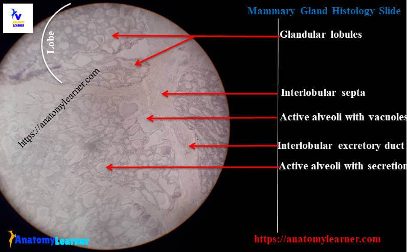
Some of the alveoli show irregular branching and lines with simple cuboidal epithelium or tall columnar epithelium (vary with active and inactive mammary gland). These alveoli of mammary gland histology section surrounded by the myoepithelial cells.
From the mammary gland slide, you might identify the following structures under light microscope –
#1. Lobules of mammary gland
#2. Secratory tubules or alveoli of mammary gland structure
#3. Interlobular connective tissue of mammary gland
#4. Intralobular areolar connective tissue of gland
#5. Interlobular ducts of mammary gland and
#6. Intralobular ducts of mammary structure
Now you might find out these structures from mammary gland slide picture.
Identification of mammary gland slide
Do you want to identify the mammary gland histology slide under light microscope? Well, I will enlist the identification points of active and inactive mammary gland.
Let’s identify the nonlactating mammary gland slide with the following important identification points –
#1. Presence of more connective tissue stroma and less glandular tissue in the provided tissue section
#2. The section shows underdeveloped alveoli in each lobule of mammary gland
#3. There is extensive branching in the duct system of the tissue section
#4. The alveoli are lined by tall columnar epithelium and no secration appearsmin the lumen of alveoli
So, this is the inactive or nonlactating mammary gland slide. Okay, now you might identify the lactating mammary gland slide under light microscope.
I am going to enlist the identification points for lactating mammary gland slide that might help you to identify this gland easily –
#1. The section shows more glandular tissue (consists of lobes, lobules and alveoli) and less connective tissue stroma
#2. There are compactly packed, well developed alveoli found in the each lobules of this tissue section.
#3. The lumen of the alveoli are distended and containing milk (pink color) with large vacuoles of fat droplets
#4. The alveoli of the provided section lines with the simple cuboidal epithelium
So, this is the slide of lactating or active mammary gland sldie.
Mammary gland histological characteristics
Hope you could understand the histological characteristics of mammary gland. Now I am going to describe the lactating and nonlactating mammary gland histology separately.
Non lactating or inactive mammary gland histology
In the stroma of non-lactating mammary gland, you will find the following characteristics.
There are more connective tissue and adipose tissue present in the stroma of non-lactating mammary gland section. The interlobular connective tissue septum of non lactating mammary gland is thick and composed with dense collagen fiber and adipose tissue.
Interlobular connective tissue are numerous and contain fibroblasts in non-lactating mammary gland structure.
The parenchyma of non lactating mammary gland contains less glandular tissue and underdeveloped alveoli. You will find thr extensive branching ducts system in the lobules of non lactating mammary gland. The lumen of these branching ducts could not easily identify and there is no secration found in their lumen.
Lactating or active mammary gland structure
The stroma and parenchyma of lactating or active mammary gland differs from the non-lactating mammary gland structure. In the stroma of lactating mammary gland histology, you will find the following histological features.
#1. There are less connective tissue and adipose tissue in the stroma of lactating mammary gland structure
#2. The interlobular connective tissue septum is thin than the interlobular connective tissue septum of non-lactating mammary gland stroma
#3. You will find less intralobular connective tissue that filtrates with lymphocytes and plasma cells.
And in the parenchyma of lactating mammary gland structure you will find the following histological characteristics.
There are more glandular tissue and numerous well developed, large and branched alveoli in the lobules of lactating mammary gland’s parenchyma. All of these alveoli of lactating mammary gland lines with simple cuboidal epithelium. You will find less developed duct system in the parenchyma of lactating mammary gland structure that lines with simple cuboidal epithelium.
Mammary gland histology drawing guide
Do you need the mammary gland histology drawing guide? Fine, I am going to share few of my mammary gland slide images drawing pictures with you so that you might understand all the structure easily.
If you find any mistake in this mammary gland slide image drawing then please let me inform. Again, if you need more pictures related to mammary gland slide then you may follow anatomy learner at social media as I regularly update the pictures.
You might read other different article related to histology of female genital organs from anatomy learner –
#1. Identifying characteristics of animal ovary under light microscope
#2. Histological features of uterus of animal
Conclusion
I know you got the best guide to learn mammary gland histology with slide pictures and labeled diagram. You might identify the mammary gland histology features from slide with proper identifying points.
If you think this article is helpful to learn histological features of mammary gland then share it with your friends who want to learn the structure of mammary gland. Stay connected with anatomy learner and never miss any article from this blog.