The sheep brain anatomy consists of 3 major parts – prosencephalon (forebrain), mesencephalon (midbrain), and rhombencephalon (hindbrain). These 3 main parts of the sheep brain again divide into specific segments.
There are also 5 different lobes in the sheep brain structure – frontal, parietal, occipital, temporal, and limbic area. The different anatomical features of the dorsal, ventral, and lateral aspects of the sheep brain make it complex.
But, I will make it so simple for you that you may easily identify all the anatomical features of the sheep brain. Here, I will describe all the 3 major parts and their sub-parts from the sheep brain anatomy with the labeled diagram.
I will show you the ventricular system of the sheep brain along with the course of cerebrospinal fluid (CSF). Finally, I will provide a simple sheep brain dissection guide so that you can easily perform dissection and explore all its anatomical features.
So, if you want to know the anatomical facts of the sheep brain (cerebrum, cerebellum, brain stem, and different parts of meninges), continue this article till the end.
Sheep brain anatomy
First, I would like to show you the different features of the sheep brain anatomy with the labeled diagram. This might help you to get a basic idea of the different parts of the sheep brain.
Before that, let’s know the subdivision of the 3 major parts of the sheep brain –
- The prosencephalon or forebrain of the sheep – divides into telencephalon and diencephalon,
- A mesencephalon or midbrain of the sheep brain – divides into dorsal tectum, middle tegmentum, and ventral cerebral crura, and
- The rhombencephalon or hindbrain of the sheep – divides into the metencephalon and myelencephalon,
The subdivision of the 3 major parts of a sheep brain is harder to remember. Don’t worry; I will show the contents of these subdivisions in Table 1 –
Here, the telencephalon includes the cerebral hemisphere, lateral ventricle, and cranial part of the third ventricle. Again, the diencephalon of the sheep’s forebrain consists of the thalamus, optic nerve, pineal body, pituitary body, interpeduncular structure, and caudal part of the third ventricle.
The metencephalon of the sheep’s hindbrain includes the pons, cerebellum, and fourth ventricle. In contrast, the myelencephalon of the sheep brain consists of the medulla oblongata.
Sometimes, you may hear the term “brain stem,” which consists of the midbrain, pons, and medulla oblongata. That means when you remove the major portion of the brain, like the cerebrum and cerebellum, the remaining portion is the brain steam.
Sheep brain features identification
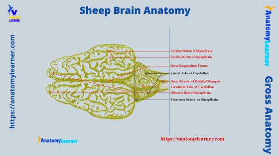
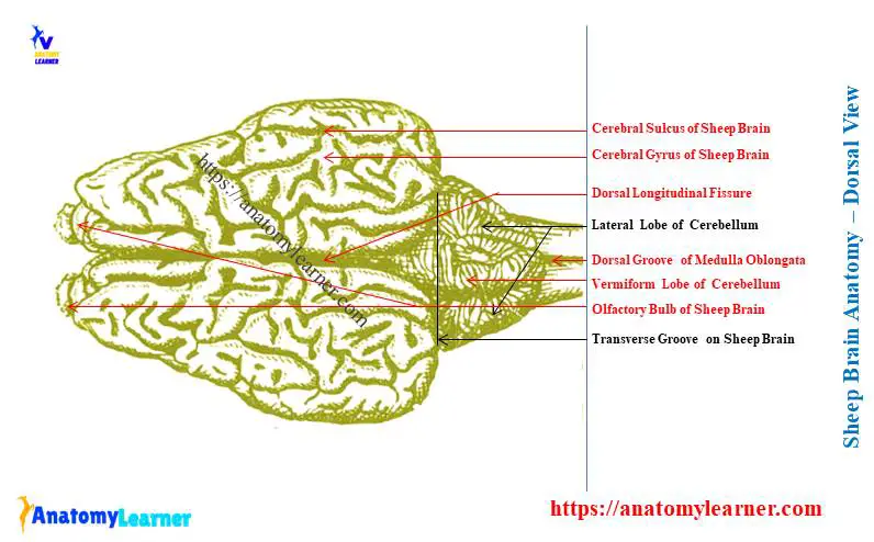
From the dorsal view of the sheep brain, you will find the below-mentioned structures –
- Cerebral hemisphere (2 lateral – right and left) of the sheep brain,
- The cerebellum of the sheep brian,
- Dorsal longitudinal groove or fissure between two lateral cerebrum hemispheres,
- Different lobes – frontal, parietal, and occipital on the cerebrum,
- The olfactory bulb in the sheep brain,
- Cerebral gyrus and sulcus in the cerebrum hemisphere of the sheep brain,
- Transverse groove in between the cerebrum and cerebellum,
- 2 lateral lobes of the cerebellum,
- A vermiform lobe of the cerebellum,
- Medulla oblongata with a dorsal longitudinal groove on it,
- Roots of different cranial nerves (shown in the separate diagram),
- Superior (rostral) and inferior (caudal) colliculus,
- Different parts of the meninges (covering of the brain) – show details in another diagram, and
- Continuation of the spinal cord of the sheep,
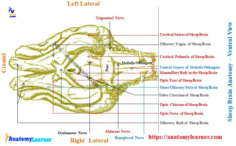
Again, from the ventral view of the sheep brain, you might identify the below-mentioned anatomical features –
- Olfactory bulb and olfactory nerves in the cerebrum region,
- Olfactory trigone of the sheep brain,
- Optic tract, chiasma, and nerve in the sheep brain,
- Tuber cinerium and opening for the hypothalamus (pituitary body),
- Mammillary body in the sheep brain,
- Interpeduncular fossa in the sheep brain,
- The root of the oculomotor nerve,
- Cerebral peduncle in the sheep brain,
- Pons of the sheep brain,
- Trapezoid body (corpus trapizoidum),
- Ventral medial groove in the medulla oblongata, and
- Roots of different nerves – trochlear, trigeminal, facial, abducent, vestibule-cochlea, glossopharyngeal, vagus, spinal accessory, and hypoglossal,
All these features from the dorsal and ventral views of the sheep brain are identified in the labeled diagram. I hope you may easily identify these features from the sheep brain with the help of the labeled diagram.
Sheep brain longitudinal and frontal sections
From the longitudinal section of the sheep brain anatomy, you might also identify the following anatomical features –
- Corpus callosum in the sheep brain,
- Genu and splenium of the corpus callosum of the sheep brain,
- Callosomarginal fissure in the sheep brain,
- Fornix (below the corpus callosum) in the brain,
- Septum lucidum in the sheep brain structure,
- Choroid plexus in the brain,
- Thalamus and hypothalamus in the sheep brain structure,
- Pineal body in the brain,
- Superior and inferior colliculus in the brain,
- Lateral ventricle (2 – right and left) in the brain,
- Third ventricle in the sheep brain,
- Aqueduct of Sylvius,
- Fourth ventricle in the sheep brain,
- Continuation of the spinal canal (in the spinal cord),
- Arbor vita in the cerebellum of the sheep brain, and
- Pons and medulla oblongata (medial view),
Again, in the frontal section of the sheep brain, you will also see the different anatomical facts. But, here, I will only focus on the main anatomical facts from the frontal section of the sheep brain (as the other facts will be enlisted before).
This section will show the hippocampus, choroid plexus, pineal body, and superior and inferior colliculus. On the other hand, you might also know the different layers of the meninges (covering of the sheep brain).
There are 3 different layers (membranous covering) in the meninges of the sheep brain – dura mater, arachnoid, and pia mater. You will find some modifications in the dura mater of the cranial meninges (meninges that cover the brain) in the sheep brain.
These 3 major modifications of the cranial meninges of the sheep brain are –
- Falx cerebri – sickle-shaped verticle fold,
- Tentorium cerebelli – folding of dura mater between cerebrum and cerebellum, and
- Diaphragm sellae – covers pituitary fossa,
Let’s find all these modifications mentioned above from the cranial meninges on the diagram.
Sheep forebrain anatomy
From the sheep forebrain (prosencephalon), I will describe the structures under the telencephalon and diencephalon. That means I will cover the below-mentioned structures from the sheep forebrain –
- Cerebral hemispheres of the forebrain,
- Lobes of the cerebral hemispheres,
- Ventricular (lateral ventricle) of the sheep forebrain,
- Thalamic and surrounding structures, and
- Pineal and pituitary bodies of the sheep forebrain,
Let’s discuss these structures from the sheep forebrain one by one. First, start with the anatomical facts of the cerebral hemispheres of the sheep forebrain.
“Anatomically, sheep brain divides into the cerebrum, cerebellum, pons, and medulla oblongata. For the description purpose, you may divide the sheep brain into the cerebrum, cerebellum, midbrain, pons, and medulla oblongata.”
A cerebral hemisphere of the sheep brain
The cerebral hemispheres are the elongated ovoid mass and the largest part of the sheep brain structure. You will find the incompletely divided into 2 halves of symmetrical cerebral hemispheres in the sheep brain.
In between these 2 halves of the cerebral hemisphere, you will find the dorsal longitudinal cerebral fissure. I don’t want to go into the deep anatomical term or features of the sheep brain; instead, I prefer to describe it very simply.
In the longitudinal fissure of the cerebral hemispheres, you will find the flax cerebrii of the cranial meninges. The longitudinal fissure of the cerebral hemisphere extends up to the corpus callosum of the sheep brain.
Each of the cerebral hemispheres of the sheep brain possesses 3 surfaces and 2 extremities –
- The lateral surface of the cerebral hemisphere – is convex,
- A dorsal or superior surface of the cerebral hemisphere – irregular and posses many sulcus and gyrus, and
- The medial surface of the cerebral hemisphere – is almost flat and forms the lateral boundary of the longitudinal fissure,
You will also find some fissures like the Sylvian fissure, suprasylvian fissure, and transverse fissure in the structure of each cerebrum hemisphere of the sheep brain. Here, the Sylvian fissure of the cerebral hemisphere originates from the middle of the base and accommodates the middle cerebral artery.
Whereas the suprasylvian fissure, locates longitudinally on the Sylvian fissure in an oblique manner. The transverse fissure is very short and transversely located in the middle. You will also find some of the posterosuperior fissures in the structure of the sheep’s cerebral hemisphere.
Sheep brain lobes
In the sheep brain cerebral hemisphere anatomy, you will see 5 lobes –
- Frontal (cranial) lobe – control the sequential job, and help in 2 works at a time,
- Parietal lobe (upper middle) – controls the planned movement and taste,
- Temporal lobe (below middle) – controls the auditory system,
- Occipital lobe (caudal lobe) – helps in visualization, and
- Limbic area (at the base of the brain) – considered as the seat of emotion,
The parietal lobe of the sheep brain is the upper middle part of the cerebral cortex (covered by the parietal bone). This area of the cerebral hemisphere has good control over the auditory, visual, motor, and sensory functions.
The parietal lobe of the sheep brain is responsible for the planned movement. It also accommodates the taste sensation in the sheep.
Let’s see the frontal lobe of the sheep brain from the diagram. The cranial part of the cerebral hemisphere of the sheep brain is associated with the motor system. The brain’s frontal lobe is responsible for performing the sequential job and two works at a time.
Now, the temporal lobe locates in the lower middle part of the cerebral hemisphere. This area (temporal) of the brain is responsible for the olfaction and equilibrium of the brain.
The occipital lobe is the caudal part of the cerebral hemisphere of the sheep brain. This part (occipital) of the brain is responsible for the visualization in the sheep.
You will find the limbic area at the base of the sheep brain, which is the area for emotion control. Normally, the limbic system of the sheep includes the olfactory pathway, pyriform lobe, hippocampus, limbic lobe, and hypothalamus.
The olfactory bulb in the sheep brain
The olfactory bulbs are the most rostral structure (ventrally) in the cerebral hemisphere of a sheep brain. There are 2 olfactory bulbs which are elongated, flat, oval structures in the sheep brain.
You will find direct contact with the cerebral hemisphere on its smooth dorsocaudal surface. Again, the rostroventral surface of the olfactory bulb is very rough as because of the presence of the olfactory fibers.
The olfactory bulb of the sheep brain continues as the olfactory peduncle. This is a short, wide fiber bundle that divides caudally into 2 distinct smaller bundles (olfactory tracts).
The olfactory tracts border the triangular area of the olfactory trigone on its rostral, medial, and lateral aspects. You will also find an olfactory tubercle in the olfactory trigone area of the sheep brain.
The medially located olfactory tract of the sheep brain appears as the main continuation of the olfactory peduncle. Again, the lateral to the medial olfactory tract lies the rostral part of the pyriform lobe of the cerebral hemisphere.
You may easily see the lateral olfactory tract of the sheep brain to the point of origin of Broca’s diagonal gyrus. But, the medial olfactory tract of the sheep brain structure is not so clearly visible.
Lateral ventricle in the sheep brain anatomy
You know there are 2 lateral (right and left) ventricles in the cerebral hemisphere of the sheep brain structure. These are two elongated irregular cavities in the cerebral hemispheres of the brain.
The sheep brain’s right and left lateral ventricles to remain separated by a thin verticle membrane. This is the septum pellucidum of the sheep brain structure.
These 2 cavities (lateral ventricles) of the sheep brain communicates with the third ventricle. I will discuss the third ventricle of the sheep brain in the next section of this article (on the description of the diencephalon or interbrain of the sheep brain).
You will find the Y-shaped opening in between the 2 lateral ventricles and third ventricles of the sheep brain. The name of this Y-shaped opening is the foramen of Monro.
The corpus callosum (CC) forms the roof of the lateral ventricle of the sheep brain. Again, the medial wall of the lateral ventricle forms with the septum pellucidum. The floor of the lateral ventricle (LV) of the sheep brain is formed by the caudate nucleus, thalamus, and hippocampus.
What is septum pellucidum?
In the lateral ventricle of the sheep brain anatomy, the septum pellucidum bounded medially on it. This bilaminar membrane works as a partition between the 2 lateral ventricles.
This lamina extends from the ventral surface of the corpus callosum to the dorsal part (1) of the body of the fornix. In the anatomical features of the septum pellucidum, you will find both grey and white matter.
A very thin slit-like cavity is present between the two layers of the septum pellucidum. If you remove the septum pellucidum from the sheep brain, the two lateral ventricles may be seen clearly.
What is the hippocampus in the sheep brain?
You will find the hippocampus at the caudal aspect of the cerebral hemisphere of the sheep brain. This structure enters the lateral ventricle of the brain.
If you want to see the hippocampus, you should remove the roof of the lateral ventricle of the sheep brain. Here, the hippocampus entirely rolled around the thalamus. And thus, it forms the almost perfect semicircle structure in the brain.
The direction of the hippocampus of the sheep brain is slightly oblique from the medial to the caudolateral. There are 2 hippocampi in the structure of the sheep brain.
The rostromedial parts of the 2 hippocampi face each other at the level of the hippocampal commissure. You will see the regular convex structure in the dorsal surface of the hippocampus.
This hippocampus surface bulges into the sheep brain’s lateral ventricle. You will see a thick layer of fibers (known as the alveus) covering the hippocampus’s bulged surface.
Now, the alveus joins with the thick fiber band rostrolaterally. This is the hippocampal fimbria that also connects with the fornix of the sheep brain.
If you want to view the ventromedial surface of the hippocampus structure, you might remove the brain stem. You will see a deep groove (hippocampal sulcus) after removing the sheep brainstem.
You may know more about the sheep brain hippocampus from another article by an anatomy learner.
Corpus callosum and fornix of sheep brain
You will find the large corpus callosum in the internal aspect of the sheep brain (cerebral hemisphere). This is a transverse commissure between the two cerebral hemispheres.
The corpus callosum of the sheep brain lies at the bottom of the longitudinal fissure. In the structure of the corpus callosum of the sheep brain, you will find the commissural fibers.
You will see 3 different parts in the structure of the corpus callosum of a sheep brain –
- Cranial thick part – known as the Genu of the corpus callosum of the sheep brain,
- Middle thinner part – which is known as the truncus of the corpus callosum, and
- Caudal thick extremity – is the splenium of the corpus callosum of the sheep brain,
Microscopically, you will see numerous fine myelinated fibers in the structure of the corpus callosum. These myelinated fibers keep the 2 cerebral hemispheres of the sheep brain working together.
You will also see a large curved structure below the corpus callosum and above the third ventricle of the sheep brain. This is the fornix of the brain and also contains numerous longitudinally directed fibers.
The septum pellucidum separates the cranial extremity of the fornix. Again, the caudal extremity of the fornix of the sheep brain structure continues with the corpus callosum.
You will find 2 cranial pillars at the cranial extremity of the fornix. They curve downward to the base of the sheep brain and form the mammillary body. Finally, this 2 pillars of the cranial part of the fornix terminate in the thalami.
Now, you will also see the caudal pillars that pass down the caudal horns of the lateral ventricles along the hippocampus. They continue as the corpus fimbriatum into the hook of sheep’s hippocampal convolutions.
Diencephalon of sheep brain anatomy
The diencephalon or interbrain of the sheep brain anatomy mainly comprises of the third ventricle, thalamus, hypothalamus, and pineal body. But, the cranial part of the third ventricle starts in the telencephalon and ends at the cranial part of the diencephalon.
Let’s discuss the following structures from the diencephalon or interbrain of the sheep –
- Formation of the third ventricle and its extension,
- Structure of the thalamus in the sheep brain,
- Location and anatomical facts of the sheep hypothalamus, and
- Anatomical facts of the sheep pineal body,
First, start with the anatomical facts of the third ventricle in the sheep brain (interbrain or diencephalon).
The third ventricle of sheep brain
The third ventricle (1) of the sheep brain is the fissure-like space between the thalami. It starts from the cranial part of the thalami and extends over this structure.
Let’s see the boundary of the third ventricle of the sheep brain –
- The lateral walls of the third ventricle – are formed by the thalami and hypothalamus,
- The roof of the third ventricle – is formed by the
- choroid plexus and a membranous fold of the pia mater, and
- Floor of the sheep’s brain’s third ventricle – is bounded by the infundibulum, tuber cinerium, and optic chiasma,
On the cranial part of the third ventricle of the sheep brain, you will find the lamina cineria. You will see the pineal gland in the caudal part of the third ventricle.
Two lateral ventricles of the sheep brain communicate with the third ventricle. In their communication, there is an interventricular foramen which is known as the foramen of Monro.
Again, the third ventricle of the sheep brain caudally communicates with the fourth ventricle. You will find the cerebral aqueduct (CA) between the third and fourth ventricles.
Thalamus and pineal glands of sheep brain
Thalami are 2 oval structures located at the top of the sheep midbrain. It forms the lateral wall of the third ventricle in the sheep brain.
The cranial end of the thalamus is narrow and located close to each other on either side of the median plane. But, the thalamus’s caudal end is wide and anterior to the rostral colliculus.
The dorsal surface of the thalamus is convex and free in the sheep brain. You will see a groove in the dorsal surface of the thalamus, which separates it from the caudate nucleus.
The thalamus of the sheep brain is responsible for the sensory relay station. It (the thalamus) acts as the perception center for some sensations.
You will find the pineal gland at the caudodorsal part of the thalamus. It is a blunt, thick, and rounded structure in the sheep brain. In contrast, you will find the elongated, pointed, and narrow pineal gland in the cow or ox brain.
This pineal gland of the sheep gets blood supply from the choroidal branches of the caudal cerebral artery. Microscopically, you will find the pinealocytes, neuroglial cells, numerous blood vessels, and piamater in the pineal gland of the sheep.
You may know more about the microscopic figure of the pineal gland from the below-mentioned article –
- Pineal gland histology slide with the labeled diagram,
Caudal to the pineal gland of the sheep brain, you will see 2 rostral and 2 caudal colliculi. The rostral colliculus (superior) is larger than the caudal (inferior) colliculus).
You may also easily identify them (colliculus) after removing the cerebral and cerebellum of the sheep brain. There are 2 medial and 2 lateral geniculate bodies present in association with the rostral colliculus.
Sheep brain pituitary gland
The sheep brain pituitary gland (hypophysis cerebrii) is an oval reddish-grey structure located in the sella turcica of the sphenoid bone. This is one of the major endocrine glands in sheep that connect at the base of the brain with a stalk.
The modification of the dura mater (diaphragm sellae) covers the pituitary gland of the sheep. This pituitary stalk passes through a small aperture of the brain.
Anatomically, you will find 2 major parts in the sheep brain pituitary gland –
- Adenohypophysis – consists of pars tuberalis, pars distalis, and pars intermedia), and
- Neurohypophysis – consists of pars nervosa,
In the microscopic slide, you may easily understand the different types of cells from the pituitary gland of the sheep brain. You may easily identify the different parts of the sheep pituitary gland under the microscopic slide.
Let’s learn more about the microscopic figure of the sheep brain pituitary gland from the below-mentioned article –
- Pituitary gland histology slide with the labeled diagram,
Sheep mid-brain anatomy
I will discuss the cerebral peduncles, interpeduncular fossa, colliculus, and cerebral aqueduct from the sheep mid-brain anatomy. You may easily find the cerebral peduncles and interpeduncular fossa at the ventral aspect of the sheep brain.
Again, you have already identified the rostral and caudal colliculus from the sheep brain. The cerebral aqueduct is the passage that communicates the third ventricle (3rd) with the fourth ventricle of the sheep brain.
What is the cerebral peduncle in sheep brain?
The cerebral peduncles or crura cerebri are the wide bundles located at the ventral surface of the sheep mid-brain. Cranially, the cerebral peduncle passes forward and enters into the body of the cerebral hemispheres.
Now, the cerebral peduncles cross the optic tract on both sides of the brain. The cerebral peduncles of the sheep brain caudally disappear into the pons.
You will see the interpeduncular fossa or groove between the right and left cerebral peduncles of the sheep brain. The medial surface of the cerebral peduncles froms the lateral boundary of the interpeduncular fossa.
You will see the root of the oculomotor nerve on the surface of the cerebral peduncles (both sides). Anatomically, the cerebral peduncles of the sheep brain structure divided into 3 segments –
- Dorsal tegmentum – continue with the pons of the sheep brain,
- Middle tectum or substantia niagra – a layer of grey mater that consists of numerous deeply pigmented nerve cells, and
- Ventral cerebral crura – consists of cortico-spinal, cortico-nuclear, and cortico-pontine fibers,
The interpeduncular fossa extends from the cranial part of the pons to the mammillary body or optic tract and chiasma. Here, the mammillary body is the small nodular structure known as the corpus Albicans.
You will see the hollow tubercle in front of the mammillary body (known as the tuber cinerium). The dorsal aspect of this hollow tuber cinerium connects with the third ventricle of the sheep brain.
In addition, the tuber cinerium connects ventrally with the conical tube (known as the infundibulum). Now, you know the infundibulum attaches to the pituitary gland of the sheep.
Cerebral aqueduct and colliculus of sheep brain
The cerebral aqueduct is the narrow passages (foramen) in the midbrain of the sheep. You know this cerebral aqueduct (CA) connects the third ventricle (3rd) with the fourth ventricle of the sheep brain.
You will find this cerebral aqueduct at the median plane of the mid-brain of the sheep. The diameter of the lumen may vary in the different portions of the cerebral aqueduct.
Here, the grey matter of the cerebral aqueduct continues with the third ventricle cranially. Again, the grey matter of this passage continues with the fourth ventricle caudally.
You already found the rostral and caudal colliculus at the dorsocaudal aspect of the thalamus. These colliculi are part of the midbrain of the sheep.
The rostral and caudal colliculus act as the relay center of the auditory pathway. They are concerned with the auditory reflexes of the sheep.
Sheep hind brain anatomy
The sheep hind brain anatomy comprises the cerebellum, pons, medulla oblongata, and fourth ventricle. Here, the cerebellum is the largest part of the hindbrain and locates on the pons and medulla oblongata.
So, in this section of the article, I will discuss the following structures from the sheep hindbrain –
- Anatomical features of the cerebellum,
- Anatomy of the pons of sheep brain,
- Anatomical features of the medulla oblongata, and
- Formation of the fourth ventricle in the sheep brain,
First, let’s start with the anatomical facts of the cerebellum from the sheep brain with the labeled diagram.
The cerebellum of sheep brain
The sheep cerebellum locates between the cerebrum and medulla oblongata. It is partly covered by the occipital lobe of the cerebrum of the sheep brain.
You will find a transverse groove between the sheep cerebrum and cerebellum. The tentorium cerebelli (modification of the cranial meninges) locates in this transverse groove.
The external surface of the sheep cerebellum possesses irregular surfaces or folds. These folds are transverse but curved fissures that have varying degrees of depth. You may also be called them (folds) the folia of the sheep cerebellum.
You will also see 3 distinct lobes in the sheep cerebellum from the external views –
- A right lateral lobe of the sheep cerebellum,
- Middle vermiform lobe of the sheep cerebellum, and
- Left lateral lobe of the sheep cerebellum,
These lobes are comparatively distinct in sheep compared to the other ruminant like the ox. The rostral surface of the sheep cerebellum is flat and smooth.
In the anatomy of the vermiform lobe of the sheep cerebellum, you will find different folia like the lingual, central lobule, culumen, folium vermis, tuber vermis, pyramid, and nodule.
Under the sheep cerebellum hemisphere, you will find the fourth ventricle (between the cerebellum, pons, and medulla oblongata).
Pons of the sheep brain
Pons is the paired transverse prominence in the front and ventral aspect of the medulla oblongata of the sheep brain. They are actually the thick transverse fiber tract that divides into bilateral symmetrical halves.
In the midline, you will find a very pronounced groove (basilar sulcus) in the structure of the pons. Caudal to the cerebral peduncle, this pons continues.
Again, at the caudal aspect of the pons, you will see the trapezoid body. The dorsal part of the pons separates from the sheep cerebellum by the fourth ventricle.
This pons of the sheep brain becomes considerably narrow at the level of the lateral edge of the cerebral crura. Then this pons continues with the middle cerebellar peduncle.
At the caudal border of the lateral and ventral surfaces of the pons, you will find the root of the trigeminal nerve. Again, you will also find the root of the facial nerve near this area of the sheep pons.
Now, the caudal round edge of the sheep pons separates from the pyramid and trapezoid body by a sulcus. This pyramid and trapezoid body is more prominent in the sheep compared to the cow or ox brain.
What are the main functions of the sheep pons? They accommodate the various nuclei and tract and take part in the control of respiration and other vital regulation.
This pons of the sheep brain is responsible for the motor relay station. It also considers the accessory breathing center of the sheep.
Medulla oblongata of sheep brain
The medulla oblongata of the sheep brain anatomy locates between the pons and spinal cord. But, the actual boundary between the pons and medulla oblongata is hard in gross view.
You will find a caudal groove in the sheep brain structure and then eminence after the pons. So, it is easy to differentiate the area of the pons and medulla oblongata in the sheep brain than in the ox or cow.
Now, the caudal part of the medulla oblongata continues with the spinal cord of the sheep. How may you differentiate the medulla oblongata from the spinal cord?
This is very simple to differentiate the area between the medulla oblongata and the spinal cord. You know a central canal (spinal canal) is present in the spinal cord of the sheep.
The dorsal part of the medulla oblongata possesses a deep groove (the fourth ventricle continues with this groove). After this groove, you will find the central canal in the spinal cord. Thus, you may easily differentiate the medulla oblongata from the spinal cord of the sheep.
The ventral surface of the sheep medulla oblongata also contains a median fissure. This fissure of the medulla oblongata continues with the median fissure of the spinal cord of the sheep.
Immediately rostral to this median fissure, you will find the root of the hypoglossal nerve of the sheep. You will also find the dorsolateral sulcus in the medulla oblongata of the sheep.
On this dorsolateral sulcus of the medulla oblongata, you will see the root of the glossopharyngeal and vagus nerves.
The pyramid is not so prominent in the sheep’s medulla oblongata. They are flattened and located at the rostral part of the medulla oblongata.
What is the trapezoid body in the sheep brain?
The trapezoid body in the sheep brain is the transverse band of fibers just caudal to the pons. This trapezoid body is more clearly visible in the sheep’s medulla oblongata than in the ox or cow.
Normally, this body lies deep into the pyramid and crosses by its fibers on either side of the midline. You will see the abducent nerve that passes through the trapezoid body just lateral to the pyramid of the sheep brain.
Again, the facial nerve passes through the craniolateral part of the trapezoid body of the sheep brain. The vestibulocochlear nerve of the sheep appears as a continuation of the lateral end of the trapezoid body.
Where is the fourth ventricle in the sheep brain?
The fourth ventricle is another passage or cavity of the sheep brain located in the cerebellum region. This is an almost elongated or quadrangular cavity in the brain of a sheep.
On the ventral aspect of the fourth ventricle, you will find the pons, medulla oblongata, and rhomboid fossa. The lateral wall of the (4th) fourth ventricle is formed by the restiform bodies in the rostral part and the caudal cerebral peduncle.
Rostrally, this fourth ventricle of the sheep brain communicates with the third ventricle. You will find the cerebral aqueduct between these 2 ventricles (third and fourth).
This structure (cerebral aqueduct) extends at the level of the caudal colliculus level to the caudal commissure of the third ventricle. Now, the fourth ventricle communicates with the subarachnoid space by 2 lateral foramina (foramina of Luschka) and one middle foramen (foramen of Magendie).
A course of CSF (Cerebrospinal fluid)
From the right and left lateral ventricles of the sheep brain, CSF (Cerebrospinal fluid) passes to the third ventricle. Here, the CSF passes through the foramen Monro to the third ventricle.
Now, with the help of a cerebral aqueduct or mesencephalic aqueduct, the cerebrospinal fluid passes to the fourth ventricle of the sheep brain.
With the help of the foramina, Luschka, and foramen of Magendie, the cerebrospinal fluid (CSF) passes to the subarachnoid space. CSF goes to the venous sinus and the jugular vein from the subarachnoid space.
Finally, CSF reaches the cranial vena cava and passes to the heart. Now, the CSF goes to the general circulation of the blood.
All these courses of the CSF (cerebrospinal fluid) are shown in the below-mentioned diagram.
The sheep brain supplies the different branches of the basilar artery. You may know more about the basilar artery anatomy that supplies in the sheep brain.
Sheep brain covering anatomy (meninges)
Meninges are the 3 layers of the membrane covering the sheep’s brain and spinal cord. So, the meninges that cover the sheep brain is the cranial meninges. In contrast, the meninges covering the sheep’s spinal cord is the spinal meninges.
In the structure of the meninges, you will find the following 3 layers –
- Dura mater – moderately thick and posses 2 folds,
- Arachnoid mater – thin and delicate membrane of the meninges, and
- Pia mater – more delicate and close contact with the external surface of the sheep brain,
You will find the modification in the dura mater of the cranial meninges, whereas the spinal meninges don’t possess any modification. There are 3 modifications in the structure of the cranial dura mater of the sheep –
- Falx cereberi (verticle fold),
- Tentorium cerebelli (transverse fold), and
- Diaphragm sellae (ventrally),
Falx cereberi is the sickle-shaped verticle fold that locates between the 2 cerebral hemispheres of the sheep brain. This structure divides the sheep cerebrum into an incomplete compartment.
Again, the tentorium cerebelli is the partial transverse partition between the cerebral and cerebellum. You will see the large venous sinus between the falx cereberi and tentorium cerebelli.
Diaphragm sellae is the fold of the cranial dura mater that extends transversely across the sella turcica. It makes the roof over the pituitary gland of the sheep brain.
The Arachoinoid mater of the sheep meninges is a very thin membrane. Between the arachnoid and pia mater (2nd and 3rd Layer) of the sheep’s meninges, you will find the subarachnoid space, which remains filled with cerebrospinal fluid (CSF).
The paimater of the meninges is a more delicate membrane and closely invests the sheep brain and spinal cord. You will see minute blood vessels and numerous areolar tissue in the paimater of the sheep’s meninges.
What is epidural space?
This is the space between the vertebral canal’s periosteum and the meninges’ dura mater. You will only find the epidural space in the spinal meninges (spinal cord region of the sheep).
This epidural space remains filled with the areolar or adipose tissue. Now, let’s know what the subdural space is.
The subdural space (identified) is the area between the dura mater (1) and the arachnoid mater (2) of the meninges. This subdural space contains a small amount of fluid.
You will also find the subarachnoid space (a) between the arachnoid (2) and pia mater (3) of the meninges that remains filled with the CSF.
Sheep brain dissection labeled diagram
Now, I will show you different labeled diagrams on the sheep brain anatomy dissection. In the first diagram, I showed you the other structures from the dorsal view of the sheep brain.
Here, the cerebral hemispheres (2), cerebellum, medulla oblongata, and colliculus are identified in the labeled diagram. Again, the diagram shows the cerebral gyrus and cerebral sulcus of the sheep brain.
The 2 lateral lobes and single middle lobe of the sheep brain cerebellum are also identified in the labeled diagram.
Now, in the second sheep brain labeled diagram, I will show you the different features from the ventral aspect. The olfactory bulb, optic chiasma, pituitary fossa, mammillary body, cerebral peduncles, and pons are the most important features of the ventral aspect of the sheep brain.
In the third labeled diagram of the sheep brain, I showed you the different internal structures of the sheep brain. This diagram also shows the different ventricles or cavities of the sheep brain (lateral, third, and fourth).
The thalamus, hypothalamus, and pineal gland from the sheep brain are also identified in this diagram. Now, let’s see the fourth diagram of the sheep brain; here, I tried to show you the hippocampus of the sheep brain.
You may also get more labeled diagrams on the sheep brain on social media of anatomy learners. Let’s check the updated labeled diagrams on the sheep brain from there.
Sheep brain anatomy dissection guide
To dissect an animal, you might follow the standard protocol. Here, I will describe only some of the processes of sheep brain dissection.
Rather I prefer to focus on the important part of the sheep brain dissection. But, you may know more about sheep brain dissection from another article by an anatomy learner.
After collecting the sheep brain sample, you might carefully separate the brain from the cavity (skull). You should remove the meninges from the sheep brain to expose the cerebrum, cerebellum, and other structures.
Now, all the structures from the sheep brain will be grossly visible to you. To view and identify the internal structures of the sheep brain, let’s perform a longitudinal section.
Conclusion
The main part of the sheep brain anatomy comprises the cerebrum, cerebellum, mid-brain, pons, and medulla oblongata. You may also describe the sheep brain structure as the fore, inter, mid, and hindbrain.
Please identify all the essential features of the sheep brain from dorsal, ventral, and longitudinal medial views. The sheep brain’s internal structures and the ventricles (cavities) are also vital features that you should know.
The meninges of the sheep brain possess 3 layers where you will find a modification in the dura mater membrane. You will now identify all the features of the sheep brain anatomy from the actual sample with the help of a labeled diagram.