The dog liver anatomy comprises five lobes, two processes, and other different accessory structures. It is the largest solid gland of the body, which is reddish brown in fresh condition. You will find the dog liver on the right side of the abdominal cavity in an oblique downward and forward direction.
The anatomical features of the dog liver primarily show the two surfaces (diaphragmatic and visceral) and four distinct borders (dorsal, ventral, right, and left). This article might help you learn the anatomy of these surfaces and borders along with lobes and processes from dog liver.
Again, you will see five more ligaments attached to the dog liver that kept fixing it with the abdomen. The diaphragmatic and visceral surfaces of the dog liver show different impressions of the internal organs and structures.
I will show you all these anatomical features from the dog liver with the labeled diagram in this article. Finally, I will describe the biliary and venous systems from the dog liver anatomy.
At the last of this article, you will find the special features of the horse, pig, and cow liver anatomical features. These might help you to identify the horse, pig, and cow liver and differentiate them from the canine liver.
Dog liver anatomy
To learn the dog liver anatomy, you might focus on the location and direction of this organ in the body cavity. This might help you to study the other different anatomical features of the dog liver so easily.
You know the dog liver is the largest gland of the body that possesses both exocrine and endocrine functions. The bile is the exocrine substance stored largely in the gallbladder before being drained into the descending part of the duodenum.
Again, the endocrine substance of the dog’s liver is released into the bloodstream. They play an important role in fat, carbohydrate, and protein metabolism.
The gallbladder is a pear-shaped structure that remains on the visceral surface of the liver in between two lobes (quadrate and right medial). But certain species (like horses) lack a gallbladder as they are not essential.
Okay, now, let’s see the exact location of the dog liver so that you may identify it from the surface approach. Again, try to identify important features (like the lobes, processes, surfaces, borders, ligaments, and impressions) from the dog’s liver with the help of below mentioned labeled diagram or video.
Dog anatomy liver location
The dog liver is located in the thoracic part of the abdomen, immediately behind the diaphragm. It lies right of the medial plane of the dog body and has a relationship with the adjacent organs.
The direction of the dog liver is oblique, downward, and forward. It extends from the last ribs to the seventh or eighth ribs obliquely.
The cranial aspect of the liver is convex and is in contact with the diaphragm. Again, the caudal aspect of the dog’s liver is concave and is in contact with the stomach, duodenum, and right kidney.
You will find the stomach and the first part of the descending duodenum on the visceral surface of the liver. But, the kidney logged with the renal impression of the dog liver that locates the caudo dorsal border of the caudate lobe. You will learn these anatomical features of the different lobes of the liver in the next section of this article.
So, how will you identify the liver from the live dog? First, you should identify the last rib from the right side of the body. Now, you may also count the seventh or eighth ribs from the external approach of the dog’s thoracic cage.
You may imagine the direction from the surface of the dog’s body. It is difficult to palpate the liver by surface approach from the liver dog (even in hepatomegaly). But, you may easily palpate the ruminant liver from the surface approach (palpating the caudal border of the last rib).
Orientation of the dog liver from removing the abdominal cavity
If you want to learn the anatomical facts of the dog liver, you might remove it from the thoracic part of the abdominal cavity. But, how will you orient the liver after removing it from the dog’s abdominal cavity?
Well, this is very simple to do; for three distinct landmarks might help you to orient the dog liver after it is removed from the abdominal cavity –
- The caudal vena cave,
- Porta hepatis of the liver, and
- Renal impression on the liver,
Let’s see how these three landmarks help you to orient the dog liver after removing it from the body cavity –
- The caudal vena cave – you may easily identify the dorsal part of the dog liver with the help of the caudal vena cava. The caudal vena cava lies dorsal part of the dog’s liver.
- Porta hepatitis – this is the area of the dog’s liver where the nerves and vessels enter this organ. You will see this porta hepatis on the visceral surface of the liver.
- Renal impression on the liver – it is the identification of the presence of the right kidney on the right side of the liver. You will find this renal impression on the thick caudodorsal border of the caudate lobe of the liver.
I hope you can orient the dog liver with these three landmarks. Let’s hold the liver in its oblique direction and start to learn the anatomical features.
Canine liver anatomy identification
I will summarize the canine liver anatomy with the essential labeled diagrams in this section. But, first, let’s see what the points or features you should cover under the anatomy of the canine liver.
- The physical characteristics of the canine or dog liver,
- Surfaces (two – diaphragmatic and visceral) and borders (four – right, left, dorsal, and ventral),
- Lobes of the liver – mainly four lobes ( Left, quadrate, right, and caudate), you will learn details from the lobes anatomy section,
- Processes of the lobes – caudate lobes possess the caudate and papillary processes,
- Peritoneal attachment of the liver (ligaments) – mainly fiver ligaments (described in the liver attachment section),
- Impression on the liver – both from the diaphragmatic (impression of ribs) and visceral surface (contain gastric, duodenal, and renal impression),
- Structure of the hepatic lobule,
- Blood supply and nerve innervation to the dog’s liver, and
- Biliary system of the dogs liver,
In this article, I will describe all these features of the dog liver one by one. But, now let’s try to identify all of the anatomical features from the dog liver with the labeled diagram.
Identification dog liver features
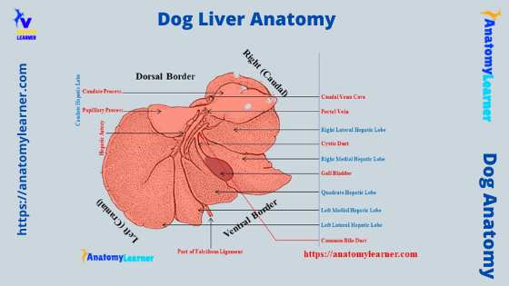
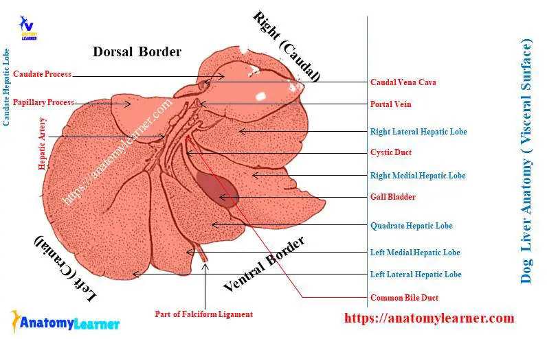
So, from the dog liver anatomy, you should identify the following features. All these features from the dog liver are identified in the labeled diagram.
- A diaphragmatic surface of the dog liver,
- The visceral surface of the canine or dog liver,
- The dorsal, ventral, right, and left lobes of the liver,
- Left lateral and medial hepatic lobes,
- A quadrate lobe of the dog liver,
- Right medial and lateral lobes of the liver,
- Caudate lobe of the canine liver,
- Caudate and papillary processes of the caudate lobe of the liver,
- Area for the caudal vena cava,
- Porta of the dog liver, and
- Gallbladder (in between the right medial and quadrate lobes),
Again, there are three important impressions on the visceral surface of the dog liver. You will find more impressions on the visceral surface of the ruminant liver (rumen, reticular, omasal, abomasal, and renal).
Let’sLet’s try to identify the following impressions from the visceral surface of the dog liver –
- A gastric impression (on the left half of the visceral surface),
- The duodenal impression on the visceral surface, and
- A renal impression on the thick caudolateral border of the caudate lobe,
If possible, try to identify the following attachments (ligaments) from the dog liver. There are five main attachments to the dog liver –
- A coronary ligament,
- Falciform ligament,
- Left and right triangular ligaments,
- Hepatorenal liagament, and
- Attachment of lesser omentum to the liver,
The porta of the dog liver comprises the hepatic artery, portal vein, and bile duct. You might differentiate these three structures from the actual sample at your laboratory.
I hope you already got the basic idea of the different important anatomical features of the dog liver. Now, you may learn more about the canine liver in the next sections of this article.
Unique features of dog liver anatomy
If you compare the anatomical features of a horse, ruminant, and pig livers with the dog, you will find some peculiar characteristics. Here, I will enlist some of the unique anatomical features of the dog liver that might help you to differentiate it from ther other species’ liver.
Mainly you will see the difference in the lobes, impression, and location of the gallbladder. Okay, let’s see what the unique feature of the dogs liver are –
- You will find the well separater four lobes in the dog liver – left, quadrate, right, and caudate,
- The caudate lobe of the dog liver divides into two distinct processes – the caudate process and papillary processes,
- The caudate process of the caudate lobe locates on the right side, whereas the papillary process locates on the left side,
- You will find the highly convex diaphragmatic surface on the dog liver, whereas the visceral surface is irregularly concave,
- The visceral surface only shows the impression of the stomach and duodenum,
- The gallbladder of the dog locates in the depression between the right medial and quadrate lobes,
- Again, the dog liver is comparatively larger in comparison to the other domestic mammals,
- The bile duct opens into the duodenum just behind the pylorus. But, if there is an accessory pancreatic duct, it may join with the bile duct and commonly opens into the duodenum.
In some dog breeds, the accessory pancreatic duct opens separately into the duodenum. Again, a few smaller hepatic ducts are present in some dog breeds, which directly join with the bile duct. They have no communication with the main hepatic duct, but this condition of the smaller bile duct is rare.
Physical properties of the canine liver
The physical properties of the dog liver, except the color and consistency, may vary with different breeds and ages. Again, the liver is much heavier in young puppies than in older dogs.
The fresh liver of a dog shows a deep red-brown color. Again, the consistency of the liver is firm, but it is friable.
The weight of the dog liver is about 2- 3% of their body weight. So, you will find a great variation in the weight of the liver in a different breed of dog.
So, if your dog’s weight is about 10 kilograms, then the weight of the liver may vary from 200 – 300 grams. Averagely, you may find the weight of the dog liver from 200 grams to 1kilogram.
The diaphragmatic surface is much convex that attaches to the diaphragm. Again, the visceral surface of the dog liver is more concave than the ruminant.
You will see the distinctly separated lobes in the dog liver where the caudate lobes show two different processes. You will also find a similar feature in the pig liver, but they lack the papillary processes (learn from the last part of this article).
The shape of the liver lobes varies in the dog. Here, you will find the different shapes in the four different lobes.
Surfaces and borders of the dog liver
Simply the outer surface or the surface that is directed to the wall of the abdomen and diaphragm is the diaphragmatic surface. Again, the inner surface or that surface that is directed to the visceral organs or contains the porta hepatis is known as the visceral surface.
Now, I will describe the different anatomical features from the following two surfaces of the dog liver –
- The diaphragmatic surface or parietal surface, and
- The visceral surface of the dog liver,
A diaphragmatic surface of dog liver
The diaphragmatic surface of the dog liver is strongly convex in all directions. It lies mainly in contact with the abdominal surface of the right part of the diaphragm.
The convexity of the dog liver is an exceptional feature that makes them unique from other. This convex surface faces bilaterally and dorsally that faces cranially and ventrally.
So, you will find two parts in the diaphragmatic surface of the dog liver –
- The right part of the diaphragmatic surface, and
- A left part of the diaphragmatic surface,
Here, the right part of the diaphragmatic surface is larger. It extends from the cranial extent of the liver at the level of the sixth intercostal space to the last intercostal space. Again, this surface’s left part lies opposite the tenth intercostal space.
What about the cranial and dorsal parts of the diaphragmatic surface of a dog liver? Okay, let’s see the cranial part of the liver; you will find a shallow, broad, and distinct cardiac impression here. This liver part is located at the left of the medial plane.
Again, the dorsal part of the diaphragmatic surface of the dog liver possesses a deep notch. It lies approximately in the middle of the median plane.
You know the caudal vena cava and esophagus form the deep notch on the dorsal surface of the liver. The caudal vena cava lies on the dorsal notch to the right of the esophagus.
The visceral surface of the canine liver
The visceral surface of the dog liver anatomy shows the irregular concave feature. It faces mainly the caudoventrally and the left. The visceral surface of the dog’s liver remains in contact with the abdominal viscera.
“The visceral and diaphragmatic surfaces of the dog liver meet ventrolaterally in a sharp edge border and dorsally by a blunt border.”
Here, the right kidney, stomach, duodenum, colon, and jejunum (little) produce impressions on the visceral surface of the liver. You may also find another impression on the visceral surface that is produced by the pancreas.
You will see the gallbladder on the visceral surface of the dog liver. This gallbladder lies on the depression made by the quadrate and right medial lobes of the dog liver.
Again, you will see different important anatomical features on the visceral surface of the dog liver. The most important one is the presence of the porta hepatis of the liver.
This is the hilus of the dog liver. You will learn more about the dog liver hilus in the next section of this article.
Another most important anatomical feature of the visceral surface is the presence of the papillary process. This process locates at the center of the caudate lobe and protrudes caudally from the dorsal part.
Porta hepatis of the dog liver
You will know the details of the papillary process in the lobes anatomy section. Now, what is porta hepatis or porta of the dog liver?
Well, you know the porta of the dog liver is the hilus of this organ. It is in the form of a linear depression which serves the purpose of the hilus of the organ.
This porta hepatis is located on the dorsal third of the visceral surface of the liver. You will find four major structures here in the porta hepatis –
- Hepatic artery,
- Hepatic nerves,
- The portal vein, and
- Bile ducts,
This porta hepatic is also termed the portal fissure. Several lymph nodes and the part of the pancreas are also attached to this portal fissure.
The hepatic nerves and arteries enter the porta hepatis dorsally. At the same time, the hepatic ducts leave the porta hepatis ventrally. In addition, the portal vein enters into the portal fissure or porta hepatis between the ducts and vein.
Impressions on dog liver – visceral surface
The visceral surface shows different impressions, but the most important is the following –
- A large gastric impression on the visceral surface of the dog liver,
- The duodenal impression on the visceral surface, and
- A renal impression of the liver,
Gastric impression: the gastric impression of the liver is larger and occupies the whole left half of the visceral surface. The gastric part of the dog’s stomach occupies this impression. Again, you will also find another impression of the pyloric part of the dog stomach.
Let’sLet’s see the gallbladder on the visceral surface of the dog liver (identified). The pyloric part of the dog stomach lies in contact with the caudal face of this gallbladder. Again, the pyloric part remains fixed in position in the abdomen.
Thus, the pyloric part of the dog’s stomach forms an oblique impression across the middle part of the gland. Sometimes you may see the coli of the small intestine that contact with the visceral surface of the dog liver (especially when the stomach is empty).
But, this small intestine does not make any impression on the visceral surface of the dog liver.
Duodenal and renal impressions of dog liver
The duodenal impression is the most cranial indentation on the visceral surface. It starts at the junction of the right and quadrate lobes of the dog liver. First, this impression runs to the right and makes an arch caudoventrally.
Finally, the duodenal impression runs caudoventrally and lying dorsal to the ventral border of the dog liver. Again, the right lobe of the dog pancreas lies dorsomedial to the cranial and descending part of the duodenum.
So, along with the duodenum, the right lobe of the dog’s pancreas is in contact with the visceral surface of the liver. But, the right lobe of the pancreas does not form any impression on the visceral surface of the liver.
On the other hand, the renal impression on the dog liver is almost hemispherical fossa. It locates in the caudodorsal part of the liver, in the caudate process of the caudate lobe. This impression is formed by the cranial pole of the dog’s right kidney.
The ventral half of the right kidney is covered by the dog’s liver. But, the right adrenal does not form any impression on the dog’s liver.
Borders of the dog liver
As you know, four distinct borders exist on the canine liver anatomy. You will see the dorsal, ventral, right, and left borders in the dog liver.
You know the dorsal border of the dog liver is thick. A deep and broad notch on the dorsal border of the dog liver contains the caudal vena cava and esophagus.
The ventral border of the dog liver is thin and sharp. The diaphragmatic and visceral surfaces of the dog liver meet the ventrolateral edge and form the thin border.
You will see other fissures (main and some accessories) that divide the dog liver into different lobes. These main and short accessories fissure cut into the border of the organ.
Let’sLet’s identify the fissure for the round ligament; it locates between the caudoventral portion of the caudate and medial lobe. It forms the right border to the caudate lobe and the left border to the medial lobe.
Dog liver lobe anatomy
The dog liver lobe anatomy possesses mainly the four lobes. You will also see the four sublobes and two processes in the dog liver lobe structure. Let’sLet’s see what the lobes, sublobes, and process are from the dog liver –
- The left hepatic lobe (main lobe),
- Left lateral hepatic lobe (sublobe),
- Left medial hepatic lobes (sublobe),
- The quadrate lobe (main lobe of the liver),
- A right hepatic lobe (main lobe),
- Right medial hepatic lobe (sublobe of the hepatic lobe),
- Right lateral hepatic lobe (sublobe of the hepatic lobe),
- Caudate lobe (main lobe of the dog liver),
- Caudate process (a process of the caudate lobe), and
- Papillary process (a process of the caudate lobe),
The dog liver labeled diagram shows the four main lobes, four sublobes, and two processes. Now, you will learn the anatomical features of these lobes, sublobes, and processes from the canine liver anatomy.
Sometimes, the right medial is the right central, and the left medial is the left central lobes of the liver.
Caudate lobe of the dog and its processes
You will see this caudate lobe on the visceral surface of the dog liver. There are two processes (caudate and papillary processes) in the caudate lobe of the dog liver that connects with the isthmus.
The isthmus between the caudate and papillary processes remains compressed between the caudal vena cava dorsally and portal vein ventrally. This is formed by the hepatic tissue approximately 0.5 centimeters thick.
Now, let’s see the anatomical features of the caudate and papillary processes from the caudate lobe of the dog liver –
Caudate process: this is the most caudal part of the dog’s liver. It extends to the right of the medial plane of the dog’s body. You may easily identify this caudate process with the help of intercostal spaces.
This caudate process extends from the last ribs (twelve intercostal spaces) on the right side of the abdomen. You will find a deep depression in the caudodorsal part of the caudate process of the dog liver.
Here, the cranial half of the right kidney is inserted into the liver. Again, the diaphragmatic surface of the caudate process forms the triangular area.
Most of the part of the parenchyma of the caudate process fuses to the right lateral lobe of the dog liver. But, in some dog breeds, you will find the completed separate lobes.
Papillary process of the canine liver
Again, the visceral surface shows a pyramidal or tongue-shaped papillary process of the caudate lobe. The papillary process lies over the left lateral hepatic lobe of the liver.
Again, this process of the caudate lobe is loosely covered by the lesser omentum. And it lies in the lesser curvature of the dog’s stomach.
One or two fissures may separate the papillary process into different incomplete parts. The more constant fissure separates the frenular part from the body of the papillary process.
Again, the parenchyma of the papillary process fuses to the left lateral lobe of the dog liver. But, you may identify two separate parts of the liver (papillary process and left lateral lobe).
The right hepatic lobe of the dog liver
The right hepatic lobe of the dog liver completely lies to the right of the median plane of the dog’s body. If you notice the dog liver labeled diagram, you will find the right hepatic lobe of the dog is smaller than the left.
You may easily mark the location of the right hepatic lobe of the dog liver from the external surface. It extends transversely between the dorsal part of the sixth to tenth intercostal spaces.
Again, the right hepatic lobe of the canine liver shows two distinct sublobes –
- A right lateral hepatic sublobes and
- A right medial hepatic sublobe,
The right medial hepatic sublobe is longer than these the lateral one. But, the width of the right lateral sublobes is more than the right medial.
Let’s see the other anatomical features of the liver’s right lateral and right medial sublobes.
Right lateral hepatic lobe: the shape of the right lateral hepatic lobe is irregular in the dog. It is compressed laterally and possesses a slightly concave base. Let’s see what structure covers or attaches with this right lateral hepatic lobe on its cranial, caudal, dorsal, and ventral aspects.
The ventral part of the right lateral hepatic lobe lies opposite the distal part of the middle third of the caudal border of the right medial lobe. Again, cranially you will also find the right medial lobe.
The caudal part of the right lateral hepatic lobe of the canine liver runs over the caudate process and fuses with it. Dorsolaterally, you will find the caudal vena cava and caudal part of the left lateral hepatic lobe.
A right medial hepatic lobe of a dog
The right medial hepatic lobe of the dog lies medial to the right lateral and connects with the quadrate lobe. But, you may find a great variation in the fusion of the right medial hepatic lobe with the quadrate lobe in a different breed of dogs.
In some dog breeds, the dorsal part of the right medial hepatic lobe fuses with the quadrate lobe. Again, some liver of the dog shows a large fusion that extends nearly to the fossa of the gallbladder.
For these livers, you will find a small fissure extending dorsally from the gallbladder fossa and separating these two lobes.
If you compare the right lateral and medial hepatic lobes, you will see – the right medial hepatic lobe is longer and triangular. In comparison, the right lateral hepatic lobe is short but wider and irregular-shaped.
The ventral border of the right medial hepatic lobes shows the extension that extends beyond the ventral part of the costal arch. But, in the right lateral hepatic lobe, you will not find any extra extension like the medial lobe.
You will see a small fissure between the right medial hepatic lobe and the quadrate lobe of the dog liver. This fissure separates these two lobes (right medial and quadrate) partly.
Again, the dorsal half of the right medial hepatic lobe (visceral surface) possesses a deep and large fossa (between the quadrate and right medial lobes). This fossa on the visceral surface of the right medial hepatic lobe is designed for the gallbladder.
The diaphragmatic surface of the right medial hepatic lobe is like a curved triangle. You will see a slightly concave and fissured medial border on the right medial hepatic lobe of the dog liver.
Left lobe of dog liver anatomy
The left lobe of the dog liver is the largest lobe among the four lobes. This left lobe is cranial and lies entirely to the left of the median plane of the dogs body.
Again, the left lobe forms one-third portion of the dog liver. You will see a fissure that completely divides the left lobe of the dog liver into two sublobes –
- The left lateral hepatic lobe of the dog’s liver, and
- A left medial hepatic lobe of the liver (smaller),
Let’s see what the unique features of these two sublobes of the left lobe of the dog liver are.
Left lateral hepatic lobe of the dog
The left lateral hepatic lobe of the dog liver almost attaches to the abdominal part of the diaphragm. It starts dorsally deep to the left crus of the diaphragm and crosses ventral to the left part of the tendinous center.
Again, this hepatic lobe passes ventral to the left part of the muscular periphery of the diaphragm. The diaphragmatic surface of the left lateral hepatic lobe becomes wider till the middle. Then it gradually narrows and ends at the last sternebra as a pointed part.
The lateral border of the left lateral hepatic lobe may protrude caudal to the ventral part of the costal arch. Again, the dorsal part of this hepatic lobe covers the body of the dog’s stomach.
Let’s see the features from the visceral surface of the left medial hepatic lobe of the dog liver. You will see the concave feature on the visceral surface of the left lateral hepatic lobe. This concave area is for the fundus and body of the dog stomach.
Again, the part of this hepatic lobe is partly covered by the papillary process of the caudate lobe. The center part of the left lateral hepatic lobe (visceral surface) shows a slightly convex area known as the omental tuber.
Left medial hepatic lobe of the dog
The left medial hepatic lobe of the dog locates between the left lateral hepatic and quadrate lobes. Usually, the shape of the left medial hepatic lobe of the dog is nearly triangular. Sometimes you may find the oval in the outline left medial hepatic lobe in some dog breeds.
You will see a deep and curved fissure that extends from the porta hepatis of the liver. This deep, curved fissure separates the left medial hepatic lobe from the left lateral lobe. You will see the different intrahepatic vessels and nerves in between the left medial and left lateral hepatic lobes of the dog.
Again, there is another deep fissure that also extends from the porta hepatis and esophageal notch. This fissure separates the quadrate lobe from the left medial hepatic lobe of the dog.
You will see the round ligament of the liver within this deep fissure. The round ligament of the dog liver has a connection with the falciform ligament on its free ends.
Dog quadrate lobe anatomy
The quadrate lobe of the liver lies between the right medial and left medial hepatic lobes. You will find this quadrate lobe in the middle of the medial plane of the body.
The diaphragmatic surface of the quadrate lobe of the dog liver is fusiform. It extends from the ventral border of the esophageal notch to the caudal vena cava.
The visceral surface of the dog quadrate lobe shows two distinct fissures. The left deep fissure separates this quadrate lobe from the left medial hepatic lobe. In contrast, the right deep fissure separates the quadrate lobe from the right medial hepatic lobe.
Again, the visceral surface of the quadrate lobe forms the fossa for the gallbladder. The cystic duct runs in the fossa between the quadrate and right medial hepatic lobes of the dog liver.
Again, the small bile duct also exists from the cranial part of the quadrate lobe of the dog liver.
Summary of the dog liver lobes
I hope you can understand and identify all these four lobes, four sublobes, and two major processes from the dog liver structure. Here, I will summarize the features of the dog liver lobes.
Caudate lobe: is located caudodorsal to the porta hepatis of the liver and consists of caudate and papillary processes. The papillary process is the part of the caudate lobe to the left of the medial plane and lies on the lesser curvature of the stomach.
Again, the caudate process is the most caudal region of the dog liver. There is a renal impression on the caudodorsal border that receive the cranial end of the right kidney.
The left lobe of the dog: lies left to the medial plane and is the largest lobe in the dog liver. It shows two distinct divisions – larger left lateral and triangular smaller left medial hepatic lobes.
A quadrate lobe of the dog: is located between the right and left medial hepatic lobes and ventral to the porta hepatis of the liver. Form the fossa along with the right medial hepatic lobe for the gallbladder.
The right lobe of the dog liver: lies right of the median plane and consists of two distinct parts – right lateral and medial hepatic lobes. The right medial hepatic lobe is somewhat triangular and longer than the right lateral.
Canine liver ligaments or attachment
While studying the canine or dog liver anatomy, you might also know how this liver attaches to the abdominal cavity. You will find the five different important ligaments and some associate structures that help to fix its position on the thoracic part of the abdominal cavity.
You will see the peritoneum covering the entire liver that forms the serous coat. Again, this serous coat of the dog liver fuses to the underlying tissue and fibrous capsule.
You know the fibrous capsule of the dog liver is a thin but denser layer. This fibrous capsule of the liver comprises the collagen fibers closely invested on the liver surface.
Again, if you see the parenchyma of the dog liver, you will find some trabeculae that divide it into different lobules. These are the interlobular trabeculae and come from the collagenous capsule.
At the porta hepatis of the liver, you will see the thicker fibrous coat that continues into the interior part of this organ. Here, they remain together with the vascular and nervous elements of the dog liver. This is the capsula fibrosa perivasculairs of the dog liver.
Let’s see the diaphragmatic surface of the liver; it attaches to the diaphragm by serous and fibrous coat structure. Here, this serous and fibrous coat forms the coronary ligament. Again, some of the small folds also arise from this coronary ligament.
These folds from the coronary ligament form the right triangular left triangular, and falciform ligaments. You will also find the hepatorenal ligament and the lesser omentum attached to the dog liver.
So, you have got the five ligaments in the dog liver. Let’s see some anatomical features of these five ligaments from the dog liver.
Coronary ligament of the canine liver anatomy
The coronary ligament of the canine liver is irregular in outline. It represents the close embryonic relationship between the diaphragm and the liver of the dog.
Some authors did not define it as the true peritoneum ligament because two parts of the peritoneum did not form one fold and remained separated.
This coronary ligament attaches to the triangular area on the diaphragmatic surface of the dog liver. Then it continues on the dorsal surface of the caudal vena cava.
Again, from the part of the coronary ligament of the dog liver, another three ligament arises. The triangular (right and left) ligaments and the falciform ligament arise from the coronary ligament’s stellate border (or folds).
Right triangular ligament of the dog liver
The right triangular ligament of the dog liver arises from the peritoneum folds. It extends between the diaphragm and the dorsal part of the right lateral lobe of the liver.
You will see a wide free border in the right triangular ligament of the dog’s liver. But, it becomes progressively narrow as it passes medially.
The peritoneal layer of this right triangular ligament continues with the right peritoneal leaf of the coronary ligament. You know, the round ligament and the origin area of the right triangular ligament bind the triangular area on the diaphragmatic surface of the dog liver.
The length of the right triangular ligament is more than its wide. This right triangular ligament includes a fold from the diaphragm to the diaphragmatic surface of the right medial lobe of the dog liver.
Left triangular ligament of canine liver
The left triangular ligament of the canine liver also originated from the peritoneal fold at the triangular area of the diaphragmatic surface. It extends from the dorsal segment of the left lateral hepatic lobe to the left crus and the tendinous portion of the diaphragm.
You will find different components in the structure of the left triangular ligament of the canine liver. Normally, there are two components in the canine left triangular ligament of the liver – cranial and caudal.
The cranial component runs from the left lateral lobe to the diaphragm. Again, the caudal component of the right triangular ligament is larger and consists of a fibrous appendix of the liver.
But, you may not find this fibrous appendix in all dog’s livers. This is only present in the liver of the older dog.
The fibrous appendix of the liver is a narrow, thin, tapering band of the hepatic tissue. This structure locates near the free border of the left triangular ligament of the canine liver.
Falciform ligament of the dog liver
The falciform ligament of the dog liver is the remnant of the primitive ventral mesentery. This ligament extends between the liver and diaphragm and ventral body wall caudally to the umbilicus.
The umbilical vein locates on the free border of the falciform ligament in early fetal life. A portion of the falciform ligament extends from the umbilicus to the diaphragm. This part of the falciform ligament possesses fat filed irregular folds.
The falciform ligament of the dog liver may disappear completely in the inner aspect. But, it may remain as a thin, avascular fold that extends from the dorsal extremity of the fissure between the right and left medial lobes to the coronary ligament.
The left peritoneal part of the dogs falciform ligament joins with the left portion of the coronary ligament. Again, the right peritoneal part of the falciform ligament connects with the ventral segment of the coronary ligament.
Again, the falciform ligament of the dog runs between the quadrate and left medial hepatic lobes ventrally.
Hepatorenal ligament of the canine liver
The hepatorenal ligament is the delicate peritoneal fold that is not constant. It extends from the medial part of the renal impression to the ventral surface of the right kidney. Again, it lies lateral to the fat that fills the hilus of the dog kidney.
The canine liver anatomy also shows the lesser omentum that helps to fix it in the position. This lesser omentum is the thin, fat-streaked, loose peritoneal fold that attaches to the liver.
The lesser omentum is the remnant of the primitive ventral mesentery of the dog. It extends from the liver to the lesser curvature of the dog stomach and cranial part of the duodenum.
Summary of the dog liver ligaments
Here, I will summarize all these five ligaments and the lesser omentum that help fix the dog’s liver in its position. Let’s see what the main facts of these five ligaments and lesser omentum are –
Coronary ligament of the liver: the reflection of the peritoneum from the diaphragmatic surface to the liver into the diaphragm. It forms a circular area of reflection around the caudal vena cava. The coronary ligament comes together in three places to form two triangular and one falciform ligament.
Right triangular ligament of the dog liver: it attaches to the dorsal part of the reight lateral lobe of the liver to the right crus of the diaphragm.
Left triangular ligament of the canine liver: possesses two-component and attaches to the dorsal part of the left lateral lobe to the left crus of the tendinous center of the dogs diaphragm.
Falciform ligament of the dog liver: connect the ventral surface of the liver to the sternal part of the diaphragm and ventral abdominal wall.
Hepatorenal ligament of the liver: connect the ventral surface of the kidney to the renal impression on the caudal part of the liver.
Lesser omentum: extends from the porta hepatis of the dog liver to the lesser curvature of the stomach and the cranial part of the duodenum. This lesser omentum divides into hepatoduodenal and gastrohepatic segments.
Structure of the dog liver
You know the free surface of the dog liver is almost completely covered by the peritoneum, which forms the serous coat. Again, this serous coat fuses with the fibrous capsule that encloses the whole organs.
The finer trabeculae originated from the fibrous capsule that divides the liver parenchyma into different small lobules. Again, the interlobular connective tissue carries blood vessels into the liver.
The gross anatomy of the dog liver clearly shows the lobules in its parenchyma. These hepatic lobules are the smallest grossly visible functional unit of the liver.
The lobules of the dog liver consist of a curved sheet of hepatocytes that surrounds the blood-filled cavities (known as the sinusoid). If you observe the histological features of the dog liver, you will find two important parts –
- A polygonal hepatic lobule in the parenchyma, which center is occupied by the central vein, and
- Triangular space between the lobule accommodates small branches of the hepatic artery, portal vein, and bile ducts,
In this article, I will not describe the details of the microscopic features of the dog liver. Rather, you may read the microscopic liver features from another article by an anatomy learner (please see the histology section).
Each hepatic lobule of the dog liver comprises the rows of hepatic cells radiating from the central vein. The arrangement of these lobules with the hepatic cells will be observed clearly in the pig liver.
A group of three tubes comprises an interlobular branch of the hepatic artery, portal vein, and interlobular bile ductile. All these structures enclose in a perivascular connective tissue in the spaces between the lobules. This structure of the dog liver parenchyma is known as the portal triads.
Dog liver vessel anatomy
The dog liver vessel anatomy mainly comprises the hepatic artery and portal vein. Through these two vessels, the dog liver receives a generous blood supply. The dog liver receives about 80% blood from the portal vein and about 20% from the hepatic artery.
Within the liver, these vessels divide to form the segmental vessels that divide into interlobular vessels. These interlobular vessels of the liver run into the portal canal.
Again, the further division of the interlobular vessels of the liver parenchyma opens into the hepatic sinusoids. Thus, you will find both arterial and venous supply to the liver sinusoid.
Now, I will discuss the two major vessels from the dog liver –
- Hepatic artery or hepatic, and
- The portal vein or porta of the liver,
Again, I will discuss the single central vein from the dog liver lobule. Let’s see the important and unique anatomical features of the hepatic artery and portal vein of the dog liver.
Hepatic artery to the dog liver
The hepatic artery is a celiac artery branch that furnishes nutritional supply to the dog liver. You know the hepatic artery enters the liver parenchyma together with the portal vein at the hepatic porta on the visceral surface.
Along with the fibrous trabeculae of the dog liver parenchyma, both the hepatic artery and portal vein gives branches together with the tributaries of the hepatic ducts.
Now, the branches of the hepatic arteries supply to the framework of the liver, the capsule, and the intrahepatic biliary duct system. Finally, they open into the portal vein into the liver sinusoid.
Thus, the parenchymal cells receive mixed blood from the portal vein and hepatic arteries. So that they receive the nutrition both from these vessels of the liver.
The hepatic sinusoid of the liver is lining with the kupffer cells. These are the phagocytic cells and present cleft between the hepatic plate.
During the active phase, each radiating column of the hepatic cells has a vascular capillary on one side. This joins with the central vein and a bile capillary on the other aspect. This bile capillary of the liver parenchyma goes out of the lobule to join the bigger bile channel.
Porta vein of the canine liver
The portal vein of the canine liver brings the functional blood to this organ from the stomach, intestine, pancreas, and spleen. About 80% of the blood entering the liver reaches it by the portal vein.
You will see three major branches of the portal vein in the dog – the splenic vein and the cranial and caudal mesenteric veins. These veins collect the blood from the unpaired organs of the dogs’ abdomen. They collect blood from the stomach, pancreas, intestine, and spleen.
And thus, the portal vein of the liver supplies the functional blood to it. You may also see some of the contributing veins of the portal vein. These veins are connected to the veins of the cardioesophageal and caudal regions of the dog’s abdomen.
In the fetus, the ductus venosus is the direct continuation from the umbilical vein to the hepatic venous system. It develops within the liver between the left intrahepatic branch of the portal vein.
The ductus venosus bypasses the hepatic circulation to join the caudal vena cava. You know the ductus venosus of the dog becomes fibrotic after birth and is known as the ligamentum venosum.
This structure extends obliquely from left to right in the liver parenchyma. Again, it lies ventral to the attachment of the papillary process of the caudate lobe of the dog liver.
The central vein of the dog liver
The venous drainage of the dog liver starts with the single central vein in the middle of the hepatic lobule. These veins collect mixed blood from the hepatic artery and portal vein. The collected blood mixed in the liver sinusoid and contact with the hepatocytes.
Here, the central vein of the liver lobule fuse to form an interlobular vein. Again, the interlobular veins fuse and form the hepatic veins. You know these hepatic veins of the dog liver open into the caudal vena cava.
You will see the spiral and circular muscle fibers in the wall of the central and intralobular veins of the dog liver. These muscle fibers of the veins possess a sphincter function that restricts venous drainage.
The hepatic veins convey to the caudal vena cava all the blood that the dog liver receives through the portal and hepatic arteries. Again, these hepatic veins enter the caudal vena cava progressing from the right lobe to the left lobe.
Important structures of the venous system of liver
Now, let’s point out some of the important structures from the dog liver anatomy that are related to blood supply –
Hepatic lobule and cells: lobule is the structural unit of the liver. The hepatic cells are the liver cells arranged radially around the central vein.
Portal vein: the large vein in the dog liver that carries the nutrient-rich blood from the stomach, intestine, pancreas, and spleen into this organ at the porta hepatis.
Proper hepatic arteries: carry the oxygenated blood (nutritional blood) to the hepatic cells at the porta hepatis. They are the branch of the common hepatic artery.
The central vein of the liver: courses from the center of each liver lobule, receiving blood from the liver sinusoid and carrying it to the hepatic veins.
Hepatic veins: are located inside the liver and open into the caudal vena cava. The central vein joins to form the hepatic vein in the dog liver.
The liver sinusoid: is a space where blood is received from both hepatic arteries and portal veins. It interacts with the hepatic cells and then exits through the central vein of the liver lobules.
Nerves of the canine liver anatomy
In the canine liver anatomy, you will find the supply of both afferent and efferent axons of the vagus nerve. Again, you will see the innervation of the sympathetic axon from the celiac plexus.
The vagus axon passes through the diaphragm with the esophagus as the dorsal and ventral vagal nerve trunk. Finally, this vagus axon reaches the dog abdomen.
Again, the vagus axon passes obliquely to the right of the lesser omentum near the porta hepatis. Then, the vagus axons enter the liver parenchyma and supply to the biliary system.
The dog liver is also innervated by both the sympathetic and parasympathetic nerves. The sympathetic axon reaches the dog liver through the splanchnic nerve, celiac ganglia, and celiac plexus.
This axon continues on the hepatic arteries and the lobular arteries. The dog biliary system also receives the afferent axons from the phrenic nerve.
Biliary system from the dog liver anatomy
Before going to the details of the biliary system of the dog liver anatomy, you might have known some terms and structures. First, let’s see what terms and structures are necessary to study the biliary system of the dog liver.
Bile: you know this is the liquid substances of the liver cells into the duct system of this organ.
So, you might know the different ducts of the dog liver. Normally, you will find the following ducts in the dog liver –
Interlobular ducts: these ducts pass in the interstitial tissue between the two lobules of the dog liver. They unite to form the lobular ducts that course to the porta hepatis and join to form the hepatic duct.
Hepatic ducts: you may see the variation in the number of hepatic ducts in the dog liver. You may find three to five hepatic ducts that leave the porta hepatis to join with the cystic duct.
Cystic duct: this structure connect the gallbladder with the hepatic ducts. Now, the hepatic and cystic ducts together form the bile duct. You know, the cystic duct is the two ways street where bile can pass through it and store in the gallbladder.
The store bile can pass through the cystic duct to the bile duct and into the duodenum of the dog. Again, the bile is discharged into the duodenum through the cystic and bile ducts.
Bile duct: this duct is formed by the junction of the hepatic and cystic duct. It runs over the lesser omentum to enter the duodenum. Sometimes it joins with the pancreatic ducts, and both open on the major duodenal papilla.
Bile passage in the dog liver
The bile is produced by the sheet of the liver cells and discharged into the minute bile canaliculi. You know these bile canaliculi lie between these cells.
Bile canaliculi: these are the small tubes immediately surrounding and collecting bile from the hepatic cells. They run to the periphery of the lobule opposite direction of the blood flow, where they join to form the interlobular duct.
The bile canaliculi join to form the biliary ductile that continues into the interlobular ductules. You know these interlobular ductules lie in the interstitial tissue between the lobules.
These interlobular ducts are found in the portal triads, where you will also find the hepatic artery and portal vein. Larger interlobular bile ducts form by the anastomosis of the interlobular ductules.
Again, the interlobular ducts unite to form the lobar intrahepatic ducts. The number of these intrahepatic ducts may vary and terminate at the origin of the extrahepatic ducts.
The extrahepatic bile ducts consist of the hepatic ducts. Again, the cystic ducts join the gallbladder with the hepatic ducts from the bile ducts.
These bile ducts (sometimes known as the common bile duct) open into the duodenum on the major duodenal papilla.
Canine gallbladder anatomy
You already identify the location of the canine gallbladder from the liver. This gallbladder will be on the visceral surface of the liver in between the quadrate and right medial hepatic lobes.
The dog gallbladder is a pear-shaped vesicle-like structure. It lies medial to the quadrate lobe and lateral to the medial hepatic lobe of the dog liver.
This may extend through the thickness of the dog liver to its diaphragmatic surface and contact with the diaphragm. The capacity of the canine gallbladder may vary with the different breeds of dogs.
Anatomically, you will find three parts in the canine gallbladder – fundus, body, and neck. The blind, rounded cranial end of the dog gallbladder is the fundus.
The large middle part of the canine gallbladder is the body. Whereas the slender, tapering caudodorsally directed part of the gallbladder is the neck.
The canine gallbladder stores and concentrates the bile secreted from the hepatic cells. Epithelium of the dog gallbladder possesses absorptive function.
They absorb lipid-soluble compounds, including cholesterol.
The bile is released into the duodenum as a suspension of fatty compounds that can be absorbed into the small intestine. Again, the gallbladder possesses a little contractile power as they have good musculature on its wall.
Cystic and bile ducts
The cystic duct begins the biliary system of the dog’s liver. It extends from the neck of the dog’s gallbladder to the junction site with the first tributary from the liver.
At this level, just distal to the duodenum, you will also find the liver’s bile duct. This bile duct is the main excretory channel that receives bile from the hepatic ducts.
Sometimes the lobar duct does not unite to form a single hepatic duct but enters into the main trunk of the excretory tree. The distal part of the bile duct enters into the dorsal wall of the mesentery.
The bile duct may open separately or along with the pancreatic ducts in a dog. You know the site of the combined opening of the bile duct and pancreatic duct on the duodenum is the major duodenal papilla.
Again, the accessory pancreatic ducts of the dog also open on the duodenum. The opening site of the accessory pancreatic ducts is known as the minor duodenal papilla.
Horse, pig, and cow liver anatomy compared to dog
So, this part is very important for you if you want to compare the anatomy of the liver of different species like horses, pigs, and cows. Here, you will find the unique features of the horse, pig, and cow liver that help you to identify them from the dog liver anatomy.
But, first, let’s see some of the important information that might help you to gather great knowledge on the liver from different species –
Lobes of liver from horse, pig, and cow (comparison)
You know the right and left lobes of the dog’s liver divide into medial and lateral sublobes. Again, the caudate lobe subdivides into a caudate process and papillary process. But what about the horse, cow, and pig?
In the horse liver, you will find the most similar structure in the liver, like the dog. But, the right lobe of the horse’s liver is undivided. Again, you will not find the papillary process in the horse liver anatomy.
The horse liver locates obliquely across the diaphragm with the left ventral and right dorsal lobe. I hope you can understand the location and main anatomical difference of the horse liver from the dogs.
Again, the lobes of the pig liver anatomy are almost similar to the canine, but they are well separated. You will not find any papillary process in the pig liver structure. The other special features will discuss in the specific part of the pig liver structure section.
In the cow or ruminant, you will not find any division on the right and left lobes of the liver. The rumen (part of the ruminant stomach) has moved the liver to the right side of the abdominal cavity. Here in the ruminant or cow, the right lobe of the liver is dorsal, and the left lobe is ventral.
Let’s see the lobes from the horse, dog, pig, and ruminant (cow) liver – (table)
| Lobes of liver | Dog | Horse | Pig | Cow |
| Left lobe | + | + | + | + |
| Left lateral lobe | + | + | + | – |
| Left medial lobe | + | + | + | – |
| Quadrate lobe | + | + | + | + |
| Right lobe | + | + | + | + |
| Right lateral lobe | + | – | + | – |
| Right medial lobe | + | – | + | – |
| Caudate lobe | + | + | + | + |
| Caudate process | + | + | + | + |
| Papillary process | + | + | – | + |
| Total Lobes | 4 | 4 | 4 | 4 |
A unique feature of horse liver anatomy
Let’s see the unique features of the horse liver anatomy –
- The liver of the horse places obliquely on the abdominal surface of the diaphragm, and the major part of the liver is at the right side of the medial plane,
- It is extensive and comparatively thin compared to the dog liver,
- The right lobe does not posees the right lateral and right medial lobes like the dog,
- You will not find any gallbladder on the visceral surface of the liver,
- Both the left and right borders of the horse liver are thin,
- You will find an extra right lateral ligament that connects the left lobe with the diaphragm,
- The hepatic and pancreatic ducts open together on the duodenum (at hepato pancreatic ampulla),
These are very few but important information on the horse liver anatomy. But, you may learn more about the horse liver from another article by an anatomy learner.
Pig liver anatomy
Except for the papillary process, the anatomical features of the pig liver are almost similar to the dog. Let’s see the unique features of the pig liver anatomy –
- The pig liver is comparatively larger compared to the dog and posses all four lobes,
- The diaphragmatic surface of the pig liver is highly convex compared to these of the dogs liver,
- You will see the gallbladder in a depression on the right medial lobe of the pig liver,
- Here, the bile duct and pancreatic ducts open separately on the duodenum just behind the pylorus,
You may learn more about the pig liver anatomical features from another article by the anatomy learner (pig anatomy learning section).
Cow (ruminant) liver anatomy
You will see a great variation in the anatomy of the cow or ruminant liver compared to the dog. The variation is normally found in the shape of the liver, lobes, and also in ligaments –
- The cow or ruminant liver is somewhat rectangular (elongated),
- You will find the four lobes in the cow liver, but the lobes are not well separated like the pig or dog liver,
- There is no division found in the left and right lobes of the cow liver,
- The parietal or diaphragmatic surface is less convex compared to the dogs liver and remains molded with the abdominal surface of the right part of the diaphragm,
- You will see the falciform ligament that extends from the esophageal notch to the umbilical fissure along the parietal surface,
- There are some impressions of ribs on the diaphragmatic surface (caudodorsally) as it is closely associated with the thoracic wall,
- The visceral surface of the ruminant liver is less concave and possesses impressions of different organs,
- There are omasal, abomasal, and reticular impressions on the left visceral surface where the omasal impression is larger,
- You will find the thick dorsal border, which presents the caudate lobe and renal impression for the right kidney,
- The portal fissure locates above the omasal impression on the visceral surface,
- You will see the long pear-shaped musculo membranous gallbladder at the visceral surface of the cow liver above the omasal impression,
- The bile duct opens into the duodenum at the second bend a few centimeters away from the pyloric end of the stomach,
But, in sheep and goats, the pancreatic duct joins with the bile duct before opening into the duodenum without forming any ampulla.
Rabbit liver anatomy
I would also like to enlist some important anatomical facts from the rabbit liver. Let’s see what the unique features of the rabbit’s liver anatomy compared to the dog –
- You will find the comparatively extensive liver in the rabbit that possesses five lobes,
- The parietal surface of the rabbit liver is comparatively convex and applied against the diaphragm,
- Again, the visceral surface of the rabbit liver shows the concave features that partially accommodate the stomach,
- You will find the renal impression at the right lateral lobe, which is more wide and deep compared to the dog,
You may read more about the rabbit liver anatomy and the other internal organs from the rabbit anatomy learning section of the anatomy learner.
Dog anatomy liver spleen and pancreas
While learning the anatomy of the dog liver, you might also have good knowledge of the spleen and pancreas. Though I will not describe the anatomy of the dog’s spleen and pancreas here. You may read the details anatomical facts about the dog pancreas and spleen in the following articles –
- Dog spleen anatomy with the labeled diagram
Now, let’s see some of the important features of the dogs spleen and pancreas.
Dog pancreas anatomy
The dog’s pancreas is located at the dorsal part of the cranial and lateral abdominal, just caudal to the liver. The pancreas also possesses both endocrine and exocrine functions like the liver.
Normally, two lobes in the dog pancreas – right and left- meet behind the pylorus and form a V-shaped structure. Here, the right lobe of the dog pancreas is slender and thin, whereas the left lobe is shorter, thicker, and wider.
The right lobe of the pancreas lies in the mesoduodenum in contact with the dorsal part of the right abdominal wall. It extends from the middle of the ninth intercostal space to the fourth lumbar vertebra.
Towards the pylorus, the right lobe runs obliquely and becomes narrow and flattened dorsoventrally. The caudal part of the right lobe is related to the sublumbar fat that contains the ureter and ventral surface of the right kidney. Again, the caudal part of the right lobe relates to the caudate process of the dog liver.
The body of the dog pancreas lies closely to the caudodistal part of the pyloric region. Here, it forms a large concave impression on the cranial part of the pancreas’s body.
The left lobe of the dog’s pancreas lies deep in the wall of the greater omentum. It begins at the body of the pancreas and runs caudodorsally.
The dorsal surface of the left lobe relates to the dog liver’s caudate process and the portal vein, caudal vena cava, and aorta. Again, the ventral surface of the left lobe relates with the ventrocaudal to the transverse colon. This surface also connects ventrocranially to the dorsal wall of the dog’s stomach.
Ducts and accessory pancreas
Sometimes the accessory pancreas may find in the dog. This accessory pancreas locates located in the wall of the gallbladder and in the caudal part of the mesentery.
You will see two pancreatic ducts in the dog’s pancreas –
- Pancreatic duct – drain the right lobe, and
- Accessory pancreatic duct – drain the left lobe,
The accessory pancreatic duct of the dog pancreas is the largest excretory duct that opens into the duodenum on the minor duodenal papilla. Again, the pancreatic duct of the dog pancreas is small and opens with the common bile duct on the major duodenal papilla.
The cranial and caudal pancreaticoduodenal arteries supply the dog pancreas. Again, the left ends of the left pancreatic lobe of the dog supply the pancreatic branch of the splenic artery.
Dog spleen anatomy
The dog spleen is the organ under the lymphatic system that lies in the left hypogastric region parallel to the greater curvature of the stomach. It is roughly tongue-shaped and slightly constricted in its middle.
The anatomy of the dog spleen shows two extremities, two surfaces, and two borders. Here, the dorsal extremity is rounded and wedge-shaped, whereas the ventral extremity is variable in shape and position.
The parietal surface is convex and faces the diaphragm. Again, the visceral surface of the dog spleen is convex and faces medially.
The other anatomical features of the dog spleen consist of the capsule, trabeculae, and parenchyma. You will find the red and white pulp in the parenchyma of the dog spleen.
Canine spleen anatomy labeled diagram
Now, I will provide again the canine spleen anatomy labeled diagram that might help you to memorize the summary of it. Here, in the dog (canine) spleen labeled diagram, I tried to show and identify the different surfaces and borders.
The labeled diagram shows the right lobe, left lobe, quadrate lobe, and caudate lobe. Again, the right medial, right medial, left lateral, and left medial lobes from the dog liver are also identified in the labeled diagram.
The diagram also shows the caudate and papillary processes of the caudate lobe of the liver. Again, the visceral surface of the liver shows the gallbladder, cystic duct, bile ducts, and hepatic ducts.
From the porta hepatis, the hepatic artery, portal vein, and bile duct are identified in the dog liver labeled diagram. The impressions for the stomach, duodenum, and kidneys are also shown in the labeled diagram (visceral surface).
You may find more dog liver-labeled diagrams on the social media of anatomy learners.
Frequently asked questions on the dog liver anatomy
Now, let’s see the most frequently asked questions on the dog liver anatomy that the veterinary anatomy learners ask. Fine, let’s see what these questions are and try to solve them with me –
Why do dogs have liver problems?
Different factors can cause problems in the dog liver. The dog liver is very much susceptible to the different types of viral and bacterial infections.
The cysts and gallstones also may obstruct the bile duct. This is another main cause to have of problems in the dog liver.
Different types of endocrine diseases like diabetes and hyperthyroidism may also cause problems in the dog liver. Again, trauma due to an accident or heat may also cause problems in the dog liver.
Ingestion of the toxic substance and infectious agents may cause acute liver failure in dogs. These are the most common factors that may cause problems in the liver of a dog.
How do you know your dog has liver problems?
Ingestion of the toxic substance, different endocrine diseases (diabetes, hyperthyroidism), and different viral and bacterial infections may cause the problem in the dog’s liver. So, you may find a different common problem with some other extra symptoms if the dog has a liver problem.
The most common symptoms of a dog with a liver problem are lethargy, weight loss, fatigue, vomition, diarrhea, increased thirst, and yellow eyes. Again, you may find your dog to be unstable to walk.
Where is the animal liver located?
The animal liver is the largest solid gland of their body which is reddish brown. It locates on the right side of the abdominal cavity in an oblique downward (ventrally) and forward (cranially) direction in the animal.
The animal’s glands (liver) normally extend from the lumbocostal angle to the level of the seventh or eighth ribs. Again, the five ligaments keep the animal’s liver attached to the abdominal wall.
But, the location of the animal liver may vary little in some species. You may find the information (location of liver in different animal species) in this article.
What organ is on the dog’s left side?
On the dog’s left side, you will find different organs that may be divided into three major parts – the left hypochondriac region, left lateral abdominal region, and left inguinal region. In the left hypochondriac region of the dog, you will find the spleen, fundus of the stomach, and the left kidney.
Again, in the left lateral abdominal region of the dog, you will see the left kidney, parts of the colon, and small intestine. Finally, the left inguinal region of the dog consists of the left ureter, part of the colon, and a small part of the female organs.
What side of a dog’s body is the liver?
The dog liver lies to the right of the median plane in the thoracic part of the abdomen, immediately behind the diaphragm. You know there are two surfaces in the dog liver – diaphragmatic or parietal and visceral surface.
Here, the diaphragmatic surface (strongly convex) of the canine liver lies mainly in contact with the diaphragm. In contrast, the visceral surface (concave) lies in contact with the dogs stomach, duodenum, pancreas, and cranial part of the right kidney.
To identify the dog liver from the surface approach, you may take help from the guide already described in this article (please see the dog liver position section).
By counting the ribs, you may easily identify the dog liver from the surface approach. But, you might know the exact direction and extension of the dog’s liver in its body cavity.
How many lobes of the liver does a dog have?
Normally, you will see the four main lobes in the dog liver anatomy – the left hepatic lobe, right hepatic lobe, quadrate lobe, and caudate lobe. But, the left hepatic and right hepatic lobes of the dog liver again divide into two segments or sublobes.
So, the left hepatic lobes subdivide into left lateral and left medial hepatic sublobes. Again, the right hepatic lobe of the dog liver also divides into right lateral and right medial hepatic sublobes. So, you will find four different sublobes in the dog liver.
In addition, the caudate lobe of the dog liver shows two distinct processes – caudate and papillary processes. So, there are two processes in the dog liver structures.
Finally, you got the four lobes, four sublobes, and two processes in the dog liver structure.
Conclusion
I hope you learn the basics of the dog liver anatomy with the labeled diagram from this article. As a veterinary student, you might know the exact location of the liver with the distribution or extension of their different lobes.
The diaphragmatic and visceral surfaces of the dog liver show different features like the impression, hilus, or porta hepatis. You might also know the different visceral organs that have a direct or indirect relationship with the liver.
Finally, you should apply the knowledge you have learned from this article. I suggest you identify the features from the dog liver anatomy from the actual sample at your anatomy learning laboratory. You may obviously take help from the dog liver labeled diagrams I have already provided in this article.