The dog saphenous vein is the vital structure in the pelvic limb that is commonly used for venipuncture. You will find the lateral and medial saphenous veins in the dog’s pelvic limb, whereas the lateral one is more convenient for venipuncture.
But, if you are a beginner in dog anatomy learning, you might have basic knowledge of the course of saphenous veins. You should precisely locate the right site of the saphenous vein in the leg region for a successful venipuncture.
Here, I will share my practical experience on saphenous venipuncture along with their details course on the pelvic limb with labeled diagrams and genuine pictures. Again, I will show you some of the other different veins from the dog’s pelvic limb at the end of this article.
After completing this article, you will ideally locate the lateral and medial saphenous vein dog anatomy with the surface approach. So, let’s go through this article to learn the anatomy of the canine saphenous vein from the different regions of their pelvic limb with diagrams.
Dog saphenous vein
The dog saphenous is the more prominent superficial vein in the pelvic limb, especially on the hind paw and leg regions. You will also find some deep veins in the dog’s pelvic limb that I will enlist and describe with the diagrams.
If you dissect the dog’s pelvic limb, you will find the lateral saphenous and superficial branch of the deep circumflex iliac veins on the lateral aspect. Again, on the medial aspect, you will find the medial saphenous vein and the proximal part of the femoral vein.
That means there are two major saphenous veins in the dog’s pelvic limb –
- A lateral saphenous vein, and
- The medial saphenous vein on the dog’s pelvic limb,
These two veins on the dog’s pelvic limb are superficial, so you may easily use them for perfect venipuncture. Again, you may use the superficial femoral vein (proximal part; medial aspect) for venipuncture or other diagnostic purposes.
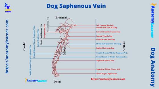
I hope the below-mentioned dog pelvic limb vein labeled diagram might help you to identify these superficial veins perfectly.
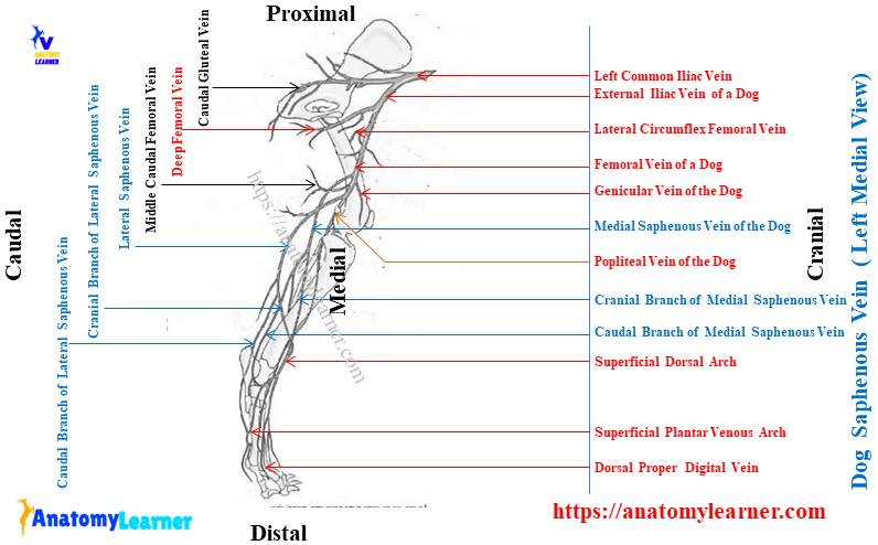
Here, the diagram shows different branches of the lateral and medial saphenous veins in the hind paw, leg, and thigh regions. The diagram also shows the lateral and medial saphenous veins continue to form the main femoral vein in the thigh region (medially).
Before going to the details description of the canine saphenous vein, let’s enlist the main deep veins from their pelvic limb –
- A cranial and caudal tibia veins in the dog’s leg,
- The popliteal vein on the popliteal region of the canine leg,
- A middle caudal femoral vein,
- A proximal caudal femoral vein in the dog’s thigh,
- The femoral vein of the canine leg,
- The lateral circumflex femoral vein in the dog’s thigh,
- Pudendoepigastric and deep femoral veins, and
- External iliac vein continue with the caudal vana cava,
Where is the saphenous vein on a dog?
The dog saphenous vein extends from the hind paw to the distal third of the femur bone. But, the lateral saphenous lies more superficial to the lateral aspect of the tibia bone (leg region). At the same time, the medial saphenous is external to the medial part of the tibia bone.
The dorsal proper and common digital veins continue to form the dorsal metatarsal veins in the hind paw of a dog. Now, the dorsal metatarsal and lateral tarsal veins continue to the distal part of the tibia bone.
Finally, it divides into the cranial branch of both lateral and medial saphenous on the dorsal surface of the tibia bone. Now, the course of the lateral saphenous vein is essential as you may easily collect blood from it.
From the distal third of the tibia bone, the lateral saphenous vein crosses obliquely and passes along the caudolateral surface of the gastrocnemius muscle. Then this branch of the lateral saphenous continues with the distal caudal femoral.
The rest course of this vein will discuss in the lateral saphenous vein description section. Now, let’s see a caudal branch of the lateral saphenous vein that comes from the planter common digital veins and superficial plantar venous arch.
It crosses the calcaneus tubercle and runs between the common calcaneus tendon and caudal flexor muscle of the dog’s leg. Finally, it joins with the cranial branch of the lateral saphenous vein and forms the main lateral saphenous.
You will also find the same branches (cranial and caudal medial saphenous) that run medially of the tibia bone. They join at the caudomedial aspect of the popliteal region (caudal proximal part of the femur) and form the main medial saphenous.
How to perform venipuncture on a dog lateral saphenous vein?
I hope now you know the exact location of the lateral saphenous vein. So, it will be easy for you to perform venipuncture on the dog’s lateral saphenous vein.
Anatomically, the lateral saphenous vein of the dogs is larger compared to the medial superficial saphenous. To perform the perfect venipuncture on the canine lateral saphenous, you may follow the below-mentioned procedure –
- First, let’s identify the distal third (cranial surface) of the dog’s tibia bone,
- Let’s hold the leg region with two paws and twist this region a little,
- Hopefully, you will see the oblique vein (lateral saphenous) on the lateral aspect of the tibia bone,
- You may create pressure on that vein that hinders the normal flow of blood through it,
- Thus, the vein becomes swell, and now you may efficiently perform the venipuncture,
But, make sure you should take the proper hygienic measurement before performing a perfect venipuncture on the canine lateral saphenous vein.
Again, you should have detailed anatomical knowledge of the dog’s tibia and different muscles directly or indirectly involved in the leg region. You will find all these anatomical features (bone and muscle) of the dog leg from the below-mentioned article –
- Dog leg bone (tibia) and muscles with the labeled diagram,
Now, I will discuss the anatomy of the dog’s saphenous veins (both lateral and medial) from the different regions of the pelvic limb.
Saphenous vein dog anatomy and location
In the saphenous vein dog anatomy, I will show how this structure forms in different locations of the pelvic limb. The lateral and medial saphenous veins begin by collecting the various branches of veins from the dorsal and plantar surfaces of the dog’s hind paw.
From this section of the article, you will come to know the details of vein anatomy from the following regions of a dog’s pelvic limb –
- Hindpaw of the dog (dorsal aspect) – dorsal proper digital veins, dorsal common digital veins, lateral and medial tarsal veins, a cranial branch of both lateral and medial saphenous veins,
- Hindpaw of the dog (plantar aspect) – planter proper digital veins, plantar common digital veins, superficial plantar venous arch, plantar metatarsal veins,
- Leg region of a dog (both lateral and medial aspects) – a cranial branch of lateral and medial saphenous, a caudal branch of lateral and medial saphenous, cranial and caudal tibia veins, and main lateral and medial saphenous veins,
- Thigh region of the dog – medial saphenous, popliteal vein, proximal, middle, and distal caudal femoral veins, lateral circumflex vein, and deep femoral vein,
- Hip region of the dog – pudendoepigastric vein, and different branches of the internal iliac vein (iliolumbar, cranial and caudal gluteal veins, lateral caudal vein, and internal pudendal vein),
But, here, I will focus (show) only on the anatomical details of the lateral and medial saphenous veins from the dog’s pelvic limb. You will find a short description of the other different veins with the labeled diagrams.
How does the lateral saphenous vein form in a dog?
If you like to know the details formation of the lateral saphenous vein in dogs, you might start with the veins of the hind paw. Different branches from the dorsal and plantar surfaces of the dog’s hind paw contribute to the lateral saphenous vein.
At the dorsal surface of the distal end of the tibia, first, you will find the cranial branch of the lateral saphenous vein. Again, on the lateral surface of the calcaneus bone, you will also find the caudal branch of the lateral saphenous vein.
You will also find the comparatively smaller cranial branch of the medial saphenous vein at the level of the distal third of the canine tibia bone (on its dorsal surface). These veins (cranial branch of medial saphenous) also come from the dorsal common digital and dorsal metatarsal veins.
Finally, you will find the caudal branch of the medial saphenous vein at the medial aspect of the calcaneus bone.
But how are these cranial and caudal branches of the canine lateral and medial saphenous veins formed? Like to know this formation; well, let’s first understand the different veins from the dorsal and plantar aspects of the dog’s hind paw.
Veins of the dog’s hind paw
So, from the dog hind paw veins diagram, you may quickly identify the dorsal and planter sets of the venous system. All the dorsal proper digital veins come from the axial and abaxial surfaces of the phalanges or digits.
You will see the anastomoses between the dorsal and plantar branches of veins (proper digital) at both proximal and distal extremities of the intermetatarsal spaces. First, let’s try to identify the below-mentioned veins from both the dorsal and plantar surfaces of the dog’s hind paw –
- Dorsal proper digital veins (II – V),
- Common dorsal digital veins (II-IV),
- Dorsal metatarsal veins (II-IV),
- Cranial branches of both lateral and medial saphenous veins,
- Plantar proper digital veins (II-IV),
- Common plantar digital veins (II-IV),
- Plantar metatarsal veins (II-IV),
- Caudal branch of the medial saphenous vein, and
- Caudal branch of the lateral saphenous vein,
Let’s learn about these veins from the dog’s hind paw. And also know how they contribute to the dog’s saphenous vein (lateral and medial).
Dorsal proper and common digital veins
The dorsal proper digital nerves in the dog’s hind paw arise from the digital venous arches. But where is this digital venous arch in the dog? Well, you will find these digital arches on the abaxial side of the distal end of the second phalanx.
How are these digital arches formed in the dog’s hind paw? They are formed by the anastomoses between the dorsal and plantar proper digital veins at the level of a distal end of the second phalanx.
Again, you will also find the plantar proper digital vein anastomoses with the dorsal proper digital veins at the level of metatarsophalangeal articulation. These proper digital veins continue forming the dorsal common digital veins.
You will find the dorsal common digital veins (II, III, and IV) in the dog’s hind paw. Here, the dorsal common digital vein III is formed by the axial dorsal proper digital veins of the third and fourth digits.
Again, the other common digital veins arise second and fifth dorsal proper veins (III) and third and fourth dorsal proper digital veins (IV). You will find the anastomoses between the dorsal common digital veins and plantar metatarsal veins at the distal end of the metatarsal bones of the dogs.
Metatarsal veins of the dog’s hind paw
You will find the small dorsal metatarsal veins (II, III, and IV) on the dog’s hind paw. They lie in the grooves between the adjacent metatarsal bones.
A dorsal deep venous arch on the hind paw crosses the dorsal surface of the proximal end of a metatarsal bone. You will also find the small branches from the canine lateral and medial saphenous veins here.
The metatarsal veins on the dorsal aspect of the dog’s hind paw enter into the deep dorsal deep venous arch. Finally, they anastomose with the proximal lateral tarsal veins at the level of the hock joint.
Dog cranial lateral and medial saphenous vein
The three common dorsal digital veins form a cranial branch of the dog lateral saphenous vein at the level of the distal part of the tibia bone. This vein continues proximally on the long digital tendon to the distal end of the dog’s tibia bone.
At the level of the hock joint, the lateral branch of the saphenous vein receives the lateral tarsal vein. The lateral tarsal vein also joins with the caudal branch of the lateral saphenous vein.
The cranial branch of the medial saphenous vein runs proximally over the flexor surface of the tarsus. It goes parallel to the tendon of the tibialis cranialis tendon.
The cranial medial saphenous vein receives the medial tarsal vein from the plantar surface of the tarsus bone. It will also receive the long anastomotic branch from the second dorsal common digital vein at the level of the hock joint.
Plantar proper and common digital veins
The plantar proper digital veins (II-IV) arise from the axial and abaxial surfaces of the respective digits. They receive small branches of the veins from the digital pad, skin, and terminal part of each phalange.
The plantar proper digital veins continue proximally with the digital venous arches at the level of each first phalanges. Now, the digital venous arches continue proximal and form the plantar common digital veins.
You will find the short plantar common digital veins on the plantar surface of the dog’s hind paw. All the short plantar common digital veins, along with some other veins, form the superficial plantar venous arch at the level of distal metatarsal bones.
The cranial branch of the medial saphenous vein also contributes to forming the superficial plantar venous arch on the plantar surface of the dog’s hind paw.
Caudal branch of lateral and medial canine saphenous veins
After the superficial plantar venous arch, the vein continues proximally along the planar surface (laterally) pes and forms the caudal branch of the lateral saphenous vein. At the proximal metatarsal bone level, this caudal branch of the lateral saphenous vein receives the deep plantar venous arch.
Again, this caudal branch lateral saphenous vein joins with the deep dorsal arch at the level of the hock joint. Finally, you will also find a connection with the lateral tarsal vein at the distal end of (laterally) calcaneus bone.
You will find the plantar metatarsal veins (II-IV) on the plantar surface of the metatarsal bone. These veins lie in the intermetatarsal grooves or on the plantar surfaces of the third and fourth metatarsals.
They anastomose with the dorsal common digital veins at the level of middle metatarsal bones. Now, the caudal branch of the medial saphenous vein starts from the more prominent medial tarsal veins (distal to the medial malleolus bone).
The caudal branch of the medial saphenous passes the tarsal joint and runs along with the caudal branch of the lateral saphenous vein. Finally, this caudal branch of the medial saphenous vein joins with the main medial saphenous.
So, now, you may know the details of the canine lateral and medial saphenous veins.
Dog lateral saphenous vein
You already got the cranial and caudal branches of the lateral saphenous veins that come from the dorsal and plantar surfaces of the hind paw, respectively. Now, these two branches run proximally and join from the main lateral saphenous vein.
Let’s see how these two branches (cranial and caudal) of saphenous veins pass along the leg (tibia) region of the dog. Anatomically, the cranial branch of the lateral saphenous is larger (diameter is more) than these of the caudal branch.
Here, the cranial branch of the canine lateral saphenous vein proximally inclines caudally at the level of a distal end of the tibia. This cranial branch of the lateral saphenous crosses the lateral surface of the tibia bone obliquely (on its distal end).
Now, this cranial branch passes over the space between a deep digital flexor muscle and the beginning of the common calcaneus tendon. In this level (distal half of the tibia), the cranial branch of the saphenous receives the small caudal branch.
You know, the caudal branch continues over the lateral surface of the calcaneus bone and passes in front of the common calcaneus tendon. This vein also receives some other small branches from the calcaneus tubercle of the hock joint.
Now the main lateral saphenous vein of the dogs continues proximally and passes subcutaneously. At the level of the popliteal lymph node (caudal to the stifle joint), this vein (main lateral saphenous) passes proximally on the caudal surface of the gastrocnemius muscle.
Now, the lateral saphenous vein runs deep to the biceps femoris and semitendinosus muscles of the dog’s thigh region. Finally, this vein terminates at the distal caudal femoral vein at the level of the popliteal fossa (distal end of the tibia).
Medial saphenous vein dog
I hope you already found the cranial and caudal branches of the dog medial saphenous vein previously. These two branches unite at the level of the stifle joint (caudally) and from the main medial saphenous vein.
Here, the cranial branch of the medial saphenous vein comes from the cranial surface of the distal extremity of the dog’s tibia bone. You know, at this level of the tibia bone, this cranial branch of the medial saphenous anastomoses with the cranial branch of the lateral saphenous vein.
Now, the cranial branch of the medial saphenous vein crosses over the tibialis cranialis muscle and the tibia bone. The longer caudal branch of the medial saphenous vein joins with the cranial branch of the medial saphenous at the caudal aspect of the stifle joint (medially).
Finally, they continue to form the main medial saphenous vein on the medial aspect of the stifle joint. This main medial saphenous runs over the gracilis muscle and passes between the caudal belly of the Sartorius and gracilis muscles.
You will find a prominent medial genicular vein that arises from the main medial saphenous vein. This prominent medial genicular vein is distributed in the craniomedial aspect of the stifle joint.
Proximally, the main medial saphenous vein continues with the femoral vein and passes through the femoral canal. Now, you will learn the anatomical details of the femoral dog vein, as this is another best vein for performing venipuncture.
Dog femoral vein
The dog femoral vein is another important structure on the medial aspect of the thigh. But, this vein is not always superficial in the medial aspect. On the femoral triangle (just the origin of the femoral vein; after medial and lateral saphenous veins), you will find the femoral vein in a superficial location.
So, this is another best option for venipuncture in the dog. Again, the diameter of this femoral vein on the medial aspect of the thigh is more than those of the lateral and medial saphenous veins.
Here, the femoral vein of the dog lies caudal to the femoral artery and cranial to the small saphenous vein. You will see different branches of the femoral vein on the medial aspect of the dog’s thigh. I will enlist the major branches from the dog’s femoral vein –
- Distal caudal femoral vein,
- A proximal caudal femoral vein in the dog thigh,
- Middle caudal femoral vein,
- A lateral circumflex femoral vein in the dog thigh,
- Middle circumflex femoral vein in the dog’s thigh, and
- A deep femoral veins with different branches,
The distal caudal femoral vein is the continuation of the main lateral saphenous vein of the dogs. Here, the diameter of the distal caudal vein is larger than that of the other different veins on the distal extremity of the tibia bone.
Some small veins come from the distal caudal thigh and proximal caudal tibia muscles (flexor muscles on the tibia). Finally, the distal caudal femoral vein continues proximally on the medial aspect and as the main femoral vein.
Dog middle and proximal caudal femoral vein
Proximal to the distal caudal femoral vein, you will find the middle caudal vein. This vein is larger and drains the biceps femoris muscle. Again, the middle caudal femoral vein crosses the medial aspect of the main medial saphenous vein.
This vein of the dog begins as the cutaneous branch from the caudolateral surface of the dog’s thigh. You will see a small muscle branch of the vein that comes from the gastrocnemius muscle and attaches to the middle caudal femoral vein.
Again, you will also find some other small veins that come from the quadriceps femoris, adductor, semimembranosus muscles, and also from caudal thigh muscle. All these small veins join with the main middle caudal femoral vein in the different levels of the thigh.
You will also find a small genicular branch of the vein that comes from the craniaomedial aspect of the distal extremity of the tibia bone. This small vein also joins with the femoral vein at the middle caudal femoral vein level.
The femoral vein continues proximally and receives the proximal caudal femoral vein at the middle of the dog’s thigh. The proximal caudal femoral vein arises from the medial surface of the proximal half of the gracilis muscle.
Again, this vein also receives a large branch from the proximal part of the adductor muscle. The main femoral vein of the dog’s thigh receives a lateral circumflex femoral vein at the level of the proximal extremity of the femur bone. It joins with the femoral vein just above the proximal caudal femoral vein.
Another name for this lateral circumflex femoral is a cranial femoral vein of the dog’s thigh. It begins on the skin of the proximal lateral surface of the thigh and the adjacent gluteal region.
Dog tibial, popliteal, and saphenous vein
The cranial and caudal tibia and popliteal are considered the deep veins in the pelvic limb of a dog. Here, the cranial tibia vein passes along the craniolateral surface of the dog’s tibia bone. In comparison, the caudal tibia vein of the dog passes along the caudal border of the leg (tibia bone).
You know, there is a dorsal pedal vein on the dorsal surface of the hind paw. It continues proximally and anastomoses with the dorsal metatarsal and lateral tarsal vein at the hock joint (proximally). Here, you will find a connection of the cranial tibia vein with the cranial branch of the lateral saphenous vein.
So, you may say, the dog’s cranial tibia is the continuation of the dorsal pedal vein that arises from the flexor surface of the tarsus bone. This cranial tibia vein receives small veins from the cranial tibia group of muscles at their proximal extremities.
Now, the cranial tibia vein of the dog leg passes between the tibia and fibula bones. It also runs deep to the popliteus muscle and unites with the caudal tibial vein (at the proximal caudal extremity of the tibia bone).
After joining the cranial and caudal tibia veins, it continues proximally and forms the popliteal vein on the popliteal region of the dog’s thigh. Now, the dog popliteal vein crosses the popliteal notch of the tibia and enters into the popliteal fossa.
Here, this popliteal vein of the dog leg receives medial and lateral branches of veins from both sides of the stifle joint. Again, the popliteal vein joins with the distal caudal femoral vein on the caudomedial aspect of the stifle joint.
You know, the distal caudal femoral vein receives the main lateral saphenous vein on the distal medial aspect of the stifle joint.
Veins in the dog pelvis
So, the dog medial saphenous vein, along with the distal caudal and popliteal veins, continue as the femoral vein in the deep of the thigh region (proximally). Finally, this femoral vein continues as the external iliac and joins with the common iliac vein.
The different branches of the internal iliac artery drain blood from the pelvic region into the common iliac artery. Here, you will find the below-mentioned veins that together form the internal iliac vein in the dog’s pelvic region –
- A caudal gluteal vein in the dog pelvis,
- An internal pudendal vein in the dog’s pelvis,
- A lateral caudal gluteal vein in the canine pelvic region,
- A cranial gluteal vein in the dog’s pelvic region, and
- Iliolumbar vein in the dog pelvis,
You will also find other small branches of the veins in the pelvis region of a dog that together form the internal iliac vein. Here, I only show the main and larger veins from the internal iliac in the dog’s pelvic region.
A gluteal and internal pudendal vein in a dog pelvis
Different small veins come from the biceps femoris, semitendinosus, and semimembranosus muscles and form the caudal gluteal vein. This vein runs proximally across the greater ischiatic notch, just lateral to the ischiatic spine and internal obturator muscle.
Again, it runs caudally to the sciatic nerve of the dog. At the level of the (dog’s) pelvic outlet, the caudal gluteal receives the dorsal perineal veins. This vein comes from the caudal half of the thigh and internal obturator muscle.
The sacral intervertebral vein and also the vein from the intercoccygeal muscle enter into the main caudal gluteal vein. Here, you will find the male internal pudendal vein that connects to the caudal gluteal vein at the greater ischiatic spine of the hip.
This internal pudendal vein receives the third and sometimes second sacral intervertebral vein. But, in a female dog, you will find some other veins that are shown here in the labeled diagram.
You will also see a lateral caudal vein, the main venous drainage from the tail. It passes through the caudal surface of the hip and enters into the internal iliac vein.
This vein of the dog also receives different segmental branches and runs cranially from the free end of the tail. It also gets a large vein from the skin of the tail and the adjacent part of the pelvic region.
The cranial gluteal vein drain from the proximal part of the middle gluteus muscle. You will find a large cutaneous vein that comes from the skin over the proximal dorsal part of the pelvic region. Again, this cranial gluteal receives the veins from a first and second sacral intervertebral vein.
Other veins in dog pelvis
The iliolumbar is the small vein in the dog’s pelvic region that crosses the cranial border of the wing of the ilium bone. This vein drains into the lateral side of the internal iliac vein at its termination. The iliolumbar vein of a dog receives the seventh lumbar intervertebral vein.
All the caudal gluteal, internal pudendal, cranial gluteal, and iliolumbar veins continue to form the internal iliac vein. Now, the internal and external iliac veins form the common iliac vein on the tendon of the psoas minor muscle.
Finally, the left and right common iliac veins form the caudal vena cava at the level of a sixth lumbar vertebra. You will also find the unpaired medial sacral vein in the pelvic region of a dog.
This unpaired medial sacral vein receives the middle caudal vein from the dog’s tail. The median sacral vein runs cranially between the right and left ventral sacrocaudal muscles.
Dog saphenous vein labeled diagram
I have already provided different diagrams of the dog’s saphenous vein along with the other different veins of their pelvic limb. Again, I will provide some of the other various labeled diagrams on the canine saphenous vein that might help you to understand and identify the lateral and medial saphenous from the actual sample.
In the first diagram of the dog leg vein, I tried to show you the different veins from the dorsal surface of the dog’s hind paw. Here, the diagram shows the dorsal proper digital veins that form the digital venous arches at the level of the first phalanges.
After the digital venous aches, the dorsal proper digital veins continue to form the dorsal common digital veins (shown in the labeled diagram). Again, the dog vein labeled diagram shows different metatarsal veins on the dorsal surface of the hind paw.
Now, the dorsal metatarsal and dorsal common digital veins form a digital venous arch on the proximal extremity of the dog hock joint along with the lateral tarsal veins. After this arches, the vein passes proximally (shown in the diagram) and divides into cranial branches of the lateral and medial saphenous veins.
Here, the cranial branch of the lateral saphenous vein passes dorsolaterally. In contrast, the cranial branch of the medial saphenous vein runs along the dorsomedial aspect of the tibia bone (shown in the diagram). The diagram shows the larger diameter in the lateral saphenous cranial branch compared to the medial saphenous ones.
Again, in the second diagram, I tried to show you the plantar proper digital veins, distal and superficial venous arches, and the caudal branches of both lateral and medial saphenous veins. Let’s find more diagrams on dog leg veins here.
Lateral and medial saphenous on the dog leg
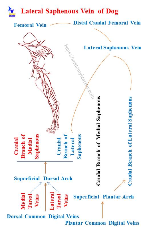
Now, let’s see the diagram below, where I tried to show the important course of both the lateral and medial saphenous veins of the dog. From the lateral aspect of the tibia bone (distal third), the cranial branch of the lateral saphenous vein of the dog is identified.
Again, the diagram also shows the caudal branch of the dog’s lateral saphenous vein just in front of the common calcaneus tendon. After joining the cranial and caudal branches of the lateral saphenous veins, the main larger lateral saphenous vein is also identified in the labeled diagram.
Again, from the dog’s leg region, the cranial branch of the medial saphenous vein is identified in the diagram. You will also find the caudal branch of the medial saphenous just in front of the caudal branch of the lateral saphenous.
These two branches of the medial saphenous will join to form the main medial saphenous (shown in the diagram) and cross the lateral saphenous vein. Finally, the medial saphenous vein joins with the femoral vein and runs proximally.
From the dog’s leg region, other veins like the cranial and caudal tibial (deep veins of the pelvic limb) are also identified in the labeled diagram.
Now, let’s see the dog thigh vein labeled diagram; here, I tried to show the different small branches that join with the main femoral vein at a different thigh level. Here, the diagram shows the different small main veins like distal caudal, middle caudal, proximal caudal, and lateral circumflex femoral veins.
Again, the diagram shows the formation of the internal and external veins in the dog’s pelvic limb. Finally, the internal and external iliac veins form the right and left common iliac veins that form the caudal vena cava.
A cephalic and jugular vein in the dog
The cephalic and jugular veins in the dog are another essential structure that you may use for perfect venipuncture. You will find the cephalic vein is more superficial to the arm and forearm of the dog.
The formation and distribution of the dog cephalic vein have been described in another article by an anatomy learner. You may read the below-mentioned article to know the details anatomical features of the dog cephalic vein –
Dog cephalic vein – course and distribution with the labeled diagram,
Here, I will show (point out) some of the essential features of the dog cephalic vein so that you may quickly identify this vein from the live sample. This vein of the dog’s thoracic limb comes from the common dorsal digital veins and the accessory cephalic veins.
Then the cephalic vein obliquely crosses the radius bone from its medial surface to the lateral surface (runs the cranial surface of the radius bone). At the elbow joint, the cephalic vein of the dog joins with the medial cubital vein and the brachial vein.
So, you may consider the elbow joint as a landmark to locate the dog’s cephalic vein perfectly. You may create slight pressure on the craniolateral surface of the radius bone and easily find this cephalic vein.
Now, the cephalic vein of the dog runs proximally over the cleidobrachialis muscle and the cranial border of the lateral head of the triceps brachii muscle. You will find the cranial tibia nerve just caudal to the dog cephalic vein.
Proximally, the dog cephalic vein continues with the axillobrachial and omobrachial veins, which also continue with the main external jugular vein.
Now, the dog jugular vein passes along the ventrolateral aspect of the neck (shown in the labeled diagram).
A saphenous vein in a cat
The basic structure of the cat saphenous vein is similar to that of the dogs. In the cat leg, you will also find both lateral and medial saphenous veins like the dogs.
Both these lateral and medial saphenous veins of the cat are formed by the union of cranial and caudal branches of the saphenous like the dog. But, the medial saphenous vein of the cat is larger compared to the lateral one.
So, you may choose the medial saphenous vein for the perfect vein puncture in the cat. Again, the lateral and medial saphenous veins form the larger main saphenous vein that passes on the medial aspect of the cat’s thigh (runs superficially).
You will also find the cranial and caudal tibial veins in the cat’s leg region. If you want to learn the details of veins from the cat’s pelvic limb, you may read the below-mentioned article –
Arterial supply to the pelvic limb of a cat with the labeled diagram,
This article might help you to identify all the arteries and veins from the cat’s pelvic limb. And you will also compare the main difference in the different veins between the dog and cat.
Frequently asked questions on dog saphenous vein.
In this section of the article, I will enlist some of the common questions on the dog saphenous vein that are asked by the dog’s anatomy learners. If you read the above information from this article, then it will be easy to understand all these enlisted topics.
Let’s see the common questions on the canine lateral and medial saphenous veins with their possible answers –
Do dogs have a medial saphenous vein?
Yes, dogs have a medial saphenous vein in their hind legs. The medial saphenous vein of the dog’s leg extends from the distal cranial end of the tibia bone to the caudomedial aspect of the femur bone (on its distal third portion).
At the cranial aspect of the dog hock joint, you will find anastomoses of different veins from where the cranial branches of the lateral and medial saphenous veins arise. If you observe the structure of the cranial branches of the lateral and medial branches of the dog’s saphenous vein, you will find the followings.
The diameter of the cranial medial saphenous vein is less than that of the cranial lateral saphenous vein. Now, the cranial branch of the medial saphenous vein runs a little obliquely on the medial surface of the tibia bone (at the level of the middle portion).
Finally, this cranial branch of the medial saphenous vein joins with the caudal branch of the medial saphenous. You know the caudal branch of the medial saphenous vein comes from the plantar veins.
This branch of the medial saphenous will run in front of the common calcaneus tendon and the caudal branch of the medial saphenous vein. The cranial and caudal branches of the medial saphenous vein join at the caudal aspect of the proximal tibia bone.
Finally, the main medial saphenous vein crosse the lateral saphenous vein and continue proximally. It joins with the femoral vein at the medial aspect of the distal extremity of the femur bone. I have already provided the detailed course of the dog’s medial saphenous vein with the labeled diagram previously.
You may take help from that diagram to understand the distribution of the canine medial saphenous vein on the hind limb.
What vein is blood drawn from in dogs?
Sometimes you need to collect blood from the dogs for research or diagnostic purposes. You may use the saphenous vein (obviously the lateral saphenous) on the leg. It is more superficial on the craniolateral aspect of the dog’s leg.
Again, you may also choose the caudal branch of the lateral saphenous vein for venipuncture. And you know, this caudal branch of the lateral saphenous runs along the common calcaneus tendon. But, this area of the dog leg is thin, and there are other different structures (like nerves and arteries) that pass along this area.
So, it is better to avoid the caudal lateral saphenous vein for venipuncture. Again, you may also use the medial saphenous vein for perfect venipuncture.
Rather than the saphenous vein, you may also use the cephalic vein (more prominent in the arm and forearm regions) and jugular vein. Some veterinarians use the superficial femoral vein (the medial aspect of the thigh) for venipuncture.
So, you may use any of the veins for venipuncture in the dog – saphenous, cephalic, femoral, and jugular veins.
What is the main reason for catheterizing the dog via the lateral saphenous vein?
The diameter of the lateral saphenous vein is larger compared to the medial one. Again, the lateral saphenous vein of the dog leg is more superficial on the lateral aspect of the leg region. These are the two main reasons for catheterizing the dog via the lateral saphenous vein.
Conclusion
The lateral and medial saphenous veins form the main saphenous vein in the dog. Here, the dog lateral saphenous vein is more superficial and passes obliquely from the tibia and at the caudal aspect or over the gastrocnemius muscle.
Again, the medial saphenous vein passes medially, receiving the cranial and caudal branches from the dorsal and plantar veins. The cranial branch of the dog’s lateral saphenous is larger than the caudal branch.
You may choose this lateral cranial branch of the main lateral saphenous vein for the perfect venipuncture in dogs. But, it would help if you observed the gross anatomy of the canine saphenous vein from the actual sample at your anatomy learning laboratory. All the provided labeled diagrams on the dog’s saphenous vein might make your learning easier.