The dog humerus bone anatomy comprises a long shaft and proximal and distal extremities. Here, the proximal extremity of the dog humerus consists of the head, neck, and tubercles. Again, the distal extremity contains condyles, crest, and fossa.
Some important muscles of the dog thoracic limb originate or insert on the dog’s humerus bone. This guide will help you learn the anatomy of the dog humerus bone (osteological features of the bone and muscles) with a labeled diagram.
Proximally, the dog humerus articulates with the scapula and forms the shoulder joint. Again, it articulates with the radius and ulna bone distally to form the elbow joint. So, in the end, I will try to summarize the anatomical features of these two important joints that are formed by the dog humerus.
Dog humerus bone
The dog humerus is the bone of the thoracic limb (arm or brachium region). You will see some peculiar osteological features in the dog humerus bone anatomy compared to the ruminant and horses.
The long shaft of the humerus bone is also known as the body. Again, the proximal extremity of the dog humerus bone shows head, neck, lesser tubercle, greater tubercle, and intertuberal groove. In contrast, the distal extremity of the dog humerus consists of a humeral condyle.
The humeral condyle of a dog comprises the lateral and medial condyles. You know the lateral condyle of the dogs humerus is the capitulum, and the medial condyle is the trochlea. Again, you will see the epicondyle in the humerus’s lateral and medial epicondyle.
Now, let’s see what the most important osteological features of the dog humerus bone are. First, you should identify the shaft and two extremities (proximal and distal).
Let’s see the long shaft of the humerus bone and try to identify the four surfaces –
- The lateral surface of the humerus,
- Medial surface of the bone,
- Cranial surface, and
- Caudal surface of the humerus,
But, how will you confirm these surfaces from the dogs humerus bone? Well, I will help you identify the different surfaces from the dog’s humerus bone.
The lateral surface of the dog humeral bone contains the brachialis or musculospiral groove. Now, let’s see the shaft of the humerus bone; one surface of this bone shows a spiral groove. So, this is the lateral surface of the humerus bone.
Again, the medial surface of the humerus bone consists of teres major and minor tuberosities. In comparison, the cranial surface of this bone is narrow and contains a crest (humeral crest; faces cranially). Again, the caudal surface is smooth in the dogs humerus and contains the nutrient foramen.
Dog humerus bone identification
Now, I will show you the osteological features you might identify from the dog humerus bone. But make sure you can identify the proximal and distal parts and the shaft of the humerus bone.
From the proximal extremity of the dog humeral bone, you might identify the following important osteological features –
- Head and neck on the proximal extremity of the humerus bone,
- Lesser tubercle on the proximal part of the humerus (medially flattened enlargement),
- Greater tubercle (large cranio lateral projection on a proximal extremity),
- An intertuberal groove (between lesser and greater tubercles), and
- Several small foramina (between head and greater tubercle),
From the shaft of the dog humerus, you will find the following important osteological features –
- Elongated deltoid tuberosity,
- Tricipital line or anconeal line (proximally),
- Brachial or musculospiral groove,
- Crest of the greater tubercle,
You will find the above-mentioned osteological features in the lateral surface of the dog humerus bone. At the same time, you will find the other different osteological features in the medial, cranial, and caudal surfaces of the dogs’ scapula.
- Crest of the lesser tubercle,
- Tuberosity of the teres major,
- Crest of greater tubercle or humeral crest (cranially), and
- Caudal nutrient foramen (caudaaly),
Finally, the distal extremity of the dog humerus bone anatomy shows the below-mentioned osteological features. The supratrochlear foramen is the unique osteological feature in the distal extremity of the dog humerus.
Okay, let’s see the other osteological features from the humerus bone –
- Lateral capitulum (condyle),
- Medial trochlea (condyle),
- Lateral and medial epicondyles,
- Lateral supratrochlear crest and fossa,
- Radial (cranially) and olecranon (caudally) fossa, and
- Supratrochlear foramen,
All these osteological features of the distal extremity are identified from the dogs humerus. Let’s know the anatomy of these features in the next section of this article.
Unique features of dog humerus bone
So, before learning the details of the dog’s humerus bone, let’s enlist some unique osteological features that help you identify it from other animals. You will see the following unique osteological features in the humerus bone of the dogs –
- The dog humerus bone is comparatively longer, and is less twisted bone compared to the ruminant or horses,
- Bicipital or musculospiral groove on the lateral surface of the humeral shaft is not so prominent,
- You will see the deltoid tuberosity in the form of a ridge on the dog’s humerus,
- The proximal extremity of the dogs humerus bone contains a more rounded and convex head,
- There are radial and olecranon fossa at the distal extremity of the dogs humerus, which are communicated by a supratrochlear foramen,
- The caudal surface of the humeral shaft possesses a nutrient foramen,
- Again, the humeral crest of the dogs humerus bone is not so prominent compared to the ruminant and horses,
So, these are the unique osteological features found in the dogs’ humerus bone. You may learn more about this humeral bone from the next part of this article.
Dog humerus anatomy
In the dog humerus bone anatomy, you will learn the location and osteological features of different extremities and shafts. Again, you will learn the anatomy of the muscles that originate or insert into the dogs humerus.
Location and relationship: you know the dogs humerus bone is the long bone locates obliquely downward and backward. It forms the shoulder joint above the scapula and the elbow joint below with the radius and ulna bones.
- The cylindrical shaft of the dogs humerus bone is less twisted and possesses four different surfaces –
- Lateral surface – spiral and posses the brachialis or musculospiral groove,
- Medial surface – rounded and possesses teres tuberosity (insert the teres major and latissimus dorsi muscles),
- Cranial surface – triangular, wide and smooth above and narrow and rough below; posses the crest of humerus and deltoid tuberosity, and
- Caudal surface – round and smooth that contains nutrient foramen,
Again, you know the proximal extremity of the dogs humerus bone is expanded and posses a head, neck, tuberosities, and groove. The head of the humerus is circular, convex, and articulates with the glenoid cavity of the dog scapula.
The neck is the constricted part that lies below the head of the humerus. There are lateral and medial tuberosities on the proximal extremity of the dogs humerus.
The lateral tuberosity is larger and prominent in the humerus bone. There is a bicipital or intertuberal groove present in between the tubercles.
The distal end of the dogs humerus is articular, expanded, and known as the modified condyle. The capitulum (lateral condyle) and the trochlea (medial condyle) are in this distal end. You will also find the lateral and medial epicondyles, radial and olecranon fossa, and supratrochlear foramen.
The lateral surface of dog humerus shaft
On the lateral surface of the dog humeral shaft, you will see the tricipital line and deltoid tuberosity at its proximal end. The tricipital line starts at the caudal aspect of the humeral head near the greater tubercle. Again, cranially it extends to the crest of the elongated deltoid tuberosity.
The proximal and distal parts of the lateral surface are inclined cranially. Again, the proximal part of the lateral surface lines between the crest of the greater tubercle (medially) and tricipital line (latarally).
Another name for the tricipital line is an anconeal line. On this tricipital line, you will find a small enlargement for inserting the teres minor muscle. Again, the remaining distal part of the tricipital line serves as the origin of the lateral head of the triceps brachii muscle.
The deltoid tuberosity is the most prominent osteological feature in the lateral surface of the dogs’ humerus bone. This deltoid tuberosity of the humerus bone form two areas –
- A narrow, slightly convex area in the distal extremity that faces craniolaterally, and
- A smooth, wide, slightly concave area that faces caudolaterally (brachialis groove),
You know this deltoid tuberosity serves to insert the deltoideus muscle of the dogs. Now, let’s discuss the brachialis groove of the dogs humerus.
You already identified the brachialis groove from the dogs humerus. This groove forms the smooth and slightly convex structure on the lateral surface of the humerus bone.
This structure (groove) starts at the neck caudally and extends laterally. Finally, the groove twists cranially and runs along the distal extremity of the humerus bone.
You will find the brachialis muscle in the musculospiral or brachialis groove of the humerus bone. This muscle originates from the proximal part of the brachialis groove of the humerus.
Medial surface of dog humerus shaft anatomy
The medial surface of the dog humerus shaft anatomy is nearly flat and triangular in its proximal fourth. Again, the distal end of the medial surface of the humeral shaft is rounded transversely.
The crest of the lesser tubercle cranially bounds the medial surface of the humeral shaft. Again, this medial surface is bent with the rounded and smooth caudal surface of the shaft.
You will see a tuberosity at the distal end of the medial surface of the humeral shaft. This tuberosity locates at the same transverse plane as the laterally located deltoid tuberosity. The tuberosity on the medial surface of the shaft is the teres major tuberosity.
Different muscles attach or originate on the medial surface of the dog humeral shaft. The teres major and latissimus dorsi muscles insert on the teres tuberosity of the shaft (medially).
Near the teres major tuberosity, the coracobrachialis muscle inserts on the crest of the lesser tubercle (medial to proximal extremity). Again, the medial head of the triceps brachii muscle arises from the cranial part of the lesser tubercle.
Again, the medial surface of the dog humerus shaft is loosely covered by the biceps brachii muscle. You know the distal part of the medial surface is almost rounded, smooth, and joins with the condyle of the humerus.
Cranial surface of the humeral shaft
The cranial surface of the dog humeral shaft is irregular, wide, and smooth proximally. This proximal part of the cranial surface of the humeral shaft starts from the crest of the greater tubercle (laterally).
The crest of the greater tubercle passes just medial to the deltoid tuberosity of the lateral surface. Again, it reaches the cranial edge of the musculospiral (brachialis) groove.
The distal part of the cranial surface of the dogs humerus shaft is narrow and roughened. You will see a prominent border on the cranial surface of the humerus shaft. This prominent border is known as the crest of the humerus (humeral crest).
Do you know how this crest of the humerus is formed in the dog? The cranial and lateral surfaces of the dog humerus shaft meet cranially and form this border or crest.
At the middle of this crest, you will see the prominence where the deltoid muscle is inserted. This prominent osteological feature on the cranial border is known as the deltoid tuberosity (listed previously at the lateral surface).
Again, proximal to the deltoid tuberosity, you will also find a small prominence. This small prominence of the humeral shaft is the teres minor tuberosity.
You will see the attachment of the two muscles on the cranial surface of the humeral shaft. The pectoralis superficialis muscle attaches to the crest of the greater tubercle (along the cranial surface). Again, the pectoralis profundus muscle attaches to the proximal part of the cranial surface of the humeral shaft.
You will learn more about these two important muscles (pectoralis superficialis and profundus) in the humerus bone muscles section of this article.
Caudal surface of the dog humerus body
The caudal surface of the dog humerus body is smooth and rounded side by side. This surface begins at the neck of the humerus and ends at the expanded distal extremity (condyle).
The caudal surface continues with the lateral supracondylar crest at the distal fourth of the bone. You will learn this supracondylar crest from the humeral condyle section of this article.
Another name for this crest is crista supracondylar lateralis. You will see different muscles of the arm or brachium of the dog that attaches to this supracondylar crest.
Let’s see what the muscles that attach to the crista supracondylar lateralis structure of the dog humerus bone are –
- Brachioradialis muscle of the dog,
- Extensor carpi radialis muscle of the dog, and
- Anconeus muscle of the dog’s thoracic limb,
You will learn the details of these muscles (brachioradialis, extensor carpi radialis, and anconeus) from the arm muscles anatomy. There is a nutrient foramen at the caudal surface of the dogs humerus. This nutrient foramen is directed distally.
Now, let’s discuss the proximal and distal extremities of the dog humerus bone. First, let’s start with the osteological features of the proximal extremity (head, neck, tuberosity, and groove) of the humerus.
Head and neck of canine humerus bone anatomy
The head and neck of the dog or canine humerus bone anatomy are the important features at the proximal extremity. A head is an oval, smooth articular surface at the medial aspect. In comparison, the neck is the constricted, well-defined structure that locates caudally and laterally.
The head of the humerus is oval but becomes elongated in the sagittal section. It possesses a large convex and smooth articular surface that articulates with the glenoid cavity of the dog scapula and forms the shoulder joint.
The cranial part of the head (articular surface) is much flatter than the caudal part. Again, the articular surface of the head continues cranially with the intertuberal groove. There you will find a ridge of the craniomedial part of the proximal extremity of the humerus bone.
In the dog shoulder anatomy structure, you will find the joint capsule on this intertuberal groove. Again, there is a bicipital tendon along with the joint capsule on the inertuberal groove.
Again, the neck of the dog humerus is a distinct constricted area that locates only caudally and a little laterally. The head and parts of the tubercles join with the shaft of the humeral body at the neck region.
Tubercles and groove of the proximal extremity
You know two tubercles (greater and lesser) are present at the proximal extremity of the dog’s humerus bone. The greater tubercle is the large craniolateral projection of the proximal extremity of the dog humerus.
The greater tubercle is a smooth and high structure on the proximal part of the bone. You will see various foramina in between the head and the greater tubercle.
Several muscles of the dog’s shoulder and arm (thoracic limb muscles) insert into the humerus bone. Let’s see what are the muscles of the dogs thoracic limb that insert on the greater tubercle of the humerus –
- The supraspinatus muscle of the shoulder region is completely inserted into the greater tubercle,
- There is a partial insertion of the pectoralis profundus muscle on the greater tubercle, and
- Distal to these insertions (of two muscles), the infraspinatus muscle is also inserted into the greater tubercle,
Again, the lesser tubercle on the proximal part of the dogs humerus bone is a medially flattened enlargement. The convex border of the lesser tubercle of the humerus bone is not so large.
The subscapularis muscle of the dog scapula inserts on the convex border of the lesser tubercle. Again, the lesser and greater tubercles of the dogs humerus separated craniomedially. Between these two tubercles, you will see the intertuberal groove that faces craniomedially.
At the same time, these two tubercles are also separated by the head of the humerus caudolaterally. I hope you can identify all the osteological features from the proximal extremity of the dogs’ humerus bone.
Now, let’s know the anatomical facts of the humeral condyle (features of the distal extremity) from the dog’s fore limb.
Humeral condyle dog anatomy
The distal extremity of the dog humerus possesses the humeral condyle. This humeral condyle articulates with the forearm bones (radius and ulna), forming the elbow joint.
The humeral condyle in the dog humerus divides into more extensive medial and small lateral parts. The trochlea is the more extensive medial part of the humeral condyle, which articulates with the ulna bone. Whereas the small lateral part is the capitulum that articulates with radius bone.
The medial articular surface of the humeral condyle further divides with the ridge. It is a pully-shaped structure that extends proximally into the adjacent fossa.
In the distal extremity of the dogs humerus, you will also find two fossa –
- An olecranon fossa – deep depression on the caudal part of the humeral condyle of the dog, and
- The radial fossa – shallow depression on the cranial surface of the humeral condyle,
The olecranon fossa of the humeral condyle receives the anconeal process of the ulna bone (when the elbow joint extends). You will see the origin of the anconeus muscle from the margin of the olecranon fossa.
The radial fossa of another animal’s humerus is also known as the coronoid fossa. But, you will not find coronoid fossa in the dogs’ humerus. This is due to the head of the radius bone entering only the depression (cranial surface) when the elbow joint flexes.
In the older dog, you may find the supratrochlear foramen at the distal extremity of the humerus bone. But, the young dog does not possess supratrochlear foramen in their humeral condyle area.
The radial and olecranon fossae communicate with each other through this supratrochlear foramen. No structure passes through this supratrochlear foramen.
Epicondyles of the dog humerus bone anatomy
On both sides of the humeral condyles is thick eminence. These are the epicondyles that serve as the origin of the different muscles of the distal part of the thoracic limb.
- A lateral epicondyle – is a small lateral prominence on the humeral condyle, and
- A medial epicondyle – is a more prominent structure on the medial surface of the humeral condyle,
The lateral epicondyle lies caudoproximal to the lateral articular margin of the lateral condyle or capitulum. This lateral epicondyle of the dog humerus bone is also known as the extensor epicondyle because different extensor muscles originate from this structure.
Let’s see what the extensor muscles that originate from the extensor lateral epicondyle of the humerus bone are –
- Extensor digitorum communis muscle of the dog thoracic limb,
- Extensor digitorum lateralis muscle of the dog, and
- Ulnaris lateralis muscle of the dog thoracic limb,
The lateral ligament of the dog elbow joint (proximal part) attaches to the articular margin and adjacent surface of the lateral epicondyle.
You will also see the supracondylar fossa and lateral supracondylar crest in the humeral condyle of the dog. The supracondylar fossa is not as deep as the horse or ruminant humerus.
Again, the lateral crest of the supracondylar fossa extends proximally from the lateral epicondyle of the dogs’ humerus. This lateral supracondylar crest of the dogs humerus is thick and slightly rounded.
The lateral supracondylar crest joins with the caudal broader distal fourth of the dog’s humerus bone. You will see the origin of two important muscles from the lateral supracondylar crest.
The brachioradialis muscle originates from the proximal part of the lateral supracondylar crest. In contrast, the extensor carpi radialis originates from the distal part of the supracondylar crest of the dogs humerus.
The medial humeral condyle and epicondyle of the dog
The medial epicondyle of the dog humerus is the medial prominence of the trochlea (medial condyle of the humerus). It locates proximal to the medial border of the articular surface of the medial condyle or trochlea.
The medial epicondyle of the dogs humerus is larger than the lateral one. Another name for the medial epicondyle of the humerus bone is flexor epicondyle, as the flexor muscles of the thoracic limb arise from it.
Okay, let’s see what are the flexor muscles of the dogs thoracic limb that arises from the medial epicondyle –
- A flexor carpi radialis muscle of the dogs thoracic limb,
- The flexor digitorum superficialis muscle,
- A flexor digitorum profundus muscle of the thoracic limb, and
- The flexor carpi ulnaris muscle of the dogs thoracic limb,
All these muscles, as mentioned earlier, from the dogs thoracic limb are identified in the dog muscle labeled diagram. The medial ligament of the elbow joint (proximal part) attaches to the articular margin and the adjacent surface of the medial epicondyle of the dog humerus bone.
Dog humerus muscles anatomy
Let’s see the dog humerus muscles anatomy with the labeled diagram. There are two groups of muscles in the humerus (arm or brachium) of the dogs – cranial brachial and caudal brachial muscle.
The cranial brachial muscle of the dog includes the biceps brachii, brachialis, and coracobrachialis muscles. Again, the caudal brachialis muscle fills the triangular space between the scapula and humerus bone.
These are the extensor group of muscles in the dogs humerus. The main muscle of the caudal group is the triceps brachii (but posses four different parts). Again, you will also find the anconeus and tensor fascia antebrachii muscle in this caudal group.
You will find some of the craniolateral and caudomedial antebrachial muscles that originated from the humeral condyle of the dog. First, let’s see what these craniolateral and caudal antebrachial muscles are –
- A radiobrachialis muscle or supinator longus,
- The extensor carpi radialis muscle,
- An extensor digitorum communis muscle of the dog,
- The extensor digitorum lateralis muscle of the dog, and
- The ulnaris lateralis muscle of the dog’s thoracic limb,
These are a craniolateral group of antebrachial muscles attached to the humerus bone. Again, you will find the below-mentioned caudal group of antebrachial muscles that also have a relationship with the humerus bone of the dogs –
- Pronator teres of brachium,
- Flexor carpi radialis muscle of the brachium,
- The flexor digitorum superificialis muscle of the brachium,
- Humeral head of flexor carpi ulnaris muscle, and
- Humeral head of the flexor digitorum profundus,
Now, I will describe these muscles from the brachium region that have directly originated or are attached to it. But for your information, some of the cranial and caudal groups of brachial muscles (biceps brachii, coracobrachialis) are already described in the dog scapula muscle anatomy.
Dog brachialis muscle anatomy
You know the brachialis or musculospiral groove of the dogs humerus occupied by the fleshy muscle with spiral fibers. This is the brachialis muscle of the dog brachium that helps flex the elbow joint.
Origin: the dog brachialis muscle arises from the humerus’s proximal part of the caudal surface.
Relationship: this brachial muscle extends laterally on the humeral crest and then medially on the medial surface of the humerus. This muscle courses from the caudolateral to the cranial surface to the musculospiral groove (distally).
Again, this muscle becomes narrow at the distal third of the humerus bone and runs over the flexor surface of the elbow joint. Laterally, you will see the biceps brachii muscle. Again, this brachialis muscle of the dog also covers the triceps brachii and extensor carpi radialis muscles.
Insertion: the tendon from this brachialis muscle inserts into the medial aspect of the proximal end of radius bone (radial tuberosity). Again, the remaining part of the brachial tendon inserts on the proximal end of the ulna bone (ulnar tuberosity).
Action and innervation: the main function of the brachialis muscle is to flex the elbow joint of the dogs. The musculocutaneous nerves from the dog brachial plexus innervate to this brachialis muscle. But, sometimes, the branches from the radial and median nerves also innervate to this brachialis muscle.
Again, a branch from the brachial artery supply to the dog brachial muscle.
Dog triceps brachii muscle – lateral and medial heads
You already know there are four heads in the dogs triceps muscle – long, lateral, medial, and accessory. But, you will normally find three heads of triceps brachii muscle in other animals. The dog possesses the extra accessory head in the triceps brachii muscle.
I have already described the long head of the dogs triceps brachii muscle (in the dog scapular anatomy article). Now, in this part, I will describe the rest three heads from the dogs triceps brachii muscle.
The lateral head of dogs triceps brachii: this is the large and rectangular muscle that lies between the long head and humerus bone. The lateral head of the dogs triceps brachii also joins with the accessory head and lies over the brachialis muscle.
This head of the triceps arises from the teres minor tuberosity and the deltoid tuberosity of the dogs’ humerus bone. It forms a short but broad tendon that inserts on the olecranon tubercle of the ulna bone.
Branches of the radial nerve innervate to the lateral head of the triceps brachii. In contrast, the deep brachial and caudal circumflex arteries supply the lateral head.
The medial head of the triceps brachii is the smallest part and spindle-shaped muscle that helps extend the elbow joint. The medial head of the dogs triceps brachii arises from the crest of the minor tubercle of the humerus bone.
It forms a thick tendon that inserts medially and independently on the olecranon tuber of the ulna bone. Again, this tendon from the medial head of the triceps also joins with the tendon of the long head.
The deep brachial artery supplies to the medial head of the triceps, whereas the branch of the radial nerve innervates to this head.
Does a dog have an accessory head in the triceps brachii muscle?
Yes, dogs have an accessory head in the triceps brachii muscle. This is the irregular rectangular muscle in the cross-section that lies on the caudal surface of the humerus between the other heads and brachialis muscle.
The accessory head of the dogs triceps brachii muscle arises from the proximal caudal part of the humeral neck. But it becomes tendinous at the distal third of the humerus bone. This tendon of the accessory head inserts on the olecranon tuber.
Anconeus from dog humerus bone anatomy
The anconeus is a small dog humerus bone anatomy muscle that is located under the triceps brachii and partly covers the olecranon fossa. This small anconeus also assists in extending the dog’s elbow joint. This anconeus muscle lies on the caudal surface of the distal half of the humerus between two epicondyles.
Origin of anconeus muscle: it arises from the lateral epicondyle crest and lateral epicondyle.
Relationship: this anconeus muscle of the dogs’ brachium and antebrachium fills the olecranon fossa and part of the medial epicondyle. It also covers the proximal surface of the dog’s elbow joint capsule. This muscle contains a high density of the muscle spindles compared to the triceps brachii muscle.
Insertion: the tendon from the anconeus muscle inserted on the lateral surface of the proximal end of the ulna (olecranon process).
Action and innervation: the anconeus muscle of the dog’s thoracic limb is important in resisting elbow flexion during standing. It also helps to tense the antebrachial fascia. Again, the branch of the radial nerve innervates to the anconeus muscle, and the caudal circumflex artery supplies this muscle.
Tensor fascia antebrachii of the dog
The tensor fascia antebrachii of the dog’s thoracic limb (brachial) is a thin, flat, and strap-like muscle. It lies the caudal half of the medial surface and caudal edge of the long head of the dogs triceps brachii muscle.
Origin: a part of the tensor fascia antebrachii arises from the caudal border of the scapula. Again, it also arises from the fascia that covers the long head of triceps brachii and latissimus dorsi muscles.
Insertion: this tensor fascia antebrachii muscle forms a common tendon with the triceps brachii muscle and inserts on the olecranon tuber. It is also inserted independently in the antebrachial fascia.
The action of dogs tensor fascia antebrachii muscle: it tenses the fascia of the forearm (antebrachial fascia) and extends the dog’s elbow joint. So, this muscle supports the function of the triceps brachii muscle.
Now, I will describe some craniolateral and caudomedial antebrachial muscles from the dog’s thoracic limb. You should learn these muscle details from the dog’s antebrachial muscle anatomy. As because they are the muscles of the antebrachial region of the thoracic limb of the dogs.
So, why will I describe these muscles in dogs’ humerus anatomy? This is because most of the extensor and flexor groups of muscles originate from the humerus bone.
Dogs brachioradialis muscle
The brachioradialis is the craniolateral antebrachial muscle. It is the long and narrow muscle in the flexor angle of the dog’s elbow joint.
This brachioradialis becomes in the cranial position between the superficial and deep antebrachial fascia. This muscle is not always present in dogs.
Origin: the brachioradialis arises from the proximal end of the lateral epicondylar crest of the humerus bone. Here in the proximal end, you will see the flexor carpi radialis muscle of the dogs.
So, it extends cranially first with the flexor carpi radialis muscle and turns more medially. Then it passes between the extensor carpi radialis and radius bone.
Insertion: the tendon of the brachioradialis muscle inserted on the periosteum of the radius bone.
Action and innervation: it helps to rotate the radius bone craniolaterally. The branch of the radial nerve innervates to the brachioradialis muscle of the dogs.
Extensor carpi radialis muscle and dog humerus
The extensor carpi radialis is also the craniolateral antebrachial muscle that lies on the cranial surface of the radius bone and medial to the extensor digitorum communis. This extensor carpi radialis muscle acts to extend the carpus of the dog.
Origin of extensor carpi radialis muscle: it arises from the lateral crest of the humeral condyle. It united with the extensor carpi communis muscle of the antebrachium.
This muscle of the antebrachium forms the belly and divides into two flat tendons at the distal third of the radius bone. They pass over the dorsal and extensor surfaces of the dogs’ carpus bone.
Insertion: finally, the tendon is separate, and one insert on the small tuberosity of the metacarpal II. At the same time, another tendon inserts on the tuberosity of the metacarpal III.
The action of the extensor carpi radialis: this muscle possesses two important functions – an extension of the carpal joint and flexion of the elbow joint.
Innervation: the deep branch of the radial nerve innervate the extensor carpi radialis muscle. Again, the branch from the brachial artery supply to this extensor muscle of the dogs.
Extensor digitorum communis muscle of the dog
This extensor muscle lies on the craniolateral surface of the radius between the extensor carpi radialis and extensor digitorum lateralis muscles. This muscle assists in extending the thoracic limb (digits) as a whole.
Origin: it arises from the lateral epicondyle, just cranial and proximal to the attachment of the lateral collateral ligament of the elbow joint. Again, a small part of this extensor muscle also arises from the antebrachial fascia.
Relationship of the extensor digitorum communis: after the origin of this muscle, it fuses deeply with the extensor carpi radialis by a common aponeurosis. The slender belly of the extensor digitorum communis divides into four bellies.
These bellies pass over the dorsal and extensor surface of the carpus and the dorsal surface of the metacarpus bones.
Insertion: you will see four digital extensor tendons (II – V) of the extensor digitorum communis. Each tendon inserts into the palmar surface of the distal end of the proximal phalanx.
Again, the tendon of the lateral digital extensor unites with the tendon of the common digital extensor of digits III – V.
Action and innervation: this extensor muscle of the dog’s thoracic limb helps to extend the joint of the four principal digits. The deep branch of the radial nerve supply to this extensor digitorum communis muscle. Again, branches of the brachial artery supply to this muscle (extensor digitorum communis).
Extensor digitorum lateralis muscle of the dog
The extensor digitorum lateralis is another craniolateral antebrachial muscle that possesses two bellies. It lies lateral to the radius bone between the extensor digitorum communis and ulanris lateralis muscle. The abductor digit I longus muscle lies under the extensor digitorum lateralis muscle.
Origin: you will see the two origins of this extensor muscle of the dogs thoracic limb (antebrachial). It arises (extensor digitorum lateralis) from the cranial edge of the lateral collateral ligament of the elbow joint. Again, another part of this muscle arises from the lateral epicondyle of the dogs humerus bone.
Relationship: you will see the distinct tendons in two muscle bellies at the distal half of the forearm. The tendons of these bellies run through the groove between the distal end of the radius and ulna bones.
It also passes over the dorsolateral border of the carpus and metacarpus bones and divides from each other.
Insertion: the tendons from the extensor digitorum lateralis muscles ends on the distal phalanx of III and IV. Again, the large tendon from the caudal belly of this muscle extends from the metacarpal V and inserts into the proximal phalanx of V.
Action and innervation: this muscle help to extend the joints of the digits III, IV, and V. You will find the branch of the radial nerve that supplies to the extensor digitorum lateralis muscle. Again, the branches from the interosseous and brachial arteries supply this extensor digitorum lateralis muscle.
Ulnaris lateralis and dog humerus bone anatomy
In the dog humerus bone anatomy, you will also find the origin of the ulnaris lateralis muscle. This is another craniolateral antebrachial muscle (extensor group) that lies caudal to the radius and at the caudolateral aspect of the ulna bone. This muscle assists in flexion of the carpus and extension of the dogs elbow joint.
Origin of ulnaris lateralis muscle of the dog: the origin of this ulanris lateralis muscle on the lateral epicondyle of the humerus bone. It is just caudal to the lateral collateral ligament of the elbow joint.
Relationship: there is a large tendinous band on the lateral surface at the middle of the antebrachium. This tendon goes to the terminal part of the large tendon. On the medial surface of the terminal tendon, fibers of the muscle radiate into a broad tendon on the carpus.
Insertion: the main tendon of the ulnaris lateralis muscle is inserted on the proximal end of the metacarpal V. Again, two muscle bundles arise from the antebrachial fascia and cross each other. Finally, they join with the tendon of the ulnaris lateralis muscle at the carpus bone.
Action and innervation of the ulnaris lateralis muscle: abduction and the lateral rotation of the carpal joint are the main functions of this ulnaris lateralis muscle. Again, this muscle supports the carpus when extended to bear the weight.
The branch of the radial nerve innervates to the ulanris lateralis muscle of the dogs. Again, the branches from the interosseous and brachial arteries supply the dogs ulnaris lateralis muscle.
Flexor muscles originate from dog humerus bone.
You know the caudomedial muscles of the antebrachium are under the flexor group. In this group you will find the flexor carpi radialis, flexor carpi ulnaris, flexor digitorum superficialis, and flexor digitorum profundus muscles. Again, you will find the small pronator teres and pronator quadratus muscles in the caudomedial or flexor group of antebraichum muscles.
If you read the anatomy of these flexor muscles from the antebrachium region of the dog, you will see the followings –
- Most of the antebrachium muscles originates from the medial epicondyle of the dogs humerus bone (flexor epicondyle),
- These muscles cover the caudal and medial part of the antebrachium (forearm),
The small pronator teres muscle locates cranially and only the proximal third of the antebrachium. You will also find the flexor carpi radialis and flexor digitorum profundus at the proximal third of the antebrachium, along with the pronator teres.
The flexor carpi ulnaris muscle of the antebrachium appears to flexor digitorum superficialis and flexor carpi ulnaris. In the diagram, I tried to show you the order of these antebrachium muscles of the dogs.
Again, the ulnaris lateralis muscle locates next to the flexor carpi lateralis on the lateral surface of the antebrachium of the dogs. Now, I will describe these flexor muscles that originate from the epicondyle of dogs humerus bone.
Pronator teres muscle of the dog
The dog pronator teres is a small muscle that crosses the medial aspect of the elbow joint. It lies deep into the skin and facia on the proximal third of the radius bone.
Origin of the dogs’ pronator teres muscle: this muscle arises from the medial epicondyle of the dogs’ humerus bone (cranial to flexor carpi radialis muscle). Again, the body of this pronator muscle runs obliquely and form a thick tendinous band.
Insertion: the thick tendon of the pronator teres muscle inserted at the upper third of the medial surface of the radius bone (near to supinator muscle).
The action of the dogs pronator teres: this muscle rotates the antebrachium of the dog. It may also help in the flex of the dog’s elbow joint.
Innervation: the branches from the median nerve innervate the dogs’ pronator teres muscle. Again, the branches of the median artery supply this muscle of the dog.
Flexor carpi radialis anatomy and dog humerus bone
The flexor carpi radialis of the antebrachium is the slender muscle that lies medial part of the antebrachium. It lies deep into the skin and antebrachial facia and covers the flexor digitorum profundus muscle.
Origin of flexor carpi radialis muscle: it arises from the medial epicondyle of the dog’s humerus bone. You will see the medial collateral ligament of the elbow joint just cranial to the origin of the flexor carpi radialis muscle.
Again, the flexor carpi radialis muscle runs between the pronator teres and flexor digitorum profundus muscle. Distally, it also runs between the pronator teres and flexor digitorum superficial muscles. At the metacarpus, the flat tendon of the flexor carpi radialis divides into two parts.
Insertion: the two distinct tendons from the flat part of the flexor carpi radialis insert on the palmar aspect of the base of metacarpal II and III.
Action and innervation of flexor carpi radialis muscle: the main function of the flexor carpi radialis is to flex the carpus. Again, this muscle also helps extend the dogs’ elbow joint.
The branches from the median nerve innervate to the flexor carpi radialis muscle. Small branches from the median artery supply this flexor muscle of the dog’s antebrachium.
Flexor digitorum superficialis muscle of the dog
The flexor digitorum superficialis is the flat and strong muscle that lies deep in the skin and antebrachial fascia. This muscle lies in the caudomedial aspect of the dogs antebrachium and is considered the flexor group of muscle. It covers the flexor digitorum profundus and humeral head of the flexor carpi ulnaris muscle.
Origin: this muscle arises from the medial epicondyle of the dogs humerus to the humeral head of the flexor carpi ulnaris and flexor digitorum profundus muscle. You know this muscle is fleshy and reaches distally, becoming tendinous to the proximal part of the carpus bone.
Insertion of the flexor digitorum superificialis muscle: the tendon of this muscle divides into four parts at the proximal third of the metacarpus. Finally, these four tendons insert into the second to fifth metacarpophalangeal joints.
Action and innervation of the flexor digitorum superficialis muscle: this muscle helps to flex the metacarpophalangeal and proximal interphalangeal joints (second to fifth). Thus it helps to flex the whole forepaw of the dogs.
Again, the branches from the median nerve (brachial plexus nerve) innervate the flexor digitorum superficialis muscle. The branches of the interosseous and median arteries supply this flexor muscle of the dogs.
Flexor carpi ulnaris muscle of the dog
The dogs’ flexor carpi ulanris is the thin and flat muscle that lies caudolaterally to the antebrachium of the dog. It possesses two heads – the ulnar head of flexor carpi and the humeral head of flexor carpi. The main function of this muscle (flexor carpi) is to flex the carpus.
The ulnar head of the flexor carpi ulanris muscle is small and most superficial, covering the flexor digitourm superficialis muscle. Again, the humeral head of this muscle is larger and lies deep to the flexor digitorum superificialis and supericialis to the flexor digitorum profundus muscle.
Origin of the dogs flexor carpi ulnaris muscle: the ulnar head of the dogs’ flexor carpi ulanris muscle is straplike. It arises (flexor carpi ulnaris) from the palmar border of the proximal end of the ulnar bone.
Proximal to the middle of the forearm, it forms the flat tendon and extends distally. Then it runs lateral and caudal to the flexor digitorum superficialis and covers the humeral head.
Finally, the tendon of the ulnar head inserts into the accessory carpal bone.
The humeral head of the dogs flexor carpi ulnaris muscle arises from the medial epicondyle of the humerus bone. It forms the thick and short tendon that inserts on the accessory carpal bone.
Insertion: so, you have already seen the tendons from the ulnar and humeral head insert on the accessory carpal bone.
Action and innervation: it helps to flex the carpus or forepaw of the dog. Again, this flexor carpi ulnaris muscle helps to extend the elbow joint. Branches from the ulnar nerve innervate to this flexor muscle of the dog forearm. Again, the brachial and median artery branches supply this flexor muscle.
Flexor digitorum profundus of the dog
In the dog humerus bone anatomy, you will also find the origin of the flexor digitorum profundus muscle. However, this is the flexor muscle group of the dogs antebrachium (forearm or radius ulnar region). The dogs’ flexor digitorum profundus muscle has three heads – humeral, radial, and ulnar.
The bellies of this flexor muscle lie directly caudal surface of the radius and ulnar bones. Again, this flexor muscle of the dog is covered with the flexor carpi radialis, flexor carpi ulanris, and flexor digitorum superificialis muscles.
The humeral head of the dogs flexor digitorum profundus muscle is the largest division of the three heads of flexor digitorum profundus and consists of three bellies. This muscle arises from the medial epicondyle of the dogs’ humerus.
It is immediately caudal to the origin of the flexor carpi radialis tendon. This part of the flexor digitorum profundus covers with the flexor digitroum superficialis and flexor carpi ulnaris muscles.
At the carpus, the tendon of the humeral head joins with the radial and ulnar head and forms the deep flexor tendon. This flexor tendon of the muscle is inserted on the tuberosities of the distal phalanges of digits II to V.
Radial and ulnar head of flexor digitorum profundus: the radial head arises from the medial border and the caudal surface of the proximal part of the radius bone. The ulnar head of the flexor digitorum profundus muscle is flat and lies caudal to the ulna bone.
This ulnar head covers by the flexor carpi ulnaris and ulnaris lateralis muscles. This part arises from the caudal border of the ulnar and distal part of the medial ridge of the olecranon process.
Action and innervation: this flexor muscle of the dog antebrachium helps to flex the whole forepaw. Branches from the median and ulnar nerves innervate to this flexor muscle. Again, the branches of the interosseous and median arteries supply the flexor digitorum profundus muscle.
Dog humerus labeled diagram
I know you already found the different labeled diagrams on the dog humerus bone anatomy. But now, I will again provide some of the other dog humerus bone-labeled diagrams.
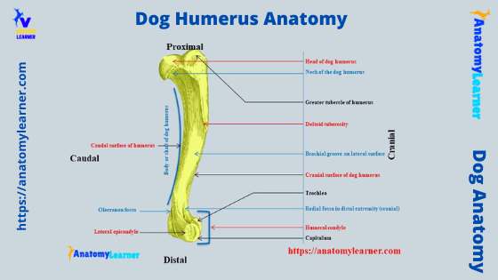
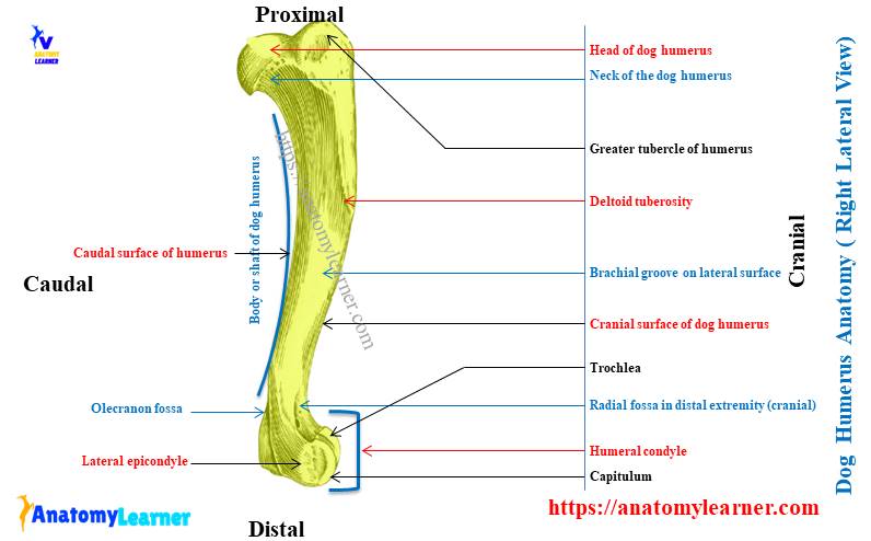
First, in the dog humerus labeled diagram, I tried to show you the longer body or shaft and two extremities. In the proximal extremity, all the important osteological features like the greater tubercle, lesser tubercle, inertuberal groove, and several foramina between the head and greater tubercle are identified in this diagram.
Again, from the distal extremity of the dogs humerus most important osteological features are also identified. Here, the labeled diagram shows the humeral condyle of the dog, which consists of lateral and medial condyles and epicondyles.
The labeled diagram also shows the radial fossa (cranially) and olecranon fossa (caudally). The supratrochlear foramen is also identified in this labeled diagram.
The shallow supracondylar fossa and the lateral supracondylar crest are also shown in the dog humerus bone labeled diagram.
Now, from the body, the four surfaces are identified with their main osteological features. Here, the lateral and cranial surfaces from the dog humerus bone are the most important.
The labeled diagram identifies the musculosprial groove and deltoid tuberosity from the humerus bone lateral surface. Again, the humeral crest is also shown from the cranial surface of the dog’s humerus.
The other osteological features like the teres major tuberosity, teres minor tuberosity, and tricipital line are also identified from the respective surface of the dogs’ humerus bone. If you need more updated dogs humerus labeled diagrams, you may find them on the social media of anatomy learners.
Muscles that originated from dog humerus bone
In the dog or canine brachium muscle labeled diagram, I will try to show the different muscles from the cranial and caudal aspects. Let’s see the diagram; here, the long and lateral heads of the triceps brachii muscles are identified.
After removing the long and lateral heads, the lateral aspect of the brachium shows the accessory head and a part of the medial head of the triceps brachii muscle. Again, the brachialis muscle from the lateral aspect of the humerus bone is identified in the labeled diagram.
The labeled diagram identifies the coracobrachialis, anconeus, and teres major muscles from the dogs’ humerus (origin or insertion). In the diagram, you will also see the insertion of the infraspinatus muscle on the proximal end of the dogs’ humerus.
The medial aspect of the dogs brachium shows the clear view of the medial head of the triceps brachii (identified). Again, most craniolateral and caudomedial antebrachial muscles originated from the epicondyles of the dogs’ humerus.
In the dog thoracic muscles labeled diagram, the craniolateral and caudomedial muscles are identified. You may find more dog muscles labeled diagrams on here.
Horse humerus anatomy compared with canine
Here I will enlist the most important osteological features from the horse humerus anatomy that differs from the canine humerus. Let’s see what the unique osteological features of the horse humerus anatomy are –
- The horse humerus is comparative longer, and rough compare to the dogs,
- There is a deep brachialis or musculospiral groove, which is more twisted compared to the dogs,
- The deltoid tuberosity of the horse humerus is more prominent than in other animals,
- There is an intertuberal groove in the proximal extremity that again divides into two parts by a median ridge,
- You will see the nutrient foramen at the distal third of the medial surface of the humeral shaft,
- The supracondylar fossa is deep, and the crest of this supracondylar fossa is more prominent,
- Again, the radial fossa and the olecranon fossa of the humeral condyle of the horse are deeper,
These are very short information on the osteological features of the horse humerus compared to the dogs. You will nerve find any infratrochlear foramen in the horse humerus.
Joints anatomy and dog humerus bone
Let’s learn about the anatomy of the two important joints formed by the dog humerus bone. You already know that the dog humerus forms the shoulder joint above the glenoid cavity of the scapula. Again, the distal extremity (humeral condyle) forms the elbow joint with the head of the radius bone.
- Suggested reading: you may read more about the shoulder joint and elbow joint anatomy from the anatomy learner.
The dog shoulder joint is formed between the scapula’s glenoid cavity and the humeral head’s articular surface. You will find different essential ligaments in the dogs shoulder joint anatomy. Let’s see what are these ligaments in the shoulder joint –
- A loose articular capsule,
- The transverse humeral retinaculum, and
- Lateral and medial glenohumeral ligaments,
Again, in the dogs elbow joint, you will find two types of joints – the humeroradial joint and the humeroulnar joint. The bone involvements on these humeroradial joints and humerounlar joints are shown in the labeled diagram.
Again, you will find different important ligaments in both the humeroradial and humeroulnar joints of the dog’s elbow. In the humeroradial joint, you will see the joint capsule, lateral collateral ligament, annular ligament, medial collateral ligament, and olecranon ligament.
Again, in the humeroulnar joint, you will see the interosseous and radioulnar ligaments. All these ligaments are described briefly in the dog elbow joint anatomy.
Some inquiries on dog humerus bone anatomy
Now, let’s see some of the learners’ common inquiries on the dog humerus bone anatomy. Here, I tried to enlist most of the questions on dog humerus bone with their answers.
What is a dogs humerus?
The dog humerus is a bone of the brachium or arm region of the thoracic limb. It is a long bone of the dog skeleton that possesses a cylindrical and long body or shaft and two expanded extremities.
The expanded extremities possess different osteological features that make them special from the other bones of the skeleton. Here, the proximal extremity of the dog humerus bone comprises the head, neck, tubercles, and grooves.
Again, the distal extremity of the dogs humerus possesses the humeral condyle (consists of the lateral and medial epicondyle), fossa, and supratrochlear foramen.
What is the humerus bone on a dog?
While studying the dog skeleton anatomy, you might find the humerus bone on the thoracic limb. This is the second bone in the thoracic limb of the dog that comprises the characteristics of the long bone.
The direction of the dog humerus is downward and backward. This humerus bone possesses four surfaces on its body and two definite extremities.
Where is the humerus in a dog?
The dog humerus is the long bone that finds in the thoracic limb of the skeleton. It locates obliquely from the shoulder joint and runs caudally.
This bone of the dog skeleton is froming the shoulder joint (proximally) with the glenoid cavity of the scapula. Again, the humeral bone of the dog forms the elbow joint (distally) with the radius and ulan bones.
Do dog dogs have a humerus bone?
Dogs have the humerus bone in their thoracic limb like other animals. But, the humerus bone of the dogs has some peculiar features that are different from other animals.
The most important osteological feature of the dogs humerus is the presence of supratrochlear foramen on the distal extremity (humeral condyle). With this foramen, the radial and olecranon fossa communicate with each other.
Again, the brachial groove is less deep than located on the lateral surface of this bone. The shaft of this bone is less twisted compared to the other animals like the horses and ruminants.
What is the function of the humerus in animals?
The main function of the humerus in animals is to bear the weight and support the other part of the skeleton. Again, different muscles originate or are inserted on the different surfaces of this bone.
These muscles that originated or were inserted from this humerus bone have diverse functions. Some of these muscles, like biceps brachii and brachialis, help to flex the elbow joint. Again, some of the muscle like coracobrachialis, triceps brachii, and anconeus helps to extend the elbow joint.
Again, you will find the origin of most of the extensor and flexor muscle group that acts differently. The main function of these muscles is to extend or flex the dogs’ elbow joint, carpus, and forepaw.
What is the strongest bone in a dogs body?
Most of the bones from the canine skeleton are strong. But, you will find the strongest bone in a dog’s skull and the hind limb. Most of the authors stated that the petrous part of the temporal bone from the dogs’ skull is the strongest bone in a dogs body.
Again, you should talk about the hindlimb bones if you think the strongest bone from the limbs. The femur of the dog skeleton is the more massive bone. Again, all other bones from the hind or pelvic limb of the dogs are stronger compared to the other bones of the skeleton.
How can the humerus of a cat be distinguished from of a dog?
Sometimes you may be asked to distinguish the cat humerus from the dog humerus bone. The basic anatomy of the dog humerus bone is almost similar to that of cats. But, the osteological features of these two species may vary in size, shape, and length.
The humeral head of the cat is less circular than the dog. The coronoid fossa is proximal to the trochlea in the cats’ humeral condyle.
The supracondylar foramen is the ovoid slit-like structure in the cat humerus that locates proximal to the medial epicondyle. This feature is exceptional in the distal extremity of the cat humerus bone if you compare it with the dogs humerus.
Again, you will see the sharp supracondylar ridge, a medial pectoral ridge, and a sharp and crest-like deltoid ridge on the cat humerus bone. Thus, you may distinguish the cat humerus from the dog humerus bone with these exceptional osteological features.
Conclusion
I hope this article might help you learn the basics of dog humerus bone anatomy. All the dog humerus bone-labeled diagrams will also help you to identify the different osteological features from the different surfaces of the body and extremities.
Again, you will identify the origin and insertion of the brachial muscles with the help of this article and labeled diagram. The dog humerus bone forms two important joints in the thoracic limb – above the shoulder and distally the elbow joint.
Now, you should identify all the important osteological features and muscle anatomy from the actual sample of dog humerus bone at your laboratory. You may again take help from the labeled diagram and video provided by the anatomy learner on dog humerus bone.