The dog throat anatomy comprises the features of the trachea, esophagus, larynx, and thyroid gland. You will also find some lymph nodes in the throat of a dog. This article will help you learn the anatomical facts of the trachea, esophagus, different parts of the larynx (epiglottis), thyroid gland, and lymph nodes from the dog’s throat.
Again, there are some essential muscles that are related to the throat of a dog (specific to different internal organs of this area). I will also describe the muscles attached to the particular structures of the dog’s throat.
There are also some common problems in the throat that I will describe at the end of this article. Even after completing this article, you will come to know how to palpate lymph nodes in dogs from their throat.
So, if you want to learn anatomical facts about the internal dog throat, let’s continue this article until the end.
Dog throat anatomy
The dog throat anatomy starts from the epiglottis and extends to the esophagus and trachea. There is no specific boundary of the throat area in the dogs. You will find the hyoid apparatus in front of the dog’s throat.
Collectively, you may find the below-mentioned organs or structures in a dog’s throat –
- Epiglottis or other parts of the larynx,
- Starting position of the trachea,
- Anatomical features of the esophagus,
- Different parts of the larynx,
- Thyroid and parathyroid glands,
- Different lymph nodes in the throat,
- Laryngeal and esophageal muscles, and
- Different external muscles that cover the throat area,
You may also describe the soft and hard palate under the throat of dogs. In the dog mouth anatomy article, I have already described the anatomical facts of these soft and hard palate features.
Again, most organs or structures continue to the dog’s neck. So, it will be better if you read the following article to get the basic idea of dog neck structure –
- Dog neck anatomy with the labeled diagram,
Okay, let’s discuss the different structures and organs from the throat region of the dogs with the labeled diagram. But, before going to start, let’s see some of the unique features of the throat.
Special features of dog throat structure
Externally, the dog throat is covered by the different muscles of the neck and face. You will see the below-mentioned unique features from the different structures or organs of the throat –
- There is an extra pair of cartilage found in the larynx of a dog which is known as the cuneiform cartilage that attaches to the arytenoid,
- The vocal cord is more prominent in the dog trachea compared to other animals like ruminants or horses,
- You will not find any apical bronchus (present in ruminant) in the dog trachea,
- The esophageal opening is small in the pharynx, whereas the orifice of the auditory tubes is narrow,
- The dog esophagus is comparatively wider and variable in length,
- You will find a constricted area at the beginning of the esophagus and a small abdominal part at the end,
- There are oval elongated, and flat thyroid glands located at the lateral aspect of the first six trachea cartilage of the dog,
- The caudal ends of the dog’s thyroid glands are generally connected by the glandular isthmus, which is inconstant,
- There are two more essential lymph nodes in the dog’s throat area – medial and lateral retropharyngeal lymph nodes (others are included under the neck anatomy),
The muscles involved in the dog’s throat structure will be described later in this article. You might know all the muscles from the neck and face region of a dog. But, there are some specific muscles in the structure of the dog’s larynx and esophagus.
Anatomy of internal dog throat
In this section, you will learn the anatomical facts of the different organs and structures of the internal dog throat. First, I will start with the other cartilages of the dog’s larynx.
Then I will describe the anatomical facts of the trachea and esophagus with the diagrams.
Before that, you might also know the anatomical facts of the hyoid apparatus and also the pharynx of the dog. Let’s see what the hyoid apparatus and different parts of the pharynx are.
There is no clear demarcation of the dog pharynx; it is a passage common for the respiratory and digestive systems.
Let’s see the anatomy of the dog pharynx labeled diagram in front of the throat. The dog pharynx is a musculomembranous junction of the respiratory and digestive tubes.
This pharynx extends between the oral and nasal cavity rostrally, esophagus, and larynx caudally. Externally, you may identify the pharynx at the level of the orbital opening to the second cervical vertebra.
Here, the dog pharynx divides into three parts –
- Nasal part – known as the nasopharynx,
- Oral pharynx – known as the oropharynx, and
- The laryngeal region – known as the laryngopharynx,
The dog pharynx is related to the base of the cranial cavity and suprapharyngeal lymph node above. Again, you will find the relationship with the pterygoid muscle, hyoid bone, mandibular salivary gland, external carotid artery, and pharyngeal lymph node ventrally.
Different pharyngeal muscles are attached to the structure of the dog’s pharynx –
- Palatopharyngeus muscle,
- Thyropharyngeus muscle,
- Hyopharyngeus muscle,
- Crico pharyngeus and arytenopharyngeus muscles, and
- Pterygopharyngeus and stylopharyngeus msucles,
All these pharyngeal muscles are responsible for sending the food bolus from the pharynx to the esophagus. But, three different successive contractions help to pass the food from the pharynx to the esophagus.
Dog nasal pharynx
The dog’s nasal pharynx or nasopharynx is the respiratory part of the laryngeal space. It locates dorsal to the soft palate of the dog mouth cavity.
This nasopharynx of a dog extends from the nasal choncae to the interpharyngeal opening of the larynx. At the dorsolateral wall of the nasopharynx, you will see a slit at the level of the soft middle palate.
This slit is the pharyngeal opening for the auditory tube. The tube opens in the tympanic cavity caudally.
The microscopic figure of the nasal pharynx shows the pharyngeal and tubal tonsils.
Overall you will find seven openings in the dog pharynx that communicate with the other structures –
- Two for the Eustachian tuber (auditory tubes),
- Two for posterior nares, and
- One for the esophagus, one for a mouth, and one for the larynx,
You know the interpharyngeal opening is the opening of the nasal cavity into the laryngopharynx. The caudal border (margin) of the soft palate and the right and left palatoglossal arches to form this opening.
The digestive tube continues caudally from the interpharyngeal opening.
The oral pharynx of a dog
This is the space between the oral cavity and the oral part of the pharynx. The palatoglossal arch bounds each side of this oropharynx.
Ventrally, you will find the tongue and soft palate dorsally to the oropharynx of a dog. Again, there are different tonsilar fossa at the lateral aspect of the oropharynx.
Practically, the oropharynx extends from the isthmus of fauces (between the oropharynx and oral cavity) to the base of the epiglottis. You will see palatomandibualr folds lateral to the palatoglossal arches.
The long, relatively thin palatine tonsil locates the lateral wall of the pharynx just caudal to the palatoglossal arch. Actually, these tonsils are located within the tonsilar fossa.
You will not find any afferent lymphatics in the structure of the palatine tonsil of a dog. The efferent vessels from the palatine tonsil drain into the medial retropharyngeal lymph node.
Later in this article, you will learn the location and structure of the medial and lateral retropharyngeal lymph nodes.
Dog laryngopharynx
The portion dorsal and lateral to the pharynx is known as the laryngopharynx. It extends from the interpharyngeal ostium and nasopharynx to the beginning of the esophagus caudally.
The caudal border of the dog laryngopharynx extends to the caudal border of the cricoid cartilage of the larynx. You will find a clear annular ridge that demarks the pharyngoesophageal junction.
The branches of the cranial thyroid and ascending pharyngeal arteries supply nutrition to the dogs’ laryngopharynx. They receive the innervation through the branches of glossopharyngeal and vagus nerves.
Hyoid apparatus of a dog
You will see the hyoid apparatus in front of the dog’s throat structure. This apparatus will act as a suspensory mechanism for the tongue and larynx.
First, let’s see how the hyoid apparatus attaches to the tongue, larynx, and skull –
- Dorsally – it attaches the skull dorsally,
- Ventrally – attaches the larynx and tongue ventrally,
The hyoid apparatus remain between the space of the bodies of dog mandibles. In a hyoid apparatus of a dog, you will see the below-mentioned segments –
- Paired thyrohyoid, ceratohyoid, epihyoid, and stylohyoid bones,
- Tympanihyoid cartilages (paired),
- Single basihyoid bone,
The basiphyoid is the unpaired dorsoventrally compressed rod. The extremities of this basiphyoid bone articulate with the thyrohyoid and ceratohyoid bones.
The thyrohyoid is the laterally bowed, sagitally compressed, slender bone in the dog hyoid apparatus. It extends dorsoventrally from the basihyoid and articulates with the cranial part of the thyroid cartilage of the larynx.
The ceratohyoid are small, short, and rod-shaped bones. They articulate with the basihyoid and thyrohyoid bones of the dog’s hyoid apparatus.
Again, the epihyoid parallels the thyrohyoid segments and articulates with the ceratohyoid bones. Here, the stylohyoid is slightly longer than articulate with the epihyoid bones.
Finally, you will find the small cartilaginous structure at the proximal extremity of the stylohyoid bone. These are the tympanohyoid cartilage that articulates with the mastoid process of the dog skull.
Dog larynx anatomy
The larynx is the musculocartilaginous organ that is considered one of the parts of the dog throat anatomy. It is guarding the entrance of anything to the trachea. Again, the dog larynx serves as the air passageway, aids vocalization, and prevents the inspiration of foreign substances.
Now, let’s see the location of the dog’s larynx in the throat region. This structure is directly located caudal to the root of the tongue, oral pharynx, soft palate, and ventral to the atlas vertebra.
The laryngeal opening is bounded by the epiglottis cartilage ventrally and cuneiform cartilage dorsally. From the dog larynx anatomy, you might identify the six (or five) types of cartilages and different muscles.
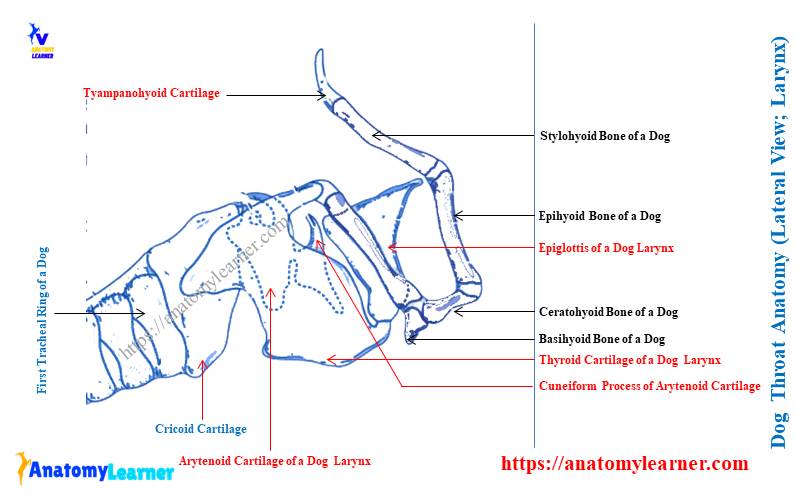
Let’s see the six (or five) types of cartilages from the dog larynx anatomy –
- Epiglottic cartilage of the dog larynx – forms the base of the epiglottis,
- Thyroid cartilage – larger and located ventrally,
- Arytenoid cartilage – irregular shape,
- Cuneiform cartilage or process – triangular shaped structure that attaches to arytenoid,
- Sesamoid cartilage – oval or dumble-shaped structure, and
- Interarytenoid cartilage of dog larynx – smaller and flat shaped,
Again, you might also learn the anatomical facts of the laryngeal muscles from the dog larynx. Let’s see what are the muscles present in the dog’s larynx –
- Cricothyroideus muscle,
- Criocoarytenoideus dorsalis and lateralis muscles,
- Thyroarytenoideus muscle,
- Vocalis and ventricularis muscles,
- Arytenoideus transversus muscle, and
- Hypoepiglotticus muscle of the dog larynx,
I will provide a short description of these laryngeal muscles from the dog. Again, you will find the five different segments in the larynx cavity – aditus laryngis, vestibule, vestibular cleft, glottis, and infraglottic cavity.
There are different ligaments in the structure of the dog larynx, but the cricothyroid, vocal, and vestibular ligaments are the most important. Now, let’s see the macroscopic (anatomical) facts of the five or six different cartilages from the dog’s larynx.
Dog epiglottis structure
The epiglottic cartilage is single and forms the base of the dog epiglottis. You will find the thin rostral margin and dorsally concave triangular apex. The lateral margins of the dog epiglottic cartilage are thick.
Here, the apex of the dog’s epiglottic cartilage is free and curved forward and downward.
There is a concave laryngeal surface on the dorsal surface of the dog larynx that faces dorsocaudally. Again, the convex lingual surface is opposite to the laryngeal surface that meets the oral pharynx.
There is a hypoepiglotic muscle in the structure of the dog larynx. This muscle attaches the lingual surface of the larynx to the middle of the hyoid body.
Again, the hypoepuglotic muscles of each side are covered by thick mucosal folds. The stalk of the dog epiglottic cartilage is made of dense fibrous tissue.
The stalk of the epiglottic cartilage attaches to the dorsocranial aspect of the thyroid cartilage.
Thyroid cartilage of dog’s larynx
The thyroid cartilage of the dog larynx is single and the largest structure that forms the lateral and ventral wall of the laryngeal cavity. Anatomically, you will find a body and two laminae in the dog thyroid cartilage.
The dorsal surface of the thyroid cartilage is related cranially to the base of the epiglottis. Again, the cranial angle of the lamina on either side is extended forward and upward.
The lamina of the dog’s thyroid cartilage possesses rostral and caudal cornua (each lamina). Here, the rostral cornua are extended, whereas the caudal cornua are blunt.
Each cranial cornua of the thyroid lamina possess a hyoid articular surface to articulate with the hyoid bone. Again, the medial aspect of each caudal cornua possesses a cricoid articular surface that articulates with the caudolateral aspect of the cricoid cartilage.
Ventral to the cranial or rostral cornua, you will find a deep fissure that forms a foramen. Within this structure, the laryngeal nerves and vessels pass.
The ventral surface of the thyroid cartilage possesses the most important anatomical features. You will find the ventral laryngeal prominence when the lamina attaches ventrally.
But, the prominence is slight in the dog’s thyroid cartilage. This laryngeal prominence is known as Adam’s apple in humans.
You will find another two structures in the thyroid cartilage –
Cricothyroid ligament – attaches the caudal border of the thyroid cartilage with the ventral arch of the cricoid cartilage, and
Thyrohyoid membrane – attaches the cranial border of the thyroid cartilage with the basihyoid and thyrohyoid bones.
Now, let’s know the anatomical (features) facts of the cricoid cartilage from the dog larynx anatomy.
Cricoid cartilage of dog larynx
The cricoid cartilage is a ring-shaped structure in front of the first tracheal ring and caudal to all other laryngeal cartilages. Here, the dorsal segment of the cricoid cartilage is wider than the ventral segment.
The expanded dorsal segment of the dog’s cricoid cartilage, known as the lamina, possesses a median crest. Sometimes you may find pair vascular foramina on the dorsal expanded segment of the dog’s cricoid cartilage.
You will also find the cricoid cartilage arch that extends from the lamina to ventrally. This arch of the cricoid cartilage help from the caudal part of the laryngeal cavity.
There are two pairs of articular surfaces in the cricoid cartilage –
Pairs of thyroid articular surface – locates at the junction of lamina and arch of the cricoid cartilage. They articulate with the apices of the caudal cornua of the thyroid cartilage.
Pairs of arytenoid articular surface – locates on the rostral border of lamina and median crest. They articulate with the arytenoid cartilage.
Arytenoid cartilage of dog larynx
The arytenoid cartilage of the dog larynx is an irregular paired structure located in front of the cricoid and forms the caudal part of the roof of the laryngeal cavity. Each arytenoid cartilage possesses three surfaces (dorsal, lateral, and medial), a base, and an apex.
Here, the base of the arytenoid cartilage is concave and attaches to the cricoid cartilage. Again, the apex is free and curved backward and upward.
But, the different authors describe the arytenoid dog cartilage in different ways. The anatomical structure of the dog arytenoid may vary in different species.
Normally, the dog arytenoid cartilage is formed by the following processes –
- Corniculate process of the arytenoid,
- A muscular process of the arytenoid,
- The vocal process, and
- A cuneiform process,
But, some authors described the cuneiform as the main cartilage of the dog larynx. The corniculate is the long process in the dog’s arytenoid cartilage. This process of arytenoid forms the dorsal margin of the laryngeal inlet.
You will see the relatively thick and rounded muscular process on the lateral surface of the arytenoid cartilage. Again, the vocal process is the caudal ventral projection of the dog’s arytenoid cartilage.
Finally, the cuneiform is another process of the dog’s arytenoid cartilage. But, as I told you before, many authors describe this process as the main cartilage of the dog larynx.
It is roughly triangular and located on the rostral part of the arytenoid cartilage. The ventral part of the cuneiform process lies on the vestibular fold, which also contributes to forming the laryngeal inlet.
Sesamoid and interarytenoid cartilages of dog larynx
Cranial to the cricoid and between the arytenoid cartilages, you will find oval, dumble-shaped cartilage. This is the sesamoid cartilage of the dog’s larynx, which may be paired or single.
The sesamoid cartilage appears to be intercalated in the transverse arytenoid muscle. There is little contact of this sesamoid with the arytenoid cartilage.
Finally, the interarytenoid cartilage is small and flat that lies cranial to the cricoid lamina and caudodorsal to the transverse arytenoid muscle. This cartilage attaches to the arytenoid cartilage and the cricopharyngeal tendon.
Anatomy of dog laryngeal muscles and throat
While studying the dog throat anatomy, you might learn the features of laryngeal muscle in detail. Here, I will show the anatomical features of the main muscle of the dog larynx with their origin, insertion, and principal action.
I have previously provided the list of dog laryngeal muscles in this article. Now, let’s discuss the macroscopic facts of the cricothyroideus muscle from the dog larynx.
Cricothyroideus muscle of the dog
This is the thick muscle on the lateral surface of the dog larynx. You may easily identify the cricothyroideus muscle from the dog’s larynx.
This muscle of the dog larynx lies between the thyroid lamina and the cricoid cartilage. It runs cranially and dorsally to attach to the caudal margin and medial surface of the thyroid cartilage.
Again, some cricothyroideus fibers are attached to the vocalis muscle. The main action of this muscle is to provide the tens of the vocal cord.
Cricoarytenoideus dorsalis and lateralis muscles
The cricoarytenoideus dorsalis muscle arises from the entire length of the dorsolateral surface of the dog’s cricoid cartilage. The fibers of the cricoarytenoideus dorsalis muscle run craniolateraly and insert into the muscular process of the arytenoid cartilage.
There are three neuromuscular compartments on the cricoarytenoid dorsalis muscle. This muscle helps to open the glottis by abducting the vocal folds.
Again, the cricoarytenoideus lateralis muscle arises from the cranial and lateral surfaces of the cricoid cartilage of the dog. The fibers from the cricoarytenoideus lateralis muscle run cranially and dorsally and insert into the muscular process of the arytenoid cartilage.
The main action of the cricoarytenoideus lateralis muscle is to pivot the arytenoid cartilage medially and close the rima glottis.
Thyroarytenoideus, vocalis, and ventricularis muscles
This is an important muscle in the dog larynx that gives rise to the vocalis and ventricularis muscles. From the midline of the thyroid cartilage, this thyroarytenoideus muscle arises.
It runs caudodorsally and inserts on the arytenoid cartilage. The aponeurosis from the thyroarytenoideus muscle attaches to the muscular processes of the arytenoid cartilage.
The main function of the thyroideus muscle is to relax the vocal cord and constrict the glottis. Now, let’s discuss the anatomical facts (macroscopic) of the vocalis and ventricularis muscles from the dog larynx.
The vocalis is the medial division of the thyroarytenoideus muscle that originates from the midline of the thyroid cartilage. Again, a few parts of the vocalis originates from the caudal part of the thyroarytenodeus muscle.
There is a vocal process on the arytenoid cartilage where the vocalis muscle inserts. This muscle draws the arytenoid cartilage ventrally and relaxes the vocal cord.
Now, the ventricualris is the cranial division of the thryoiarytenoideus muscle and also originates from the thyroid cartilage. Again, the ventricularis muscle also receives some fibers from the craniodorsal surface of the thyroarytenoideus muscles.
The main action of the ventricularis muscle is to constrict the glottis and dilate the laryngeal ventricle.
Dog’s arytenoideus transversus and hyoepiglotticus muscles
The arytenoideus transversus is the broad muscle on the muscular process of the arytenoid cartilage. This muscle inserts on the expanded lateral end and dorsal surface of the interarytenoid cartilage. It also constricts the glottis and adducts the vocal folds.
You will find the small, spindle-shaped hyoepigotticus muscle in the structure of the dog larynx. It arises from the medial surface of the ceratohyoid bone of the hyoid apparatus.
The fibers from the hyoepuglotticus muscle are inserted into the ventral midline of the dog’s epiglottis. This muscle draws the epiglottis ventrally.
Dog laryngeal cavity and innervation
So, there are five segments in the dog laryngeal cavity – aditus laryngitis, vestibule, vestibular cleft, glottis, and infraglottis cavity. The aditus laryngis or laryngeal inlet lies caudal to the interpharyngeal ostium.
The margin of the epiglottis forms the lateral boundaries and apex of the laryngeal inlet. You will find the aryepiglottic folds at the caudal part of the aditus laryngis or laryngeal inlet.
The vestibule of the dog larynx extends laryngeal aditus to the vestibular folds. It is a funnel-shaped cavity that opens freely at the dorsocranial aspect. The vestibule of the dog laryngeal cavity opens caudally into the rima glottis.
Here, the cuneiform process and the vestibular folds form the vestibular cleft in the dog larynx. Again, the ventral boundary of the vestibular cleft is formed by the thyroid cartilage.
The vestibular fold is a short but wide mucosal structure that extends from the ventral margin of the cuneiform cartilage to the craniodorsal surface of the thyroid cartilage.
Between the vocal cord and arytenoid cartilage, you will find the cleft of the glottis. Here, the glottis comprises the vocal folds, arytenoid cartilage, and rima glottis.
A vocal fold in the larynx extends from the vocal process of the arytenoid cartilage to the dorsocaudal part of the thyroid cartilage. Here, you will also find a distinct vocal ligament that supports the structure of vocal folds.
There is a small mucosal sac between the vestibular and vocal folds known as the laryngeal ventricle. The sound produced in the vocal and vestibular folds may vibrate into the cavity of the glottis.
Finally, the infraglottic cavity of the dog larynx extends from the rima glottis to the tracheal cavity.
The cranial and caudal laryngeal nerves innervate the laryngeal cavity and larynx of a dog. Here, the cranial laryngeal nerve comes from the vagus nerve at the level of distal ganglia and divides into internal and external segments.
Dog throat and trachea anatomy
The trachea is the second most important component in the dog throat anatomy. It is a relatively noncollapsable tube that extends from the cricoid cartilage to the level of the base of the dog’s heart.
The dog trachea is made of cartilage, muscles, and mucosa that divides into two primary bronchi at the dorsocranial part of the base of the heart. Anatomically, you will find almost 35 C-shaped hyaline cartilage in the dog trachea.
These are the cartilaginous tracheal rings that remain incomplete (open) at the dorsal part. You will see the smooth muscle and connective tissue fibers on the dorsal surface of the dog trachea that complete the dorsal open structure.
The C-shaped trachea ring connects in the longitudinal direction with the help of the annular ligament. This annular ligament provides considerable intrinsic movement of the trachea without breakage or collapse.
The diameter and thickness of the dog trachea are not the same throughout this structure. You will find the smallest diameter and thickness in the dog trachea at the level of the thoracic inlet.
Due to the less diameter and thickness, the dog trachea is very much susceptible to become a collapse. For description purposes, you may divide the dog trachea into two distinct segments –
- The cervical part of the dog’s trachea and
- The thoracic part of the dog trachea,
If you only describe the anatomical features of the dog throat, there is no need to describe the details of the trachea. It would help if you only focused on the starting part of the tracheal ring (the first two or three). But, if you have a great interest in learning the details anatomical facts of dog trachea, you may continue this part of the article.
The cervical part of the dog trachea
The cervical part of the dog trachea starts from the cricoid cartilage of the larynx and continues at the level of the thoracic inlet. You will find different organs and structures that have a close relationship with the dog trachea in the cervical region.
You will find the esophagus on the dorsal surface of the dog trachea at the level of a third thoracic vertebra. Then the esophagus goes to the left lateral aspect of the dog trachea (I will describe the course of the esophagus later).
You will find the longus colli muscle on the dorsal surface of the trachea after at the level of the third cervical vertebra (3rd) and continue it at the fifth cervical vertebra. Ventrally, you will find the Sterno-thyrohyoideus muscle on the dog trachea.
On the lateral aspect of the dog trachea, you will see some important vessels and nerves – the carotid artery, jugular vein, vagosympathetic trunk, and recurrent laryngeal nerve. You will also find some of the lymph nodes that are also attached to the lateral and dorsal surface of the dog trachea.
A thoracic segment of dog trachea
The thoracic segment of the dog trachea starts from the thoracic inlet and continues to the cranial and middle mediastinum. Now, the trachea is related dorsally to the esophagus again and the longus colli muscle.
Left lateral to the dog trachea (thoracic segment), you will find the left aortic arch, left axillary artery, and thoracic duct. Again, to the right lateral surface of the dog trachea, you will see the right vagus nerve.
Finally, you will see the relationship between the anterior vena cava, brachiocephalic trunk, and recurrent laryngeal nerve at the ventral aspect of the dog trachea.
The dog trachea divides into right and left principal bronchus at the middle mediastinum. Again, each principal bronchus divides into lobar bronchi, which is the basis for identifying the different lobes of the dog lung.
If you wish, you may learn the anatomical facts of the different lobes of dog lungs from their respiratory system –
- Dog lung anatomy with the labeled diagram,
Within the lobe of the dog lung, the lobar bronchi divide into the segmental bronchi. The segmental bronchi and the dog lung tissue they ventilate are known as the bronchopulmonary segment.
If you see the anatomical figure of the segmental bronchi, it again divides into the small bronchi. These are the respiratory bronchioles and give off alveolar duct, alveolar sacs, and pulmonary alveoli.
Dog throat anatomy and esophagus structure
The esophagus is the first (1st) segment of the alimentary canal that is also an important component of dog throat anatomy. This structure of the dog acts as a connective tube between the laryngeal part of the pharynx and the stomach.
So, the dog esophagus starts from the larynx, passage transverse most of the neck and thoracic cavity. Finally, they end up entering the abdominal cavity.
Thus, you will find the three segments in the dog esophagus (but two distinct segments in the ruminant esophagus) –
- A cervical segment of the dog esophagus,
- The thoracic segment of the canine esophagus, and
- A small abdominal segment of the dog esophagus,
But for the description of the dog’s throat, you might learn the starting segment of the esophagus. The dog esophagus begins opposite the middle of the axis dorsally and the caudal border of the cricoid cartilage ventrally.
There is a limen pharyngoesophageal (prominent ventral ridge) between the larynx and esophagus of the dog. So, this prominent ventral ridge might be considered the landmark in identifying the starting point of the esophagus from the pharynx.
The dog esophagus ends at the cardia of the stomach at the level of the last thoracic vertebra. But, the termination of the dog esophagus may vary in different species of dogs.
Dog esophagus anatomy
So, in the dog esophagus anatomy, I will describe the different segments (cervical, thoracic, and abdominal) along with the coats and innervation. But, the coats (layers of the esophagus) will be well understood in the microscopic figure of the animal’s esophagus.
The below-mentioned article is recommended to learnt the different coats or layers of the dog esophagus –
- Esophagus histology slide with the labeled diagram (details of four different layers),
Okay, now, start with the anatomical facts of the cervical segment of the dog’s esophagus.
The cervical part of the dog esophagus
So, the cervical part of the dog esophagus starts from the pharynx and extends up to the thoracic inlet. Let’s see the relationship of the cervical esophagus with the other different internal organs on the dog’s throat and neck.
Dorsally – the cervical part of the esophagus is related to the left longus colli muscle and longus capitis muscle.
Ventrally and right – it relates to the trachea on the ventrally and right aspect.
But, in the thoracic inlet, the dog esophagus lies left lateral to the trachea. Again, at the left lateral aspect of the dog esophagus, you will find the following structures or organs –
- Left common carotid artery,
- The vagosympathetic trunk of the dog,
- Left internal jugular vein of the dog, and
- A tracheal duct of the dog,
All these structures and organs run at the angle between the esophagus and longus capitis muscles. You will also find these structures on the right lateral side of the dog trachea (described previously).
The thoracic part of the dog esophagus
The thoracic part of the dog esophagus starts from the thoracic inlet and extends up to the esophageal hiatus on the diaphragm. At the cranial mediastinum, the esophagus lies on the little left of the trachea.
But, it obliquely crosses the left face of the trachea and becomes dorsal on it. This condition continues up to the tracheal bifurcation at the fifth and sixth thoracic vertebrae levels.
Then it crosses the right face of the aortic arch and runs ventral to the right and left longus colli muscle. The esophagus separates from these muscles and the neck by the prevertebral fascia.
Then the dog esophagus passes through the middle mediastinum and lies mid on the medial plane of the body. At the fifth to ninth thoracic vertebrae level, the abdominal aorta crosses the left side of the esophagus.
Again, the dorsal branches of both the right and left vagus nerve run dorsoventrally and across the sides of the dog’s esophagus. They unite with each other on the dorsum of the dog esophagus cranial to the dorsal part of the esophageal hiatus.
So, the dorsal vagal trunk continues and passes through the dorsal part of the esophageal hiatus. Again, the right and left ventral branches of the vagus nerves to unite immediately caudal to the root of the lung. Thus, they form the ventral vagal trunk in the dog.
The ventral vagal trunk lies in contact with the esophagus. It arches ventrally in the caudal mediastinum and passes through the esophageal hiatus with the esophagus.
Abdominal part of the dog esophagus anatomy
The abdominal part of the dog esophagus is very short and starts from the esophageal hiatus. It ends on the cardia of the dog’s stomach. The abdominal part of the dog’s esophagus is the wedge-shaped terminal part.
Let’s see the relation of the abdominal part of the dog’s esophagus with the other different internal dog structures or organs –
Dorsally – it immediately joins with the cardia of the stomach,
Ventrally – attaches with the thin, dorsal border of the caudate lobe of the liver,
There is a full guide (article) on the anatomical facts of the dog liver here in anatomy learner. You may learn the different lobes with their peculiar anatomical facts from the below-mentioned article –
- Dog liver anatomy with the labeled diagrams (with the lobes, impressions, and ligaments),
The diameter and thickness of the dog esophagus are not uniform throughout its length. You may find the average diameter and thickness in the lumen of the cervical part. But, the thickness and diameter of the esophageal lumen reduce in the thoracic part.
Again, you will find the highest diameter and thickness in the wall of the abdominal esophagus of a dog. There are numerous longitudinal folds in the mucosa of the dog esophagus that are capable of great dilatation.
Coats or layers of the canine esophagus
In the canine esophagus, you will find distinct four layers or coats – mucosa, submucosa, muscular layer, and adventitia. You will find clear views of the different canine or dog esophagus layers under the microscope.
The mucosa of the dog esophagus comprises superficially keratinized stratified squamous epithelium. They contain the opening of the dog esophageal glands. You will find more cardiac glands in the distal part of the dog esophagus.
The submucosa coat of the dog esophagus contains loosely arranged inelastic connective tissue. This layer of the canine esophagus connects the mucosa and the muscular coats.
The submucosal layer will help to form the longitudinal folds in the wall of the dog esophagus. You will find the blood vessels, nerves, and glands in the submucosa coat of the dog esophagus.
Again, you may find the thin layer of smooth muscle (the muscular layer of the submucosa) in the caudal part of the dog esophagus segment. But, it is very hard to grossly identify the smooth muscle layer from the submucosa layer of the esophagus.
The muscular coat of the dog esophagus is thick and possesses two oblique layers of smooth muscle. Here, the external muscle layer of the muscular coat arises on the ventral side of the esophagus from the medial dorsal crest of cricoid and arytenoid cartilages.
The main musculature of the dog esophagus caudal to the cricoesophageal fibers is in the form of a spiral appearance. Here, the inner coat of the muscle becomes transverse in direction, whereas the outer coat becomes more longitudinal.
The cervical part of the dog esophagus possesses few longitudinal muscle fibers along with these two oblique coats.
Adventitia of dog esophagus
The adventitia of the cervical part of the dog esophagus will blend with the deep cervical fascia dorsally. Again, it also blends with the fascia that comes from the carotid covering on the left.
Again, the adventitia of the thoracic and abdominal parts of the esophagus blend with the endothoraic and transversalis fascia. You will find the coverage of the pleura on the thoracic segment of the esophagus.
Again, the peritoneum covers the small abdominal part of the dog esophagus. Then this adventitia of the abdominal esophagus becomes the serosa. You will learn more about the difference between the adventitia and serosa from the following article –
Histological features of different four layers of a tubular organ with the labeled diagram,
I hope you will also understand the dog esophagus’s structure from this article.
Vessels and nerves of dog esophagus
The cranial and caudal thyroid arteries are supplied in the cervical part of the dog esophagus. Again, the limen pharyngoesophageal is supplied by the cranial carotid artery.
The left caudal thyroid artery will anastomose with the ascending branch of the bronchoesophageal artery. From these anastomoses, a small branch supplies to the thoracic segment of the dog esophagus.
Again, the esophageal part of the bronchoesophageal artery is directly supplied to the cranial two-thirds of the thoracic part of the dog’s esophagus. Another remaining segment of the dog esophagus is supplied by the esophageal branch of the aorta or dorsal intercostal arteries.
Finally, the terminal segment of the dog esophagus is supplied by the esophageal branch of the left gastric artery.
The pharyngeal plexus (nerves) is formed by the pharyngeal branches of the glossopharyngeal and vagus nerves. These pharyngeal plexus innervate to the cricopharyngeal muscle and the cervical segment of the dog esophagus.
Again, the left and right recurrent laryngeal nerves are also supplied to the thoracic segment of the dog esophagus. Finally, the abdominal segment of the dog esophagus is supplied by the right and left vagal trunks.
Dog throat anatomy lymph nodes
You know, the lymph nodes are the structural and functional unit of the dog’s lymphatic system. Here, I will describe these lymph nodes related to the dog throat anatomy. Let’s see the anatomical facts of the following lymph nodes that have a direct or indirect relationship with the dog’s throat –
- Medial retropharyngeal lymph node and
- Lateral retropharyngeal lymph node,
But, you will also find other different lymph nodes from the oral cavity and neck to the thoracic region of the dog –
Parotid lymph node – at the rostral base of the dog ear,
Mandibular lymph nodes – at the ventral angle of mandible and lingofacial vein,
Buccal lymph nodes – dorsal to the buccinators muscle,
Superficial cervical lymph nodes – on a lateral surface of serratus ventralis and scalenus muscles,
Deep cervical lymph nodes – at the cervical part of the trachea on each side,
Cranial deep cervical lymph node – at the caudal end of media retropharyngeal lymph node and the thyroid gland,
Middle and caudal deep cervical lymph nodes – rarely find in the dog neck,
Axillary lymph node – caudal to shoulder joint, and
Other different lymph nodes in thoracic and abdominal areas,
I have already described all these lymph nodes in the dog neck anatomy article. You may read the anatomical facts of the lymph nodes from the neck region of the dog from that article.
Now, let’s see the anatomical facts (features) of the medial and lateral retropharyngeal lymph nodes that directly relate to the dog throat.
Medial retropharyngeal lymph node of a dog
A dog’s medial retropharyngeal lymph node is the largest node found in the head and neck region. It is an elongated and transversely compressed lymph node in the dog head and neck.
You will see the pointed caudal end and wider cranial end in the structure of dog’s medial retropharyngeal lymph node. The weight of the medial retropharyngeal lymph node of the dog increase with the body weight. Again, the weight of this lymph node decrease in older dogs.
The medial retropharyngeal lymph node (two) of the dog lies ventral to the wing of the atlas. You will see the digastricus muscle cranially, longus colli muscle dorsally, and pharynx and larynx ventromedially.
Again, the medial retropharyngeal lymph node covers by the mastoid part of both the cleidocephalicus and sternocephalicus muscles. The cranioventral part of the retropharyngeal lymph node is related to the mandibular salivary gland.
The medial surface of the retropharyngeal lymph node also has a relationship with the hypoglossal nerve and terminal part of the carotid covering.
The afferent lymph vessels of the dog’s medial retropharyngeal lymph node come from the lymph vessels of the head region. Again, this medial retropharyngeal lymph node receives afferent vessels from the larynx and esophagus.
The efferent lymph vessels from the parotid, mandibular, and lateral retropharyngeal lymph nodes drain into the medial retropharyngeal lymph nodes. Again, the deep lymph vessels of the dog’s nasal cavity also drain into the medial retropharyngeal lymph nodes.
Lateral retropharyngeal lymph node of the dog
These are the smaller lymph nodes in the head and neck regions of the dog. You will find a small diameter in these lateral retropharyngeal lymph nodes.
The dog’s lateral retropharyngeal lymph node lies at the dorsal border of a horizontal part of the external acoustic meatus. Again, this lymph node is partially covered by the caudal part of the parotid salivary gland.
The afferent lymph vessels of this dog’s lateral retropharyngeal lymph node come from the structures near it. Again, you know the efferent vessels of the dog’s lateral retropharyngeal lymph node drain into the medial retropharyngeal lymph node.
How to palpate lymph nodes in dogs?
Sometimes you need to palpate the lymph node of the dogs. There are numerous deep and superficial lymph nodes in the dog’s body. In different viral and other infections, there is a huge possibility of swollen lymph nodes in different specific areas.
So, your veterinarian doctor will palpate the lymph nodes from the different regions of the dog’s body. Normally, the superficial lymph node can be easily palpated from the dog’s neck and head region.
To palpate the lymph node, you might know the exact location of that specific lymph node of the dog. Suppose you want to palate the superficial cervical lymph node of the dog.
So, you should know the exact location of that lymph node; here, the superficial lymph node of a dog locates on the lateral surface of the serratus ventralis and scalenus muscle. Again, the part of the superficial cervical lymph node locates on the cranial surface of the supraspinatus muscle.
Thus, you can locate the position and direction of the superficial cervical lymph node. Now, you should palpate this lymph node through the fingers.
As there is more adipose tissue in the neck and thoracic region of the dog, it is not so easy to palpate the superficial lymph node of the dog. But, you may easily palpate this superficial lymph node with the finger from the ruminant (cattle or goat).
Again, the mandibular lymph node from the dog can be easily palpated from the surface approach.
How to feel lymph nodes in dogs?
How to feel the dog’s lymph node cannot be expressed in words. But, in normal conditions, you may feel it as a slippery structure in a specific body area. Mostly, you may feel the mandibular, superficial cervical, and prefemoral lymph nodes by the external approach.
To feel these lymph nodes from the dogs, you might know their exact location with the anatomical facts. The size and shape of these lymph nodes of the dog vary. So, you may find the various size of lymph nodes in the different species of dogs.
In the case of swollen lymph nodes in your dogs, your dog may show pain on palpation. It is not recommended to palpate these swollen lymph nodes by the pet owners.
Is it normal to feel a dog’s lymph nodes?
It depends on the condition of the dog’s health. It is very hard to feel the dog’s lymph node if you have a healthy dog. Again, you can only palpate some of the specific superficial lymph nodes from the dogs.
Make sure you have enough knowledge of the location and other anatomical facts of these superficial lymph nodes of your dogs. Now, let’s come to the specific answer to your question – is it normal to feel the dogs lymph node?
Yes, it is normal to feel the dog’s lymph node by surface approach (but this is for some of the limited superficial lymph nodes). You may easily feel the mandibular, superficial cervical, and prefemoral lymph nodes from the dogs.
Can a dog live without lymph nodes?
The dog can not live without the lymph nodes because it is the main organs of the lymphatic system. And you know, the lymphatic system of the dog’s body directly regulates the immune functions.
You will find different lymph nodes in the course of lymph vessels, considered the structural and functional unit of the dog’s lymphatic system. These lymph nodes of the dog’s body serve different important functions.
They maintain the body fluid and drain the lymph fluid nearby organs or structures. Again, they protect the dog body from the illness-causing foreign materials. The lymph nodes also absorb the fat from the digestive tract and remove the cellular waste.
So, leading a dog’s life without the lymph nodes is very hard.
Old male dog throat problem symptoms
Both older and young dogs are very susceptible to throat problems. They will show different symptoms in various throat problems. I will not provide the details of these problems in the dog’s throat.
Rather I prefer to provide a basic idea of the different types of throat problems in young and older dogs. As you have already learned the dog throat anatomy, you will quickly understand these conditions.
But, I always recommend you learn the different problems of the dog throat in detail from other articles for anatomy learners. Let’s see the common throat problems in the dog –
- Pharyngitis or disorders of dog pharynx,
- Congenital abnormalities in the dog pharynx,
- Pharyngeal mucoceles,
- Traumatic injuries in the dog pharynx,
- Oropharyngeal dysphagia in the dog,
- Laryngeal paralysis of the dog,
- Acute and chronic laryngitis in the dog,
- Dog epiglotic entrapment,
- Traumatic injuries in the dog’s larynx,
- Dog tracheal collapse,
- Reverse sneezing or pharyngeal gag reflex, and
- Choke in the esophagus and trachea of the dogs,
Now, let’s know a little about the symptoms of these problems in the dog’s throat. But, it is impossible to describe all dog throat problems from the list.
Cleft lip and palate is a very common congenital malformation in the dog. This condition commonly affects the pharynx, but it leads the chronic rhinitis.
Different surgeries are also performed on the dog’s throat conditions like a tonsillectomy, surgical correction of pharyngeal dysplasia, marsupialization of the mucoceles, ventral laryngotomy, and arytenoid lateralization.
You will get the details guides on these surgical corrections here in anatomy learner in the veterinary surgical anatomy section.
Dog pharyngitis and tonsillitis
In the dog, acute pharyngitis and tonsillitis are very common that affect the throat. Different viral infections like the canine distemper, canine adino, and others may play a great role in a dog’s acute pharyngitis.
What are the primary symptoms of the dog’s pharyngitis? Okay, you will find the pain, fever, oral pain, and extreme discomfort in the affected dogs.
Your veterinarian doctor will diagnose acute pharyngitis based on history, physical examination, and findings upon inspecting the dog’s throat. The pharynx shows an inflamed area, edema, ulceration, and small abscesses.
In non-brachiocephalic dogs, you may also find chronic pharyngitis. Then the dog shows chronic vomition, food allergies, gagging, and retching independent of taking food.
Pharyngeal mucoceles of the dog throat
Salivary mucoceles are very common in dogs, a disease of a salivary gland. You know, the mucocele refers to a collection of saliva in the subcutaneous tissue near the site of the salivary duct or gland.
It may occur in the subcutaneous, sublingual, and pharyngeal locations. When it occurs in the pharyngeal location, it may cause discomfort to the throat.
The pharyngeal mucoceles are more common in the small breed of dogs, showing the typical clinical symptoms. You will find the obstruction in the airways, dyspnea, and snoring caused by the voluminous cyst in the pharynx.
Traumatic pharyngeal injuries in the dog
These traumatic pharyngeal injuries are also very common in dogs and are caused by the penetration of the stick. Again, other trauma also causes pharyngeal injuries that may cause edema and hematoma in the pharynx.
The clinical symptoms of the pharyngeal injuries include –
- Drooling of the blood mixed mucosa,
- Increased swallowing,
- Pain in the pharyngeal area, and
- Dyspnea with a snoring sound,
If you find pharyngeal injury symptoms, immediately bring your dog to a professional veterinarian.
Oropharyngeal dysphagia in the dog
There are different causes of oropharyngeal dysphagia in dogs. Some common causes of oropharyngeal dysphagia are – anatomical abnormalities, foreign bodies, chronic pharyngeal pain, different neuromuscular disease, and systemic problems like rabies and distemper.
The diagnosis of oropharyngeal dysphagia requires a thorough evaluation of the entire dog. A different physical and radiographic evaluation may confirm this oropharyngeal dysphagia by the veterinarian.
You may find difficulties with prehending food, dropping food from the mouth, exaggerated head movement during eating, repeated attempts to swallow food, and others.
Acute and chronic laryngitis in the dog
Infectious agents like the kennel cough mostly cause acute laryngitis in the dog. Clinical symptoms of the acute phase of dog laryngitis show a harsh, dry cough and hoarse voice. Sometimes you may find your dog has decreased appetite and lethargy.
Chronic laryngitis in the dog may be caused after the kennel cough and is associated with chronic tracheobronchitis. Clinical symptoms include recurrent coughing and retching. Again, you may find the rasping sound during panting and a roughening in the bark.
Laryngeal paralysis and other injuries
The dog laryngeal paralysis is the partial or complete failure of the arytenoid cartilage and vocal cord to abduct during inspiration. This results in obstruction in the upper airways, which may also cause aspiration pneumonia.
You may see unilateral and bilateral paralysis in the dog’s larynx. But, it is very difficult to diagnose unilateral pharyngeal paralysis in the dog.
There are mainly two types of laryngeal paralysis found in the dog – congenital and acquired. But, organophosphorus toxicity, retropharyngeal infection, and rabies may also cause laryngeal paralysis in dogs.
Clinical symptoms are more common in the larger dog breed than the smaller one. Again, middle-aged older dogs become more susceptible to laryngeal paralysis than the young.
You will find the progressive inspiratory stridor, decreased exercise intolerance, voice change, coughing, gagging, excitement, stress, and obesity in the affected dog.
Again the injuries in the dog’s larynx are very common and may be caused by a blunt or penetrating object. These injuries may lead the laryngeal contusion and obstruction. And thus, edema and hematoma may occur in the larynx.
Dog tracheal collapse
While studying the throat, you already know the anatomy of the dog’s trachea. The anatomical features of the dog trachea show the incomplete C-shaped cartilaginous ring, which is weak in structure.
These supportive cartilages of the trachea and the small airways degenerate and soften. But, this is a progressive process that requires more time to degenerate or soften the dog’s tracheal cartilage.
When the degeneration occurs in the trachea, the C-shaped cartilaginous ring becomes flattened. Thus, it obstructs the normal airways.
Now, the cartilage becomes weak, and they contact each other during coughing. Thus it causes trauma on the inner surface of the tracheal ring and stimulates more coughing.
Tracheal collapse is very much breed susceptible; the small dog breed is more susceptible than the larger breed.
Clinical symptoms of the dog trachea collapse may show at an early age or in middle age. If there is an initial stage of the tracheal collapse in your dog, it may cause partial blockage of the airways.
Veterinary management can correct the initial stage of the dog’s tracheal collapse. But, in a chronic cage of tracheal collapse, the dog needs more veterinary care.
Reverse sneezing in the dog
This is a problematic event in the dog that make unpleasant respiratory sounds. Irritation on the dog’s soft palate and throat may cause this reverse sneezing.
Different causes may irritate the soft palate and throat of the dog –
Excitement, exercise intolerance, eating, drinking, mite, pollen, foreign bodies, and different chemical may irritate the soft palate and dog’s throat.
There is no need for any veterinary management for reverse sneezing in the dog.
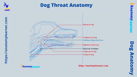
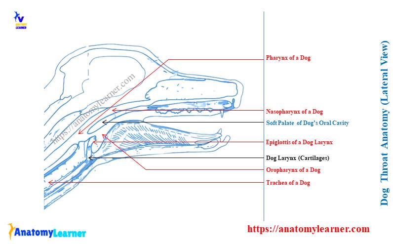
Dog throat and mouth anatomy labeled diagram
Now, I will show you the dog’s throat and mouth anatomy with the labeled diagram. You may also find the details structures or parts of the dog mouth cavity in this article.
In the labeled diagram, I tried to show you the pharynx, different cartilages of the larynx, and the relationship between the trachea and esophagus. Again, the diagram also shows the hyoid apparatus from the dog’s mouth cavity.
The diagram shows the different segments of the pharynx – oropharynx, nasopharynx, and laryngopharynx. The starting point of the esophagus and trachea are also identified in the labeled diagram.
Here, the diagram also shows the five different cartilages from the dog larynx with their different processes. The thyroid gland from the dog’s neck is also identified in the diagram.
Again, the diagram shows the C-shaped tracheal ring throughout the trachea.
The courses of the dog’s esophagus are shown in the labeled diagram. Different lymph nodes in the head and neck regions of the dog also identified in the labeled diagrams.
Common inquiries in the dog throat anatomy
In this section, I will enlist some common questions on the dog throat anatomy and its problems. You will learn some of the extra information on the anatomical features of the dog’s throat as well as the common injuries and others.
Let’s see what the most common questions related to the dog’s throat (pharynx and larynx) –
Do dogs have lumps in their throat?
The lumps in the dog’s throat are not common. But, sometimes, your veterinary doctor may find different types of lumps in the throat of your dog.
But, you may find different types of growth in the oral cavity of a dog which may be in two forms. These are very difficult to diagnose without diagnostic tests like the microscopic evaluation and imaging technique.
You know the lumps are abnormal growth in the dog mouth and also in the throat region (rarely occur). Different types of irritation and infection on the mouth and throat will cause swelling and redness.
Thus these conditions may lead the lumps in the dog’s throat or mouth cavity.
What does it mean when a dog keeps swallowing?
The dog keeps swallowing due to different problems like esophagitis, dental problems, laryngeal paralysis, and oropharyngeal dysphagia. So, to diagnose the right problem of the dog, you might care about the other different symptoms along with swallowing.
Suppose your dog suffers from oropharyngeal dysphagia, then it will show repeated swallowing attempts. Along with this, it will also show the difficulty in prehending food, dropping food from the mouth, and exaggerated head movement while eating.
Again, esophagitis, pharyngitis, and even dental problems cause the dogs to swallow.
What does a dog’s throat look like?
The exact appearance of the dog’s throat can not express in the word. You may see the labeled diagrams of the dog’s throat that I provided previously.
In the dog’s throat, you will find the two main components – larynx and pharynx which serve important functions in the digestive and respiratory systems. Again, it is a relatively unprotected area caudal to the dog mouth cavity proper, where different types of trauma may occur.
How do I know if something (objects) is wrong with my dog’s throat?
Your dog will show different typical symptoms if there is something wrong with its throat. Mainly the problems occur in the pharynx and different parts of the dog’s larynx (laryngeal cartilage).
Again, you may also find stenosis, blockage, or obstruction in the esophagus and trachea of the dogs. The clinical symptoms of the different problems in the dog’s throat are specific.
In pharyngitis, your dog will show severe pain with fever and discomfort. Again, the pharyngeal mucoceles cause pharyngeal airway obstruction and cause dyspnea and snoring.
The dog shows a harsh, dry cough and unpleasant voice in laryngitis. Again, in the laryngeal paralysis, your dog will show obstruction in the airways.
The partial obstruction of the rima glottis may occur in the dog’s epiglottis entrapment. But, hopefully, this occurrence is rare in dogs.
The dog also may show severe gagging, sneezing, and intermittent head movement during eating or other activities. So, the symptoms will be the problem specifically in the dog. You may learn more about the different problems of the dog from here.
What could be stuck in my dog’s throat?
In the young dog, their throat could be stuck by sharp objects, pieces of bone, toys, and other indigestible feed particles. All these agents may cause trauma to the dog’s throat (also known as the traumatic injury to the throat).
When the drooling of blood mixed mucosa with increased swallowing find in the dogs, there may be stuck in the pharynx or mouth cavity. You may also find emphysema and pain in the specific area of the throat and mouth cavity of the dogs.
Again, some foreign objects may cause penetrating injuries in the pharynx. Then you will find dysphagia, drooling, depression, and severe oral pain in your dog.
The stick or pieces of bone also causes damage to the epiglottis and cuneiform process of the arytenoid cartilage of the dog larynx. Thus, it leads the traumatic injuries to the dog’s larynx.
What does the epiglottis do in dogs?
The epiglottis is the spade-shaped rostral cartilage in the dog’s larynx. You will find the two surfaces (laryngeal and lingual), a pointed apex, and a base (stalk) in the structure of a dog’s epiglottis.
The laryngeal surface is concave, whereas the lingual or oral surface is convex in the dog. Here, the lingual surface of the dog’s epiglottis attaches by a short, stout hypoepiglotticus muscle to the middle of the hyoid bone.
So, the epiglottis covers the larynx or windpipe of the dogs. It prevents food (or others) from entering the trachea during swallowing.
The epiglottis of the dog is also known as the laryngeal flap. You will see the laryngeal muscles that attach to the epiglottis also help to close the opening of the windpipe during swallowing.
What is the flap is a dog’s throat called?
The epiglottis of the dog throat or larynx is also known as the flap. You have already seen that the flap of the dogs throat is a spade-shaped structure.
This flap has a different attachment with the hyoid apparatus, thyroid cartilage, and cuneiform process of the arytenoid cartilage. The dog’s flap or epiglottis prevents aspiration of the food or water during swallowing.
Conclusion
So, the main components of the dog throat anatomy are the pharynx, larynx, and trachea. Again, the starting part of the esophagus is also included under the structure of the dog’s throat. There are two important lateral and medial retropharyngeal lymph nodes in the dog’s throat.
The labeled diagrams show all the structures from the dog’s throat with little information. But, you may know more about these structures from a different dedicated article on these specific structures or organs. I hope all the diagrams and little information might help you to get the basics of dog throat.