The dog femur is the massive long bone in the skeleton. It articulates with the os coxae proximally to form the hip joint. Again, the dog femur bone articulates with the tibia distally and forms the stifle joint.
But, it is comparatively shorter than the tibia and humerus bones. As it is a long bone from the dog skeleton, you will find all the typical osteological features.
This article will help to learn the dog femur bone anatomy with the labeled diagram. Again, I will share the anatomical facts of these muscles, nerves, and vessels attached to this bone.
As a veterinary practitioner, sometimes, you may handle dog femur fractures. So, I will also share a little information on the dog’s femur fracture surgery and recovery, along with other facts.
So, if you want to learn the details of the anatomical facts of the dog’s femur, you may continue (read) this article till the end.
Dog femur bone
You will see the right and left femurs bone in the hind limb of a dog. These two femurs lie in the parallel sagittal plane in the standing condition. Now, let’s see the direction of the dog’s femur bone which is very important to identify from externally with the surface approach.
This bone is directed downward and forward in an oblique manner. The inclination on the horizontal plane is about 70 – 80 degrees.
As I told you before, this femur bone joins with the os coxae (hip) proximally and forms the hip joint. This hip joint is another important structure in the hind leg of a dog. Again, the femur bone joins with the tibia, fibular, and patella distally to form the stifle joint in the hind leg.
The dog femur bone present –
- A long cylindrical body, and
- Two extremities (proximal and distal),
The long cylindrical body of the femur bone possesses 4 surfaces (cranial, caudal, medial, and lateral) and 2 borders (medial and lateral). But, the surfaces of the dog’s femur are continuous and convex side to side.
So, performing a cross-section of the dog’s femur bone will show a round appearance. Thus, this bone of the dog leg is also known as the round bone.
Again, the proximal and distal extremity of the canine femur possesses different important osteological features. You will see the head, neck, and greater trochanter from the proximal extremity of the femur.
Again, the distal extremity of the canine femur shows a trochlea and condyle. I will discuss all these features from this bone with the labeled diagram.
But now, let’s see the most essential and unique osteological features of the dog femur bone that you might identify at your laboratory.
Dog femur identification
Following are the osteological features that you might identify from a canine dog’s femur bone –
- Cylindrical body with four different surfaces and borders,
- Proximal and distal extremities of the femur,
- Head (medially), neck, and greater trochanter (laterally) from the proximal end,
- Fovea capitis femoris (small cavity) on the head of the femur,
- Trochanteric fossa dn trochanteric ridge (at caudal surface),
- Lesser trochanter at the medial border (proximally),
- Supracondyloid fossa (on the lateral border; but indistinct),
- Lateral supracondyloid tuberosity (at the lateral border),
- Trochlea from the distal extremity (cranially),
- Lateral and medial condyles (caudally) with their epicondyles, and
- Intercondyloid fossa (caudally between the lateral and medial epicondyles),
I hope these osteological features might provide the basics of a canine femur bone. Now, you may describe all these osteological features from the femur in detail.
Unique features of dog femur anatomy
Here, I will enlist only the osteological features from the canine femur. But, you will learn the details of muscles, vessels, and nerves related to the dog femur bone anatomy.
Okay, let’s see what the unique osteological features presented by the canine femur –
- The bone is less massive and larger compared to the other animal’s femur (let’s see the anatomical facts of the ruminant femur bone),
- You will see the greater trochanter at the lower level than that of the femur’s head (but it remains in other species at the hight level of the head),
- The proximal part of the medial border possesses a small tuberculous type lesser trochanter,
- You will not find any third trochanter at the proximal end of the canine femur (this feature is only well developed in horse femur),
- The trochanteric fossa on the proximal extremity is moderately deep, and the trochanteric ridge (caudally) is oblique (but verticle in horse femur),
- You will find a small, almost indistinct fovea capitis femoris on the head of the femur bone (but larger and distinct in both ruminant and horse),
- There are medial and lateral supracondyloid tuberosities at the distal extremity (caudally), but no distinct supracondylar fossa is present,
- You will find lateral and medial condyles (caudally visible) and a trochlea (cranially) in the distal extremity of the canine femur bone,
- Above the medial and lateral condyle (caudally) possesses facet for the sesamoid bone (also known as the fabella),
- There are lateral and medial (prominent) epicondyles that serve for the proximal attachment of the lateral and medial collateral ligaments of the dog stifle joint,
So, these are the unique features of the canine femur bone. Other anatomical features from muscles, nerves, and vessels will be discussed later in this article.
A proximal end of dog femur anatomy
So, you know the important osteological features from the proximal end of the dog femur bone anatomy. Here, I will discuss the anatomical facts of the head, neck, greater trochanter, trochanteric fossa, and the trochanteric ridge from the proximal extremity of the canine femur.
Let’s find the head of the canine femur bone. It is smooth and nearly hemispherical in structure. But, in ruminant, you will find the almost rounded smooth head in the medial aspect of their femur bone.
Here, the femur head of a dog locates the dorsomedial aspect of the bone. This head joins with the constricted neck portion on its dorsocaudal and medial aspects.
The most important feature of the femoral head is the presence of a small circular pit or cavity. This is the fovea capitis femoris that is nearly indistinct compared to the ruminant or horse. You will find more distinct fovea capitis femoris in the head of the horse femur bone.
Sometimes in some dogs, you may find a depressed, moderately rough, nonarticular strip in the fovea capitis femoris. This nonarticular strip extends from the fovea capitis femoris to the nearest caudoventral nonarticular margin of the head.
This fovea capitis femoris of the canine femur serves a great role in the structure of a hip joint. It serves for the attachment of the ligament of the head of the dog’s femur bone.
The anatomical name of this ligament is ligamentum capitis ossis femoris. Many authors also called this ligament the round ligament of the femur bone.
The neck is the constricted part that is about long as the diameter of the head. It is slightly compressed craniocaudally and is reinforced by a bone ridge extending from the head to the greater trochanter.
Trochanter of the dog’s femur bone
You will see two trochanters at the proximal extremity of the canine femur bone – the greater and lesser trochanters. The greater trochanter is the larger eminence of the femur bone on its proximal extremity.
You will see the larger greater trochanter directly lateral to the head and neck of the canine femur bone. It possesses a free and pyramidal-shaped apex that extends nearly to the dorsal plane on its head.
At the caudal aspect of the greater trochanter, there is an elongated bony elevation known as the trochanteric ridge. You will see a deep fossa between the greater trochanter and the head of the femur (neck). This is the trochanteric fossa of the canine femur bone.
You will find differences in the direction of the trochanteric ridge of dogs and other animals like a horse. Here, the direction of the trochanteric ridge of the dog is oblique. But, in a horse, you will find an almost vertically directed greater trochanteric ridge.
Three major muscle of the hind limb – gluteus medius, gluteus profundus, and piriformis insert on the greater trochanter of the dog femur. Again, the gemeli, obturatorius externus and internus muscles also insert on the trochanteric fossa of the canine femur.
At the caudomedial surface of the proximal extremity of the dog femur (near the junction of the body with the head), you will find the lesser trochanter. It is also a slightly distinct, pyramidal–shaped eminence on the caudomedial aspect of the dog femur.
This lesser trochanter is also connected with the greater trochanter by a wide arciform crest. The name of this arciform crest is an intertrochanteric crest of the femur.
You will see the insertion of the quadratus femoris muscle on the intertrochanteric crest of the femur bone. Again, another important muscle – the iliopsoas, also inserts on the lesser trochanter of the canine femur.
The distal extremity of the dog femur bone
The distal end of the dog femur is expanded and becomes triangular. You will find three important osteological features in the distal extremity of a canine femur bone.
These three important features are – two condyles and one trochlea. Again, these three features possess the three articular surfaces to form the stifle joint with the tibia, fibula, and patella bones.
- Lateral and medial condyles – posses epicondyle,
- Cranially trochlea – posses an articular groove,
You know condyles are the knuckle-shaped paired articular surface on any bone or bony prominence. Again, trochlea is the pully-like grooved articular surface on the bone or bony eminence.
Let’s see the anatomical facts of the lateral and medial condyles and epicondyles with the labeled diagram from the canine femur bone.
Condyles of the dog femur
The lateral condyle of the dog femur is larger than the medial one. Again, the lateral condyle is convex in both sagittal and transverse sections.
But, the medial condyle is smaller and less convex in both sagittal and transverse sections. Each of these medial condyles possesses the articular surface that articulates medially directly with the proximal part of the tibia bone.
Again, a small portion of each condylar surface articulate with the menisci of the tibia bone. If you compare the articular surface between the medial and lateral condyle, the lateral one possesses the larger surface.
You will also find the extensor fossa at the epicondylar border of the lateral condyle. The extensor ddigitorum longus muscles arise from the extensor fossa. Again, the popliteus muscle also arises from the lateral condyle of the dog femur.
There are two sesamoid bones at the distal extremity of the canine femur bone. They are located on the caudodorsal surface of the respective condyles of the femur.
These small sesamoid bones on the distal extremity of the femur are also known as the fabellae. They are associated with the lateral and medial heads of the gastrocnemius muscle.
Both lateral and medial condyles of the dog femur possess a small projection adjacent to the respective condyles. These are the lateral and medial epicondyle of the dog femur.
These two epicondyles of the canine femur serve for the proximal attachment of the medial and lateral collateral ligament of the dog’s stifle joint.
The lateral and medial condyles of the canine femur are separated by the intercondylar fossa. Here, the caudal part of the intercondylar fossa locates further laterally than the cranial part. Thus, the direction of this intercondylar fossa of the dog’s femur is oblique.
Trochlea of the dog femur
You will find the trochlea on the cranial aspect of the distal part of the canine femur. This is the femoral trochlea which is smooth and possesses a wide articular surface.
Again, this articular surface of the femoral trochlea continues with the articular surface of the condyles. You will see the medial and lateral ridges on the femoral trochlea. Again, the medial ridge is thicker and more prominent than the lateral one.
The femoral trochlea is also known as the patellar surface. Now, the caudomedial surface of the patella attaches to the trochlear surface of the femur bone.
Body of dog femur bone anatomy
Now, let’s discuss the anatomical facts (osteological features) of the body from a dog femur bone. You know, the body of a dog femur is cylindrical and straight proximally and arched distally.
Anatomically, there are four different surfaces in the body of a dog femur – cranial, caudal, lateral, and medial. But, all these surfaces are rounded side by side, and thus it is complicated to identify these surfaces separately.
But, you may identify the caudal surface of the dog femur so easily as it is flat. The lateral surface of the canine femur is smooth and possesses a small bony eminence. Some of the authors thought that this was the third trochanter of the dog’s femur.
But, some authors stated that there is no third trochanter in the canine femur bone. You will find a well-defined third trochanter on the lateral surface of the horse femur bone. Again, the ruminant femur bone also lacks this third trochanter.
So, you may find a small third trochanter at the lateral surface of the canine femur near the lesser trochanter. The gluteus superficialis muscle of the dog hind limb insert near the lesser trochanter (third trochanter).
The lateral and medial surfaces possess a slightly roughen line, known as the lateral and medial lips. These lips run proximally into the greater and lesser trochanter of the canine femur. Again, these lips run distally but become obscure in the medial and lateral epicondyle.
You will see the proximal cranial nutrient foramen in the canine femur bone. On the caudal surface, you will find the finely rough area, which is wide at the middle part of the body and become narrow in two extremities.
The distal part of the lateral and medial surface possesses two tuberosities.
Muscles attach to the femur body
I already mentioned the insertion of the superficial gluteus muscle on the lesser trochanter of the canine femur. Now, let’s see the other hind limb muscles attached to the different parts of the femur bone.
The caudal surface of the canine femur is covered by the quadriceps femoris muscle. Most of the segments of the quadriceps femoris muscle (vastus lateralis, medialis) arise from the proximal part of the body of canine femur bone.
The adductor muscle inserts on the lateral lip of the femur and also extends up to the lesser trochanter. Again, the Brevis muscle inserts on the whole lateral lip, popliteal surface, and lesser trochanter.
In the medial lip of the dog femur, you will find the insertion of the pectineus and semimembranous muscles.
I hope you know about the lateral and medial tuberosities (supracondylar) of the femur bone at their distal caudal aspect. From these two tuberosities, the gastrocnemius muscle originated; but a small part (both lateral and medial) also arises from the two sesamoid bones.
In addition, the flexor digitroum superficialis muscle also arises from the lateral supracondylar tuberosity of the dog’s femur bone.
How to identify right and left dog femur
This is very important for first-year veterinary students learning the dog skeleton anatomy. They need to identify the right and left bones (especially the hind limb and fore limb) from the canine skeleton.
Now, I will show you how you will confirm the right and left femur bones from the dogs. You may follow the below-mentioned method to identify the right and left canine femur bone –
- First, consider the landmark from the proximal extremity,
- Let’s consider the special features of the body (from any surface), and
- Finally, consider the landmark or any special features from the distal extremity,
But, you might have a good piece of knowledge on the anatomical facts of the canine femur (general anatomy) to identify the right and left ones. Again, you might know the direction of the different bones of the hind limb of dogs.
This will work in two ways – you will identify and confirm the direction of the femur bone compared to the other bones of the hind limb. And again, you will identify the proximal extremity and distal extremity of the femur bone.
Now, let’s see the bones from the dog’s hind leg (limb) with their directions.
Dog leg bone anatomy
As you will identify the femur bone from the hind limb of the dogs, let’s see and identify the direction of the following bones –
- Hip or os coxae bone – consist of ilium, ischium, and pubis bone; they remain parallel to the long axis of the body,
- Canine femur – massive bone that directs cranially and ventrally (downward and forward),
- Tibia and fibular of the dog – longer bone in hind limb and directed caudally and ventrally (downward and backward),
- Tarsal and metatarsal bones – directed almost vertically,
Again, you should know the joint formed by these bones of the hind leg of the dogs. So, here, you will find the hip, stifle, hock, fetlock, pastern, and coffin joints. All these joints are identified and described in the below-mentioned article.
- Animal joint anatomy with the labeled diagram,
Let’s see the femur bone from the labeled diagram. The proximal part of the femur bone shows externally or lateral bony prominence. You know this is the greater trochanter which may be considered the landmark from the proximal extremity of the femur bone.
Again, the lateral surface of the body is almost smooth and possesses a few finely lateral lips. You may avoid the lateral surface as there are no special features on the lateral surface.
Now, let’s see the distal extremity; there are many important features that you may consider the landmark. The trochlea locates cranially, and you may consider it a landmark.
So, the landmark for identifying the dog femur –
- Proximal extremity – greater trochanter (will face laterally),
- Distal extremity – trochlea (will face cranially),
You should hold the bone according to the direction. Now, try with the following bone to identify the right or left femur from the canine hind limb.
The provided bone possesses the greater trochanter on my right lateral aspect and the trochlea cranially (not shown). So, this is the right femur from the dog’s right hind limb.
Dog femur bone muscles anatomy
Different cranial, lateral, and caudal muscle attaches (originate or inserted) are in the dog femur bone anatomy. But, I will show (identification) you some of the muscles that directly attach to the proximal and distal extremities of the dog’s femur.
Reading all the muscles from the dog’s hind limb with their origin and insertion is recommended. For that, you may read the below-mentioned article where I already described all these muscles from the hind limb of dogs with the labeled diagram –
- Dog leg muscle anatomy with the labeled diagram,
Let’s see the list of muscles from the proximal extremity, body, and distal extremity of the canine femur –
In the proximal extremity of a canine femur, show the following muscles attachment –
- Middle, deep, and superficial gluteus muscles of the dog,
- Gemeli, obturatoris externus and internus muscles of the dog,
- Articularis coxae muscle,
- Quadratus femoris muscle (different heads; especially vastus lateralis and medialis),
- Iliopsoas and adductor longus msucles of the dog,
Again, in the body of a canine femur, you will find the following muscle attachment –
- Vastus lateralis and intermedius muscles of the dog,
- Adductor Brevis muscle of the dog, and
- Pectineus muscle,
In addition, the distal extremity of the canine femur shows the below-mentioned muscles attachment –
- Lateral and medial heads (belly) of the gastrocnemius muscle,
- Semimembranosus muscle of the dog,
- Flexor digitroum superficialis muscle,
- Extensor digitorum longus muscle, and
- Popliteus and articularis genus muscles,
Now, I will describe (show) the anatomical features of some of the essential muscles from the dog’s femur bone.
Gluteus muscles that attach to the canine femur
There are three major gluteus muscles that are attached to the dog’s femur bone. The canine gluteus superficialis muscle is the most superficial of the gluteal muscle. It is the almost flat, small, and triangular muscle in the dog’s thigh region.
This gluteus surpficialis muscle extends between the sacrum and first caudal vertebra proximally and the greater trochanter distally. It arises from the gluteal fascia, deep facia, and sacral tuber.
This muscle runs over the greater trochanter of the femur and inserts on the small ridge on this trochanter. This muscle covers the part of the gluteus medius and piriformis muscles.
A gluteus medius is a more significant muscle that lies on the gluteal surface of the ilium bone. It arises from the iliac crest and the sacral tuber of the dog’s hip. Some of the gluteus medius muscle fibres also arise from the dorsal part of the sacroiliac ligament and deep gluteal fascia.
The gluteus medius extends over the gluteus profundus and forms a short and thick tendon. This tendon of the dog’s gluteus medius muscle inserts on the free end of the greater trochanter.
The gluteus profundus is the broad, fan-shaped deepest muscle covered by the gluteus medius and piriformis. It arises (origin) from the lateral surface of the dog’s ilium body.
This gluteus muscle of the dog also forms the short and thick tendon that inserts on the cranial part of the greater trochanter.
All these gluteus muscles of the dog will help in the extension of the hip joint. In addition, the gluteus profundus help with the abduction of the pelvic limb of a dog. Thus, they provide medial rotation of the hip joint and prevent the lateral rotation on weight bearing.
Piriformis, gemeli, and obturator muscles
The piriformis arises on the lateral surface of the third sacral and first caudal vertebrae. It lies just caudal and medial to the gluteus medius muscle. Again, this muscle of the dog is entirely covered by the gluteus superficialis.
The tendon of the piriformis muscle joins with the tendon of the gluteus medius and inserts into the greater trochanter of the dog’s femur. This muscle also acts with the gluteus muscle to extend the hip joint.
The dog’s Gemelli muscle lies between the terminal part of the obturator externus and internus. Again, this muscle also lies caudal to the gluteus profundus muscle.
The dog gemeli muscle arises from the lateral surface of the body of ischium bone. Finally, they form a strong tendon that inserts into the trochanteric fossa of the dog’s femur.
This muscle performs the lateral rotation of the hip joint. Again, the gemeli muscle also prevents the medial rotation on weight bearing.
The dog obturator externus muscle is fan-shaped that arises from the ventral surface of the pubis and ischium. This muscle covers the obturator foramen externally.
The quadratus femoris muscle covers the caudal border of the dog obturator externus muscle. Again, the adductor muscle covers the cranial part of the obturator externus.
A tendon from the dog’s obturator externus muscle inserts on the trochanteric fossa with the tendon of gemeli.
The obturator internus of a dog is a large, fan-shaped muscle that covers the obturator foramen internally. It arises medially from the foramen on the pelvic surface of the pubis and ischium bone.
Like the obturator externus muscle, the tendon of the internus is also inserted into the trochanteric fossa of the dog’s femur.
Dog’s quadratus femoris and quadriceps muscle
The quadratus femoris muscle also attaches to the dog femur bone. It arises (origin) from the ventral surface of the ischium and the lateral angle of the ischial tuberosity.
This muscle reaches the trochanteric fossa and runs between the obturator externus and adductor Brevis. Finally, it forms the thick tendon that inserts on the trochanteric crest of the femur bone.
The dog quadriceps femoris consists of four bellies – cranial, lateral, medial, and intermediate. I will discuss only the lateral and medial bellies of the dog’s quadriceps femoris muscle.
The medial belly (vastus medialis) of the dog’s quadriceps arises from the proximal extremity of the femur. It is laterally compressed and covers the distal part of the cranial belly of the quadriceps femoris.
The tendon of the medial belly insert on the cranial surface of the patella and joins with the bellies of the Sartorius and tensor fascia latae muscles.
Again, the lateral belly of the quadriceps femoris muscle is larger and arises from the craniolateral of the canine femur. You will see a small intermediate belly in the dog’s quadriceps femoris muscle.
This muscle arises with the lateral belly and covers the lateral surface of the proximal femur.
Articularis genus and coxae muscles in the dog
The articualris coxae is a small, spindle-shaped muscle in the hip joint area of a dog. This muscle locates laterally and caudally from the ilium bone. It runs over the capsule of the hip joint and reaches the neck of the canine femur.
Finally, it attaches to the common ridge between the origin of the lateral and medial bellies of the dog’s quadriceps femoris muscle.
As this is a tiny muscle, it contributes little to hip flexion. This muscle also adjusts the position of a hip joint capsule.
In addition, the articualris genus is also a short and small muscle in the stifle joint region of a dog. The tendon of the articualris genus is inserted on the deep surface of the femoropatellar joint capsule.
This muscle runs parallel to the distal attachment of the intermediate belly of the quadriceps femoris muscle. A delicate intermuscular septum separates the articualris genus muscle from the quadriceps femoris.
The main action of the articularis genus muscle is to extend the dog’s stifle joint.
Dog pectenus and adductor in femur bone
The pectineus and adductor are under the group of the adductor muscle in the hind limb of a dog. Here, the pectineus is a spindle-shaped muscle that lies cranial to the gracilis and is covered by the medial fascia of the thigh.
You will see the adductor brevis caudally of this muscle. In addition, the femoral fascia separates this muscle from the cranially lying Sartorius’s muscle.
There are two origins for the dog’s pectineus muscle –
- Fleshy origin on the iliopubic eminence of the hip, and
- Tendinous origin on the prepubic tendon and from the abdominal muscle,
This muscle becomes flattered after passing deep to the Sartorius’s muscle distally. Again, it fills the narrow space between the medial belly of the quadriceps femoris and adductor muscles.
It forms a long thick tendon that inserts along the distal end of the medial lip of the rough caudal surface of the canine femur bone. Again, you will see the common insertion with the cranial belly of the semimembranosus muscle.
The dog adductor is the deep group of muscles just next to the pectineus. You will find two types of adductor muscles in the dog hind leg (thigh) –
- Adductor longus muscle of the dog, and
- Adductor Magnus brevis muscle,
The fleshy part of the pectineus covers the adductor longus partly. It originates from the pubic tubercle and extends laterally cranial to the obturator externus muscle.
Finally, it inserts on the proximal part of the lateral lip of the rough surface of the dog femur. This muscle also covers the insertion of the quadratus femoris muscle.
Again, the adductor Magnus brevis arises from the entire pelvis symphysis and inserts in the whole length of the rough lateral surface of the femur.
Dog semimembranosus muscle
This is one of the essential muscles in the caudal aspect of the dog’s thigh. It is a fleshy muscle that crosses the medial aspect of the semitendinosus.
Again, it passes between the biceps femoris and semitendinosus muscle laterally and adductor and gracilis muscles medially.
The dog’s semimembranosus muscle arises from the ventral surface of the tuber ischia (just caudal and medial to the semitendinosus). This muscle divides into two equal bellies (cranial and caudal) and extends to the medial aspect of the stifle joint.
Here, the cranial belly of the dog’s semimembranosus muscle is thick and joins with the adductor magnus brevis. This part of the cranial belly forms a short and thick tendon that inserts into the medial lip of the dog femur.
The caudal belly of the semimembranosus muscle covers the cranial belly on its lateral aspect. It extends over the medial belly of the gastrocnemius muscle and ends at the collateral ligament of the stifle joint.
The cranial belly of the semimembranosus muscle extends the hip. In contrast, the caudal belly flexes the stifle joint and contributes to hip extension as well.
Sartorius and gracilis muscles of the dog
The dog’s Sartorius is a long, flat muscle that extends from the tuber coxae to the medial surface of the stifle joint. Thus it passes over the thigh (femur bone) and divides into cranial and caudal parts.
The cranial part of the Sartorius muscle arises from the iliac crest and the thoracolumbar fascia. This belly is related to the quadriceps femoris and tensor fascia latae muscle.
The tendon from the cranial part of the Sartorius muscle ends in the medial femoral fascia just proximal to the patella bone.
Again, the caudal part of the Sartorius muscle lies adjacent to the cranial part and arises on the tuber coxae of the dog’s ilium bone. This muscle passes over the medial surface of the medial belly of the quadriceps femoris muscle.
Finally, the tendon of the caudal Sartorius muscle inserts on the cranial border of the tibia bone.
The gracilis is an extensive and broad muscle that find in the superficial layer of the caudal part of the dog’s thigh. Aponeurosis comes from this gracilis muscle that covers the medial surface of the adductor magnus.
The dog’s garcilis muscle arises from the pelvic symphysis and passes over the medial surface of the adductor magnus brevis. It also covers both bellies of the semimembranosus muscle.
Finally, the tendon from the dog’s gracilis muscle inserts on the entire length of the cranial border of the tibia bone.
The main action of the gracilis muscle is to adduct the hind limb. Again, it extends the hip joint and flexes the stifle joint of the dog.
Gastronemius and dog femur bone
The gastrocnemius is the important and one of the largest muscles in the dog hind leg. It divides into two distinct heads – medial and lateral.
The lateral head (belly) of the gastrocnemius muscle arises from the large tendon on the lateral supracondylar tuberosity of the dog femur bone. Again, a small part of the lateral head also originated from the sesamoid bone that is located dorsocaudal aspect of the lateral condyle.
Again, the medial head of the dog’s gastrocnemius muscle arises from the medial supracondylar tuberosity and the medial sesamoid bone. The two heads (bellies) of the gastrocnemius muscle enclose the flexor digitorum superficialis muscle.
Now, the two head fuses to form a flat muscle in the leg region of the dog’s hindlimb. You will see the biceps femoris muscle on the proximal extremity of the gastrocnemius muscle. Again, this muscle is medially related to the semitendinosus, semimembranosus, and gracilis muscles.
This muscle helps to form the common calcaneus tendon along with the tendons of semitendinosus and semimembranosus muscles. The primary function of the dog’s gastrocnemius muscle is to extend the tarsal joint and slightly flex the stifle joint.
Flexor digitorum superficialis muscle of a dog
It is a flexor group of muscles that have a direct connection with the distal part of the dog’s femur bone. This muscle is greatly enclosed by the gastrocnemius muscle of the dog.
The flexor digitorum superficies muscle of the dog became wider and flattened distally, but the proximal section is narrow. Again, the proximal part of this muscle is firmly attached to the gastrocnemius muscle.
It arises from the lateral supracondylar tuberosity along with the lateral head of the gastrocnemius muscle. In the middle of the tibia bone, it winds around the tendon of the gastrocnemius muscle and reaches the caudal surface of this bone.
The tendon from the flexor digitorum superificlais muscle ends on the plantar surface of the cancaleus tuber. Do you know the main function of the dog’s flexor digitorum superficial muscle?
The main action of the dog’s flexor digitorum superficial muscle is to flex the digits. Again, it also provides a little extension and fixation of the tarsus and flexion of the stifle joint.
Dog’s extensor digitorum longus and femur
This is a spindle-shaped muscle on the extensor group that lies on the cranial surface of the tibia bone. You may quickly identify this muscle between the tibialis cranialis and fibularis longus muscle.
Again, the proximal extremity of this muscle is covered by the tibialis cranialis muscle.
The extensor digitorum longus (dog) muscle arises from the extensor fossa on the lateral aspect of the articular surface of the lateral condyle. Then this muscle passes through the sulcus extensorius of the dog’s tibia.
The tendon of the extensor digitorum longus of a dog divides into four branches near the tarsus. This tendon extends along the dorsal surface of the metatarsus bone and digits II, III, IV, and V.
The main action of the extensor digitorum longus muscle is to extend the digits and also contribute to the flexion of the tarsus bone.
Dog popliteus muscle
This is a small and triangular muscle that also has a direct connection with the dog’s femur. This muscle lies in the space on the proximocaudal surface of the tibia bone, just distal to the popliteal notch.
The popliteus muscle of a dog covers the caudal aspect of the stifle joint capsule. It also covers the medial half of the proximal third of the dog’s tibia bone.
You will find the gastrocnemius and flexor digitorum superficialis muscle on the caudal aspect of the popliteus muscle. Again, you will see a relationship with the fascia of the semitendinosus muscle on the medial aspect.
Now, let’s see the origin of the dog popliteus muscles –
This muscle arises from the caudal aspect of the articular surface of the lateral condyle of the femur bone. Again, a small part of the popliteus muscle also arises from the lateral sesamoid bone that locates above the dorsal surface of the lateral condyle.
You will see an attachment of the tendon of the popliteus with the capsule of the stifle joint. It crosses deep to the lateral collateral ligament of the stifle joint of the dog.
The tendon of the dog’s popliteus muscle will insert into the medial aspect of the tibia bone. These popliteus muscles contribute to flexing the dog’s stifle joint.
Vessels in dog femur bone anatomy
The branches of external and internal iliac arteries mainly supply different muscles that are attached to the dog femur bone. A femoral artery that comes from the external iliac is considered the major vessel in the dog thigh.
At the same time, you will also find the femoral vein in the thigh region (femur) of a dog. I will not describe all the branches and courses of the external and internal iliac arteries that supply the different muscles and structures of the dog’s hind limb.
Instead, I prefer to enlist and describe the significant vessels (arteries and veins) that are only passed along this femur bone or thigh. But, it will be better to read the full branches and courses of different arteries that come from both the external and internal iliac arteries.
So, first, let’s see the dog hind leg artery labeled diagram and try to identify these below-mentioned arteries (also veins) from the actual samples –
- Superficial circumflex iliac artery and vein of the dog leg,
- Lateral cicumflex iliac artery,
- Proximal caudal femoral artery and vein in a dog,
- Femoral artery and vein in the dog thigh,
- Middle caudal femoral artery and vein in the distal part of the dog thigh,
- Descending genicular artery and vein in the distal extremity of the dog femur,
- Saphenous artery and vein in the distal portion of the dog thigh,
Again, you will also find the following arteries and veins at the hip and thigh regions of a dog –
- Medial circumflex femoral artery and vein in the dog hip and thigh regions,
- Ascending and transverse branches of the medial circumflex,
- Deep branch of the (identified) medial circumflex femoral artery and vein, and
- The popliteal artery at the caudal aspect of the stifle joint,
Dog femoral artery anatomy
The external iliac artery is the largest peripheral branch of the dog’s abdominal aorta. This vessel runs caudoventrally and is related to the common iliac vein, psoas minor, and iliopsoas muscle.
The external iliac artery of the dog continues as the femoral artery and gives off different branches in the different regions of the hind leg. But, I will discuss only the different branches of the dog’s femoral artery in the thigh (femur) region.
You will also find a deep femoral artery that arises from the caudomedial surface of the external iliac artery. It runs obliquely over the medial surface of the external vein and leaves the abdominal cavity. Then it passes through the caudal part of the vascular lacuna.
Again, you will find other small branches like the pudendopeigastric, caudal epigastric, external pudendal, medial circumflex femoral artery, and obturator branches.
Let’s know a little about the medial circumflex femoral artery from the dog.
Medial circumflex artery of the dog
The medial circumflex femoral artery of the dog is a continuation of the deep femoral artery behind the vascular lacuna. This vessel obliquely passes the iliopsoas muscle and medial belly of the quadriceps femoris.
The medial circumflex femoral artery provides small branches into the deep part of the adductor muscle in the thigh. You will find a nutrient artery at the caudal surface of the greater trochanter of the dog’s femur.
The medial circumflex artery removes the obturator, deep, ascending, transverse, and acetabular arteries.
The obturator branch of the medial circumflex artery courses dorsally through the cranial part of the obturator foramen and lateral to the obturator nerve. This artery supplies to the levator ani, coccygeus, internus, and externus obturator muscles.
Again, the ascending branch of the dog’s medial circumflex femoral artery supplies the proximal part of the adductor muscle. Small branches also supply to the semimembranosus, quadriceps femoris, pectineus, and obturator externus muscles.
You will find a small acetabular branch of the dog’s medial circumflex femoral artery that extends laterally to the trochanteric fossa. This vessel supplies to the caudal part of the hip joint capsule and the neck of the dog femur.
Finally, the transverse branch of the medial circumflex femoral enters into the semimembranosus muscle and anastomoses with the caudal gluteal artery. You also fina an extra deep branch of the medial circumflex femoral artery near the femur bone that anastomoses with the lateral circumflex femoral artery.
Branches of dog femoral artery
As you know, the dog femoral artery is the continuation of the external iliac that start from the vascular lacuna. These vessels pass along the length of the thigh (femur bone) of the dog and give off different branches.
It continues caudal to the stifle joint and continues as the popliteal artery. Here, the femoral artery covers by the thin skin and deep medial femoral fascia.
At the popliteal surface of the dog’s femur, the femoral artery continues between the lateral and medial heads of the gastrocnemius muscle. The diagram already shows the different main branches of the dog’s femoral artery.
Now, let’s discuss the anatomical facts of these femoral branches in the thigh region of a dog –
- Superficial circumflex iliac,
- The lateral circumflex femoral artery,
- Muscular branches of femoral artery,
- Saphenous artery,
- Descending genicular,
- Middle caudal femoral, and
- Distal caudal femoral artery,
Superficial and lateral circumflex femoral artery
The superficial circumflex femoral artery is the small branch that runs dorsocranially over the medial surface of the cranial belly of the quadriceps femoris muscle. This vessel arises from the lateral surface of the femoral artery close to the lateral circumflex femoral artery.
The superficial circumflex femoral artery is mainly supplied to the tensor fascia latae muscle. Again, this muscle is also supplied to the Sartorius and cranial belly of the quadriceps muscles.
Again, the lateral circumflex femoral artery (cranial) comes from the caudolateral surface of the femoral artery. It immediately disappears between the cranial and medial heads of the quadriceps femoris muscle.
Different smaller branches form the lateral circumflex femoral artery supplies to the iliopsoas, cranial belly of quadriceps, and tensor fascia latae muscles. Three minor branches are found in the dogs’ lateral circumflex femoral artery – ascending, transverse, and descending.
Here, the ascending branch of this artery sends small branches to the cranial part of the stifle joint capsule. A transverse branch passes lateral to the head of the quadriceps femoris muscle. Finally, the descending branch of this muscle bends distolaterally around the caudal surface of the cranial belly of the quadriceps femoris muscle.
A muscular branch of the femoral artery lies adjacent to the cranial border of the vastus medialis muscle. This vessel terminates into the pectineus and also in the Sartorius’s muscles.
Proximal, middle, and distal caudal femoral artery
The proximal caudal femoral artery also arises from the caudal aspect of this main artery. It crosses the pectineus and adductor muscle and supplies them. It will also enter the cranial border of the gracilis muscle of the thigh region of a dog.
The middle caudal femoral artery arises at the level of the semimembranosus muscle. This muscle runs caudodistally and divides into the adductor and semimembranosus muscles. It will also anastomose with the distal caudal femoral artery of the dog.
Finally, the distal caudal femoral artery arises from the caudolateral aspect of the femoral artery of the dog. This distal caudal femoral artery passes between the two heads of the dog’s gastrocnemius muscle, and it continues as the popliteal artery.
The distal caudal femoral artery sends branches to the biceps femoris, the quadriceps cranial belly, and two gastrocnemius muscle heads.
Dog saphenous artery
The dog saphenous artery arises from the medial surface of the femoral artery. It runs distally across the medial surface of the semimembranosus muscle. Again, this vessel will provide the small branches to the gracilis muscle in the thing region of a dog.
Now, it passes over the medial surface of the stifle joint and sends a single or paired genicular branch to the skin and superficial fascia. Again, this artery divides into small cranial and larger caudal branches at the tibial condyle.
The cranial branch of the dog saphenous artery obliquely crosses the subcutaneous medial surface of the tibia. In addition, the larger caudal branch of the saphenous artery is the direct continuation of the main artery.
You will also find the descending genicular artery that arises from the femoral artery distal to the origin of a saphenous artery. It runs between the vastus medialis and semimembranosus muscles to the medial surface of the dog’s stifle joint.
I will not describe the details of the popliteal artery here as I have a unique article on this. This popliteal artery is the direct continuation of the dog’s femoral artery through the popliteal fossa.
Dog femur bone and nerve anatomy
The branches of the sacral and lumbar (lumbosacral plexus) nerves supply to the different muscles and structures in the dog femur bone area (thigh region). Here, the ischiatic or sciatic is the main nerve that passes along the thigh region of a dog.
You may learn the anatomical facts (features) of the different nerves from the lumbosacral plexus of a dog. For that, I would like to suggest you read the below-mentioned article to get a great idea of the different nerves from the dog’s lumbosacral plexus –
- Dog lumbosacral nerves anatomy with the labeled diagram,
Again, if you want to understand the basic structure of the lumbosacral plexus of any animal, it is recommended to read (know) the following article from start to end –
Nerves in the lumbosacral plexus of a dog with the labeled diagram,
In this article, you will find (get) the basic concept and all the short courses of different nerves from the animal’s lumbosacral plexus.
Here, I will only show you the nerves from the lumbosacral plexus that are associated with the thigh region of a dog. Let’s see what these nerves that are directly associated with the dog’s thigh region are –
- The caudal cutaneous femoral nerve of the dog,
- Pudendal and pelvic nerves,
- Femoral and saphenous nerve,
- A sciatic or ischiatic nerve in the dog’s thigh,
- Common fibular nerve, tibial nerve (origin), and
- Caudal cutaneous sural nerve,
Now, from the diagram, let’s find the distribution of these nerves in the dog thigh region.
Distribution of nerve in dog thigh (femur)
The dog’s lateral cutaneous femoral nerve is primarily formed by the ventral branch of the fourth and fifth lumbar nerves. It runs caudolateral to the psoas minor muscle and sends different small branches into the different muscles near its origin.
Again, this nerve reaches the thigh region and supplies to the lateral surface of the stifle joint.
The dog’s femoral nerve arises from the ventral branches of the fourth and fifth lumbar spinal nerves. This nerve innervates the psoas major and iliopsoas muscles of the dog’s hip.
The femoral nerve of the dog also supplies the four heads of the quadriceps femoris and articularis coxae muscle. Here, the saphenous nerve is the superficial branch of the femoral nerve. You will also find a long, slender cutaneous branch of the dog’s saphenous nerve.
From the dog’s sacral plexus, you will see the sciatic nerves and its branches. The caudal cutaneous femoral nerve (identified) arises from the ventral branches of the first and second sacral spinal nerve. This caudal cutaneous femoral nerve courses caudally on the caudal attachment of the sacrotuberous ligament.
Again, the sciatic or ischiatic nerve is the largest peripheral nerve in the dog’s body. It arises mainly from the lumbar and sacral plexus, which runs along the caudolateral aspect of the dog femur.
The sciatic (ischiatic) nerve divides into common fibular and tibial nerves at the distal third of the canine femur bone. The courses of the fibular and tibial nerve are complicated in the hind leg region and the hind paws.
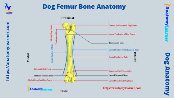
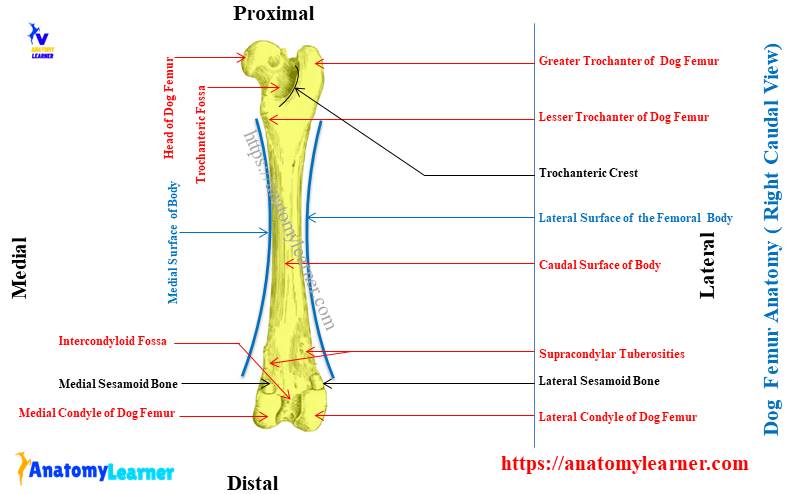
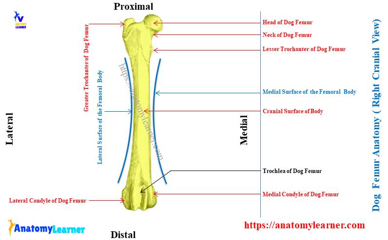
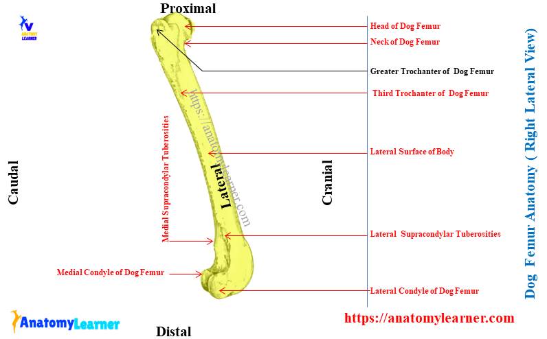
Dog femur bone anatomy diagram
Throughout this article, you already got the different diagrams on the dog femur bone anatomy. But, now, I will also provide some of the other labeled diagrams on the canine femur that might also help you to learn this bone easily.
In the dog femur labeled diagram, I tried to show you the two extremities (proximal and distal) and the cylindrical body. I tried to show you the elongated oval head and constricted neck from the dog’s femur from the proximal extremity.
Again, the diagram shows a less shallow fovea capitis femoris in the middle part of the head. The prominent lateral greater trochanter of the dog’s femur is identified in the labeled diagram.
Here, the dog leg labeled diagram also shows the lesser trochanter and the third trochanter (indistinct, you may also call it the tuberosity). The caudal aspect of the femur bone shows the trochanteric fossa (less shallow than the ruminant).
Here, the oblique-oriented trochanteric crest is also identified in the canine femur labeled diagram. Now, let’s see and identify the other different osteological features from the body and distal extremity of the canine femur bone.
The distal extremity of the canine femur shows the condyle and trochlea in the diagram. Here, the dog’s femur labeled diagram shows the more prominent lateral condyle.
The cranial surface of the trochlea is grooved, which is articulated with the caudomedial surface of the patella. The diagram shows the intercondyloid fossa and the lateral and medial supracondylar tuberosity from its distal extremity.
You will get more dog hind leg or thigh labeled diagrams on social media of anatomy learners.
Horse and pig femur compare to the dog
If you compare the anatomical features of the horse and pig femur with the dog, you will find some differences. A detailed article on the ruminant femur anatomy might help you understand the difference between the dogs and ruminants like cattle and sheep.
You may read this article – animal femur anatomy with the labeled diagram (full osteological features of the femur bone in ruminant),
Again, you may know the significant difference between the femur among the horse, cattle, pigs, and dogs from table 1 –
So, in the horse femur, you will find the below-mentioned osteological features compare to the dogs –
- In the horse, the femur is a more massive bone compared to the dog,
- There is a well-developed third trochanter at the upper part of the lateral border of the horse femur,
- The head of the horse femur is circular and possed a deep fovea capitis with a notch,
- The trochanteric ridge on the proximal extremity is verticle in position, whereas it is oblique in a dog and ruminants,
- In the distal extremity (laterally), you will see a deep supracondylar fossa, whereas there is no supracondylar fossa in the dog’s femur,
Again, in the pig femur, you will find the below-mentioned osteological features (exceptional) –
You will find a more curved head in the proximal extremity of a pig femur. The prominent greater trochanter does not extend above the head. There is no third trochanter on the lateral aspect of the pig femur bone.
The femur of rabbit and goat
The femur of a rabbit is comparatively longer and possesses some special osteological features. Here, you will find a circular head with indistinct fovea capitis in a rabbit’s femur.
Again, the larger lateral greater trochanter possesses an obliquely oriented trochanteric ridge. You will also see the third trochanter at the lateral aspect, just below the proximal extremity of the rabbit’s femur bone.
There is no supracondylar fossa in the femur of a rabbit.
Again, the goat femur possesses all typical osteological features compared to dogs. But, all these osteological features are less developed than the horses.
The proximal and distal extremities of a goat femur show the same osteological features as the cow. But, it is hard to identify the osteological features from the cylindrical body of a goat femur bone.
You will not find any third trochanter at the lateral surface of the goat’s femur bone like the horse or dog. Again, the trochanteric fossa is deep and possesses an obliquely oriented trochanteric crest or ridge in the goat femur bone.
Other inquiries from the dog femur bone anatomy
Here, I will also enlist the most common inquiries on the dog femur bone anatomy with their possible answers. Make sure you learn all the osteological features of the dog’s femur from previous sections.
Let’s see the other common inquiries on the canine femur bone anatomy –
What is a femur on a dog?
The dog femur is the bone of the pelvic limb that articulates with the hip bones (ileum, ischium, and pubis) above and the tibia bone below. Again, the femur of a dog is directed obliquely (cranially and ventrally).
The femur bone forms two major joints from the dog’s pelvic limb – the hip joint (with os coxae) and the stifle joint (below, with tibia bone).
The femur is a long (clyndircal) bone in the dog’s skeleton so that you will find all the typical osteological features. This canine femur consists of a long cylindrical body and two expanded extremities (proximal and distal).
The proximal extremity generally contains the circular (medially located) head, constricted neck, and greater trochanter (laterally). In comparison, the distal extremity of the dog’s femur possesses the cranial trochlea and caudal condyles (lateral and medial).
Can a dog recover from a broken (fracture) femur?
Yes, your dog can recover from a broken femur quickly if proper management should be provided. But, the recovery time may vary according to the intensity of the broken femur.
Normally, in some cases, it takes more than 2 weeks to bear the body weight. Again, finally, it needs 1 or 2 months to get full recovery from the broken femur.
But, the time may differ in various conditions of the dog’s femur. Ensure you provide better management immediately after breaking your dog’s femur bone.
Where is the femur bone in a dog?
The dog femur is located in the hind limb after the hip bone. This is also called the thigh bone in an animal. It locates in the thigh obliquely and goes downward and further forward.
It forms the hip joint with the os coxae bone at the proximal extremity. Again, it creates a stifle joint – (an essential joint in the hind limb) with the tibia and fibula at its distal extremity.
Different important muscles and vessels also pass along this femur bone. I have already described all these structures related to the dog’s femur bone or thigh region.
How long does it take (need) for a broken femur to heal?
Typically, it takes 3 – 5 months to head a broken femur completely. But, this number is relative and depends on various factors.
First, it varies with the fracture or breakage in the femur bone or the breakage area. Second, how quality management is provided to heal this condition.
Third, this healing of the broken femur also depends on another important thing: the intensity of the damage to blood vessels and nerves. Minimum damage in the blood vessels and nerves in the femur region will take less time (typically 2 months).
But, your dog has great damage to blood vessels and nerves along with the long fracture; it will take more than 4 – 5 months.
Can a dog recover from a broken femur without surgery?
It depends on the type of fracture in the femur bone. If there is a minimum fracture in the femur, then only proper management will recover this situation. But, in the case of a complete or partial broken femur, it is recommended to perform the surgery.
Without surgery, a wholly or partially broken femur can not recover perfectly. If your veterinary doctor does not perform any surgery, it may develop poor weight-bearing capacity. And obviously, your dog will show lameness in the future.
Can a dog survive a broken femur?
Yes, your dog can survive a broken femur if you provide proper management. Immediately after breaking the dog’s femur bone, you should bring your dog to a professional veterinarian.
They will provide better management or surgery (if required), and it will usually take 2 – 5 months to recover fully from this condition.
What is thigh in a dog?
Generally, the femur bone is known as the thigh in a dog or other animal. It is a typical long bone in the pelvic limb of a dog that possesses two expanded extremities and a cylindrical body.
The dog femur obliquely passes from the hip joint to the stifle joint. Biceps femoris, semimembranosus, and quadriceps femoris muscles are attached to the femur bone (in the thigh region of a dog). Again, the clinically most important vein – the femoral vein, also passes along the medial aspect of the thigh bone (femur) in a dog.
Conclusion
So, the dog femur bone consists of different clinically significant osteological features in both extremities and the body. Again, the muscles and vessels associated with the dog femur bone anatomy are also equally important.
As a veterinary student, you should also know the distribution of the sciatic nerve along with the length of a dog’s femur. Now, it’s time to practice all the osteological features and other different structures from the actual sample of the dog femur.
Make sure you take advantage of the dog femur labeled diagrams I have provided here in this article. These might help you perfectly identify all the osteological features from a canine bone.