The dog tibia anatomy comprises a long body and two expanded extremities. It is a long and thick bone in the pelvic limb that articulates proximally with the femur and distally with the tarsals.
The proximal part of the dog’s tibia is triangular and more massive than these of the distal extremity. Here, the distal extremity of the dog’s tibia is nearly cylindrical.
You will see a long, thin, and laterally compressed fibula bone that passes along the lateral surface of the tibia bone. In a dog, the fibula bone is wholly separated and possesses a head, neck, body, and two extremities.
In this article, I will show you the details osteological features of the dog tibia anatomy with the labeled diagram. Again, you will also find the anatomical features of the dog fibula bone at the end of this article.
I will also show you the important muscles, vessels, and nerves that pass along the tibia or leg bone of a dog in a short form. So, if you want to learn the anatomical facts about the canine tibia, let’s read this article until the end.
Where is the tibia on a dog?
The tibia is a long bone on the medial aspect of the dog leg (pelvic limb). Proximally, it articulates with the femur bone to form the stifle joint in a dog. Again, the tibia articulates with the tarsus distally and forms the hock joint.
So, this tibia bone of a dog extends obliquely downward and backward from the stifle to the hock joint. The long, thin fibula bone lies on the lateral aspect of the dog leg.
That means the fibula lies along the whole length of the dog’s tibia bone. Now, the dog leg labeled diagram will help you to identify the direction and location of the tibia bone from the pelvic limb.
The diagram shows every bone from the dog pelvic limb (hip, femur, tibia and fibula, tarsal, metatarsal, and phalanges). Now, it is time to know the anatomical details of the tibia bone from the dog pelvic limb.
Dog tibia anatomy
As a long bone, the dog tibia anatomy shows a long body and proximal and distal extremities. In the long body of this dog tibia possesses different surfaces and borders. Again, the proximal and distal extremities include various important osteological features.
Now, I would like to identify the different osteological features of the long body and the two expanded extremities of the dog tibia bone. First, let’s try to identify the following features from the body of a dog tibia bone –
- Lateral, medial, and caudal surfaces of the dog’s tibia bone,
- Cranial, lateral, and medial borders of the dog’s tibia bone,
If you notice the body of the tibia bone, you will see the triangular appearance in the proximal part of the body. So, you will only find these typical features (both surfaces and borders) on the proximal extremity of the body of a dog tibia.
But, the distal end of the tibia’s body is quadrilateral and more massive than the adjacent part of the body. So, you will not find all these three surfaces and borders on the distal extremity of the body of a tibia bone.
Now, let’s try to identify the below-mentioned osteological features from the proximal part of the dog tibia bone –
- Lateral and medial condyles,
- Intercondyloid eminence (spines) on the proximal extremity,
- Cranial intercondylar area,
- Extensor groove (craniolateral) and popliteal notch (caudally),
- Tibial tuberosity,
Finally, from the distal extremity of both dog’s tibia and fibula, you might identify the following –
- Distal articular surfaces for the tarsal bones,
- Lateral and medial malleolus bones,
I hope you can identify all the above-mentioned osteological features from the dog tibia so quickly with the help of the labeled diagram.
Unique features of the dog tibia bone
From the above discussion, you may easily point out the unique osteological features of the dog tibia bone. You will find unique osteological features, especially on the body of a dog tibia bone, compared to the ruminant or the horses.
The below-mentioned article might help you to know the details of anatomical features from the ruminant and other animal’s tibia bone –
Tibia and fibula of an ox – details osteological features of the animal’s leg bone,
Some of the unique osteological features of the dog tibia are enlisted below –
- The long body of the dog’s tibia is convex medially at the proximal part, again, convex laterally at the distal portion,
- You will find a prismatic or triangular-shaped appearance on the proximal part of the body, whereas the distal portion of the body is quadrilateral and cylindrical,
- The tibial crest of the dog’s tibia is more prominent compared to these of the ruminant and horses,
- Caudaolateral aspect of the lateral tuberosity (proximal to the tibia’s body), there is a facet for the head of the fibula bone,
- Again, the cylindrical distal part of the dog’s tibia presents a lateral facet for articulation with the distal portion of the fibula bone,
- Here, the fibula is a wholly separated thin, modified long bone that lies in the lateral aspect of the dog tibia,
You will not find any separated fibula bone in the ruminant or horse’s leg. The divided, thin, and modified long tibia extends along the whole length of the dog’s tibia bone.
The proximal end of the dog’s fibula is flat and articulates with the lateral condyle of the tibia. You will see the interosseous spaces in the whole length of the bone in a dog. But, the proximal interosseous space is larger compared to the distal one.
Dog tibia bone structure – canine leg
In this part, you will learn the details of dog tibia bone anatomy with the labeled diagrams. Here, the features from the proximal and distal portions of the dog tibia are essential as they form the stifle and hock joint, respectively.
Here, I will discuss the osteological feature from the –
A proximal end of dog’s tibia (form a stifle joint with a distal end of the femur bone),
A long body of the dog’s tibia (triangular in proximal and quadrilateral in distal), and
A distal end of the dog’s tibia bone (expanded massive extremity),
Suggested reading for you – you may know the details of the anatomical facts of the dog hock and stifle joint from the below-mentioned articles of anatomy learner –
- Dog hock joint anatomy with the diagram, and
- Details anatomical facts of the dog stifle joint with a labeled diagram (along with their muscle, vessels, and nerves),
That’s fine; now, let’s discuss the anatomical facts from the proximal extremity of a dog tibia bone.
A proximal end of the dog tibia bone
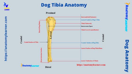
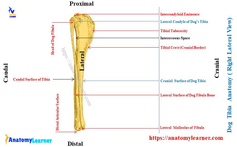
As you see in the diagram, the proximal end of the dog tibia bone is relatively flat and triangular. The most important anatomical features from the proximal end of a dog tibia are – two condyles with their articular surfaces and intercondylar eminence or spines.
These two condyles, with their articular surfaces and the intercondylar eminences of the dog’s tibia, have a significant contribution to the stifle joint. Let’s see the overview from the proximal extremity of the tibia in a minute –
The proximal extremity of a dog tibia – large and three-sided, bears an articular surface with lateral and medial condyles. A popliteal notch separates these lateral and medial condyles; you will see a tubercle in the middle of the popliteal notch. This tubercle on the popliteal notch is designed to attach the caudal cruciate ligament.
Again, the proximal extremity of the dog tibia – bears central prominence, known as the intercondyloid eminence or spines.
Now, let’s known the details of these structures from the proximal extremity of the dog tibia (if you wish).
Condyles from the proximal extremity
The proximal extremity of the canine tibia anatomy labeled diagram shows two distinct condyles – lateral and medial. Both these lateral and medial condyles of the tibia possess the articular surface for the distal end of the femur.
Here, the medial condyle of the proximal extremity of the tibia is oval, whereas the lateral condyle is almost circular. They are convex in the sagittal plane and concave transversely.
You will also find the intercondyloid eminence in between the lateral and medial condyles. This intercondyloid eminence help to attach the ligament of the stifle joint.
The fresh tibial sample from the dog shows articular cartilage that has a small contact with the articular cartilage of the femoral cartilage. Again, you will find two menisci (lateral and medial) that also attach to the articular surfaces of the condyles.
The lateral and medial menisci practically separate the articular surfaces of the tibial condyle from the femoral condyles. These cartilages are fibrocartilage in nature and biconcave.
Both the intercondyloid eminence of the proximal extremity is low and possesses a tubercle in the middle. So, you will find the lateral and medial tubercle in the lateral and medial intercondyloid eminence, respectively.
Here, the medial tubercle on the medial intercondyloid eminence is cranial to the lateral tubercle. You will see an oval, depressed area cranial to the intercondyloid eminence, which is known as the area intercondylar cranialis.
Again, there is a small, depressed area caudal to the intercondyloid eminence, which is known as the area intercondylaris caudalis. If you notice the structure of the canine stifle joint, you will see the meniscal ligaments attach to these areas.
Groove, notch, and tuberosity in the proximal extremity
You will also find groove, notch, and tuberosity in the proximal extremity of the dog tibia bone. The extensor groove is located at the craniolateral aspect of the lateral condyle of the tibia (cuts the lateral articular surface).
Again, you will clearly identify the popliteal notch from the tibia bone from the caudal aspect. This popliteal notch locates between the lateral and medial condyle caudally.
There is an obliquely placed articular surface on the lateral aspect of the lateral condyle of the dog’s tibia bone. This is the articular surface for the head of the dog fibula bone.
Again, the cranial aspect of the canine tibia (proximal extremity) shows important features, and that is the tuberosity. This is the dog tibial tuberosity, which is a large, quadrangular, and proximocranial process from the tibia bone.
Now, the tibial tuberosity extends distally in the cranial border of the tibia and forms the tibial crest. I will again discuss this tibial crest in the description of the dog tibia body.
Now, let’s see some of the leg muscles that insert or originate from the different structures of the proximal part of the dog’s tibia bone –
- Tibial tuberosity – provide insertion for the quadriceps femoris, biceps femoris, and Sartorius’s muscles,
- Tibial crest – provides the insertion for the gracilis, semitendinosus, and also the part of Sartorius and biceps femoris muscles,
- The proximal part of the lateral condyle of the tibia – serves as the origin of tibialis cranialis muscle, and
- The caudal part of the medial condyle of the dog tibia serves to insert the semimembranosus muscle.
You may learn the anatomical details of these leg muscles (origin, insertion, and fibre direction) from another article by an anatomy learner.
Body of dog tibia anatomy
The body of the dog tibia bone shows some peculiar features compare to these other animals. You already know that the body is triangular (three-sided) throughout the proximal part of the bone. In contrast, the portion of the dog tibia bone is essentially quadrilateral or cylindrical.
The body of a dog tibia anatomy shows three surfaces and three borders with some important osteological features. These features from the surfaces and borders are equally important to identify the right and left tibia from the dog skeleton.
You may easily identify the right or left tibia from the dog pelvic limb with the help of some landmark. I will describe how you will identify the right or left tibia bone from a dog skeleton at the last of this article. But, ensure you know the different features from the various surfaces and borders of the dog tibia bone.
Now, let’s discuss the following surfaces and borders from the dog tibia bone –
- The lateral surface of the canine tibia – smooth and concave proximally, and narrow and convex distally,
- Medial surface of the canine tibia – wide and nearly flat proximally,
- Caudal surface of the body of a canine tibia – posses oblique popliteal line,
- The lateral border of the tibial body – form the interosseous spaces with the medial surface of the dog’s fibula bone,
- The medial border of the body of a dog tibia bone – is smooth and has no distinct osteological feature, and
- Cranial border of the body of a canine tibia bone – articulate proximally with the tibial tuberosity and from a tibial crest,
Now, I will discuss other anatomical features from these surfaces and borders of the tibial body.
The lateral surface of a dog tibia
The proximal, middle, and distal part of the lateral surface of a dog tibia shows different features. Here, you will find the smooth, broad, and concave appearance in the proximal lateral surface of the dog tibia bone.
But the middle part of the lateral surface of the dog tibial body is almost flat. In comparison, the distal portion of the lateral surface of the body is narrow and convex.
The biceps femoris muscle of the dog leg covers the lateral surface of the tibia bone. But, this biceps femoris muscle inserts on the medial surface of the cranial border of the tibia bone.
You will find some other muscles in the lateral surface of the dog tibia that arises from it –
The flexor digitorum lateralis muscle of the dog leg – arises from the proximal end of the lateral surface of the dog tibia, and
Fibularis brevis muscle of the dog leg – arises from the lateral surface of the distal two-thirds of the fibula and tibia bone of the dog,
You may learn more about the flexor digitorum lateralis and fibularis brevis muscles from another article by anatomy learner.
Medial surface of the dog tibia
The medial surface of the body is wide and nearly flat in the dog. The medial and lateral surfaces join together to form the cranial border.
Few large but low muscular lines are present on the cranial border of the tibial body. Different muscles of the dog leg insert into these muscular lines of the dog tibia.
You will find the insertion of the gracialis, Sartorius, and semitendinosus muscles on these muscular lines on the tibia. The medial surface is slightly concave at the distal end of the tibia (toward the distal articular surface of the tibia).
Caudal surface of the canine tibia
The caudal surface of the dog tibia bone (body) is flattened but rough. You will see an oblique popliteal line that divides the flattened caudal surface into two parts.
This popliteal line of the canine tibia crosses from the proximal part of the lateral border to the middle of the medial border. You will also find a nutrient foramen on the caudal flattened surface of the dog’s tibia bone.
It locates at the junction of the proximal and middle third of the lateral border of the dog tibia. You will see the different vascular grooves that run obliquely distolaterally across the distal part of the caudal surface of the dog tibia.
There are different muscles that arise or insert on the caudal surface of the dog tibia bone –
Popliteal muscle – inserts on the proximal, middle third of the caudal surface. It will also insert on the proximal part of the medial surface of the tibial body. Finally, a small part of the popliteal muscle will be inserted on the adjacent medial surface of the tibia proximal to the popliteal line.
Tibialis cranialis, flexor digitroum lateralis and medialis muscles – arise from the proximal part of the caudal surface. These muscles of the caudal surface of the tibia bone arrange in a lateral to a medial sequence.
Borders of dog tibia bone anatomy
From the proximal part of the body of a dog tibia, you may easily identify three borders. But the distal part of the body does not show these three borders perfectly.
The cranial border of the tibial body is more important and possesses essential osteological features. Here, the lateral and medial surfaces of the body form the prominent cranial border (cranially).
It forms an elongated elevation on the proximal extremity (towards the tibial tuberosity). This structure of the dog tibia is known as the tibial crest.
You may easily locate the fibular nerve to consider this tibial crest as the landmark (not for anesthesia, only for study purposes). Again, this structure is important when you will identify the right and left tibia from the dog skeleton.
On the tibial crest of the dog’s tibia, you will find rough prominence in its middle. Here, the tendon of the semitendinosus muscle is inserted.
The medial border of the dog tibia bone is smooth and nearly straight. But, the lateral border is slightly concave at the proximal and distal extremities (more). This lateral border of the dog tibia forms interosseous space throughout the length of the dog’s fibula bone.
Now, let’s see the summary of the surfaces and borders from the body of a dog tibia from table 1 –
| Surfaces or borders | Main features |
| Lateral surface | Smooth and spiral |
| Medial surface | Posses rough prominence |
| Caudal surface | Flattened and divided by popliteal line |
| Cranial border | Prominent and form tibial crest |
| Medial border | Rounded and smooth |
| Lateral border | From interosseous spaces |
The distal extremity of dog tibia
The distal extremity of a dog tibia anatomy is different and possesses some unique osteological features. You know this distal extremity of the tibia is quadrilateral and more massive.
The distal articular surface is the most unique and essential feature of this extremity of the dog tibia bone. This distal articular surface of the tibia is designed for adapting the trochlea of the talus (tibial tarsal).
You will see two grooves in the distal articular surface, separated by the oblique intermediate ridge. A synovial fossa on the distal articular surface is located transversely and extends from one groove to another. Thus the synovial fossa crosses the intermediate ridge transversely.
The medial part of the distal articular surface is bounded by the extended bony prominence (known as the medial malleolus). On the cranial part of the medial malleolus, you will find a pyramidal-shaped process. Again, there is a semilunar notch just caudal to this pyriform process.
Here, in the semilunar notch, you will see the tendon of the flexor digitorum medialis muscle. Again, the caudal aspect of the distal extremity of a tibia is wider to hold the tendon of the flexor digitroum lateralis muscle.
If you see the hock joint structure of a dog, you will find the attachment of the medial collateral ligament with the medial malleolus.
As the fibula lies lateral to the dog’s tibia and is part of a dog leg anatomy, I would like to discuss the osteological features of this bone. Let’s find some of the osteological features of the dog fibula bone.
Do dogs have a tibia and fibula?
Yes, dogs have both the tibia and fibula in their leg anatomy. Here, the fibula is a thin, laterally compressed, and long-modified bone in the pelvis limb of a dog. Together the tibia and fibula are the prominent bones in the dog leg.
The fibula of the dog articulates with the caudolateral part of the lateral condyle of the tibia bone on its proximal extremity. Whereas the distal extremity of the fibula also articulates with the lateral aspect of the distal part of the tibia and also with the talus (tibial tarsal).
Thus, along with the distal end of the tibia, the fibula also contributes to the distal articular surface in the dog leg. I will discuss these features in the specific part of this article (next section – in osteological features of the distal extremity of a dog fibula bone).
Dog fibula anatomy
If you want to describe the osteological features of the dog fibula bone, you might point out the features from the following segments –
- The proximal extremity of the dog fibula – possesses a head, neck,
- A long modified body of the dog fibula, and
- The distal extremity of the dog fibula – consists of the medial malleolus and distal articular surface for the talus,
Now, let’s see the details of these osteological features from the dog fibula bone with the diagram.
Body of the dog fibula
The osteological features of the dog fibula are not like the tibia bone. Here, the body of the dog fibula bone is slender and irregular.
The proximal part of the dog fibula is thin, and the distal part is flattened transversely. You will see a slightly concave appearance on the proximal part of the dog’s fibula bone that faces medially.
Thus, the proximal part of the fibula is separated from the tibia bone and forms a considerable interosseous space. But, the interosseous space disappears in the middle of the bone.
Again, the proximal part of the dog fibula shows a twisted appearance, whereas the distal part of the tibia becomes wider, thinner, and more regular than the proximal part.
There are two identifiable surfaces on the body of a dog fibula bone – lateral and medial. Here, the lateral surface of the dog fibula bone is smooth and faces the outer side of the leg.
The medial surface of the dog fibula is rough and closely lies to the lateral surface of the tibia bone. On the medial surface of the dog fibula, you will see a proximally directed nutrient foramen.
The proximal extremity of the dog fibula bone
In the proximal extremity of the dog fibula, you will find a small head, neck, and some other osteological features. Here, the head of the dog fibula bone is flattened transversely and expanded.
You will see a small articular tubercle on the medial surface of the head of a fibula bone. This structure faces proximomedially and articulates with the lateral condyle of the dog tibia bone.
Obviously, you will find a small tubercle-like articular surface on the lateral surface of the proximal extremity of the dog’s tibia bone. There is no clear demarcation between the neck and body of the dog’s fibula. The neck is short and blends with the long slender body of the tibia bone.
The distal extremity of the dog fibula
The distal extremity of the dog fibula is thick and possesses an extended portion on its lateral aspect. This is the lateral malleolus of the dog fibula bone.
Medially, this lateral malleolus of the dog’s fibula contains the articular surface. This articular surface of the lateral malleolus articulates with the distal lateral surface of the tibia bone.
Again, this articular surface also articulates with the lateral surface of the trochlea of the talus. You will find a little articulation with the craniolateral surface of the calcaneus bone of the dog’s hock joint.
The lateral malleolus is thin and flat on its distal border. You will find two distinct grooves on the distal part of the lateral malleolus in a dog –
- A distinct groove on the caudal angle – runs the tendon of extensor digitroum lateralis and fibularis brevis, and
- Another groove lateral to the malleolus – passes the tendon of fibularis longus muscle from this groove,
You will find different muscles that attach to the various portions of the dog’s fibula bone (already enlisted in the previous article). The flexor digitorum medialis muscle attaches to the head of the dog’s fibula bone.
Again, the extensor digitorum lateralis and fibularis longus muscles attach to the head and adjacent body part of the dog’s fibula bone. On the medial part of the proximal end of the dog’s fibula, you will find the tibialis caudalis muscle.
Fibularis brevis and extensor digitorum I longus muscle attaches to the cranial border of the dog’s fibula bone.
I hope all this information might help you to identify the different osteological features of the dog fibula bone quickly. Now, you will learn the different osteological features of the various animal’s tibia and fibula bones compared to the dogs.
Horse tibia anatomy compared with dog
Here, I will show some osteological features from the horse tibia anatomy, which you may compare with the dog. Let’s see the main osteological features of the horse tibia and fibula bones –
- The tibia of a horse is comparatively more extensive and longer than that dogs,
- There is a larger facet below and lateral to the lateral condyle for the head of the fibula bone (on its proximal extremity),
- The anterior and tibial tuberosity of the horse tibia is grooved, but you will not find any groove on the dog’s tibial tuberosity,
- The muscular groove (that contains the tendon of different muscles) is wide in the horse tibia compared to the dogs,
- Both the lateral and medial malleolus fused with the distal end of the tibia,
- You will find a strong oblique groove at the distal end (articular end) for the ridge of the talus bone,
- The fibula bone of a horse is not well-separated like the dog but well developed than the ox,
- A proximal end of the horse fibula possesses a large and flat head that articulates with the lateral condyle of the horse tibia,
- But, the distal end is pointed and extends to the distal third of the horse tibia bone,
- You will see a more extensive interosseous space in between the horse tibia and fibula on their proximal extremity,
This is a very short information on the osteological features of horse tibia and fibula bones. But, you may learn more about these osteological features from the specific article of anatomy learner (in the horse anatomy learning section).
Pig tibia and fibula anatomy
Now, you may also know the unique features of the pig tibia and fibula bone anatomy. There is a details guide on the anatomical features of the different bones from the pig skeleton here on anatomy learner. So, you may also learn these features from the below-mentioned article –
- Pig skeleton anatomy with the labeled diagram – osteological features of the pig bones,
Now, I will enlist some of the essential osteological features from the pig tibia and fibula that might help you to differentiate them from the dog’s tibia and fibula bones –
- The osteological features of the pig tibia bone are similar to the ox,
- You will find a grooved anterior tuberosity in the pig tibia bone (but not grooved in the dog and ox tibia),
- The groove for the tendon of different muscles (muscular groove) are narrow,
- You will find the proximal and distal facets for the ends of the fibula on its lateral aspect,
- The pig fibula is an elongated thin bone that forms a wide interosseous space in the pig leg,
- Again, the body of the pig fibula is flat from side to side and extends along the lateral border of the tibia bone,
In the rabbit tibia, you will also find some peculiar osteological features compare to the dog’s tibia bone. The distal end of the rabbit’s tibia is completely fused with the distal end of the fibula bone. You will find the blunt intercondyloid spine or eminence on the proximal extremity of a rabbit’s tibia bone.
The tibia crest of the rabbit tibia is sharp and prominent compared to the horse or dog. Again, the fibula of a rabbit is thin, forming an elongated interosseous space.
Dog tibia anatomy and muscles
While studying the dog tibia anatomy, you might also learn the features of different muscles that have a direct or indirect attachment to this bone. Different craniolateral and caudal muscles have an origin or insertion on the various part of the dog tibia and fibula bones.
In a previous article, I have already described the different craniolateral (extensor) and caudomedial (flexor) muscles of the dog leg. You may read these anatomical features of the extensor and flexor muscles of the dog leg from that article.
But, here, I will also enlist the different muscles from the craniolateral (extensor) and caudomedial (flexor) from a dog leg –
Here the cranial lateral muscle of the dog leg includes – tibialis cranialis, fibularis longus, extensor digitorum longus and lateralis, and extensor digitorum I longus muscles. Again, the caudomedial (flexor) muscles of the dog leg include – gastrocnemius, flexor digitorum superificialis, profundus, lateralis, and medialis.
Again, you will find the deep digital flexor, tibialis caudalis, and popliteal muscles on the caudomedial aspect of the dog leg.
Here, I will provide some information on these craniaolateral and caudomedial muscles from the dog tibia and fibula bone (canine leg region).
Craniolateral muscles of the dog tibia
The tibialis cranialis is a flattened superficial muscle that arises from the cranial surface of the articular margin of the lateral tibial condyle. This tibialis cranialis muscle of the dog leg flexes the tarsus and rotates the paw laterally.
The extensor digitorum longus is a spindle-shaped muscle that arises from the extensor fossa on the lateral aspect of the articular surface of the lateral condyle of the femur. But, this muscle passes through the muscular sulcus of the dog’s tibia bone.
The extensor digit I longus is a small muscle that directly lies on the tibia. This extensor muscle of the dog leg anatomy arises from the cranial border of the fibula bone between the proximal and middle third.
The extensor digit I longus muscle extends obliquely deep to the long digital extensor, fibularis brevis, and finally, on the tibia bone. It becomes a fine tendinosus strand at the distal fourth of the dog’s tibia bone. Then this muscle continues between the long digital extensor tendon laterally and tibialis cranialis caudally.
The fibularis longus is another small muscle on the lateral surface of the proximal half of the dog tibia bone. This muscle of the dog leg runs between the tibialis cranialis and flexor digitorum lateralis muscles.
Again, the fibularis longus muscle covers the proximal part of the extensor digitorum lateralis and fibularis brevis muscles. The tendon of the fibularis longus runs through the groove of the lateral malleolus.
Again, the extensor digitorum lateralis is a small muscle between the fibularis longus and flexor digitorum lateralis muscles. This muscle of the dog leg arises from the proximal third of the fibula bone.
Finally, the fibularis brevis arises from the lateral surface of two third of the tibia and fibula.
Caudal muscles of the dog tibia anatomy
You will find the flexor group of muscles in the caudomedial aspect of the dog tibia. The main extensor group of muscles from the dog leg are gastrocnemius, deep and superficial digital flexors. In addition, you will find the tibialis caudalis and popliteus muscles on the caudal aspect of the dog tibia bone.
Here, the gastrocnemius is the largest muscle of the caudal aspect of the dog leg bone, which possesses two heads – lateral and medial. The lateral head of the dog’s gastrocnemius muscle arises from the lateral supracondylar tuberosity of the dog’s femur.
The medial head of the dog’s gastrocnemius muscle arises from the medial supracondylar tuberosity of the femur bone. Again, each tendon or muscle also has a little origin from the sesamoid bones on each side.
These two heads of the dog’s gastrocnemius muscle cover the flexor digitorum superficialis muscle. Again, these two heads of this muscle fuse to form a flat distal muscle.
At the proximal extremity of this gastrocnemius muscle, you will find the biceps femoris, semitendinosus, semimembranosus, and garcilis muscles. Deep into the gastrocnemius muscle, you will find the popliteus, tibialis caudalis, and two heads of flexor digitorum profundus that directly attach to the tibia and fibula bones of the dog leg.
The gastrocnemius, along with some other muscles, form an important structure in the dog hock joint. And that is the common calcaneus tendon which inserts into the tubercle of the calcaneus bone of the dog tarsus.
Flexor digitorum muscles from the dog tibia
In the caudal aspect of the dog tibia anatomy, you will find 4 well-developed flexor digitorum muscles. First, let’s see and identify these 4 flexor digitorum muscles from the caudomedial aspect of the dog tibia or leg region –
- Flexor digitorum superficialis muscles,
- Digitorum profundus flexor muscle,
- Flexor digitorum lateralis muscle, and
- Digitorum medialis flexor muscle of the canine leg,
The flexor digitorum superificialis is the flexor muscle of the dog leg that lies on the flexor digitorum profundus, tibialis caudalis, and popliteus muscles. Here, the heads of the gastrocnemius muscle cover the part of this flexor digitorum superificialis muscle.
The tendon from this caudal muscle of the dog leg winds medially at the middle of the tibia bone. Again, it becomes broadened like a cap on the tuber calcaneus. And finally, it inserts on this tuber calcaneus and unit with the tibia fascia.
The gastrocnemius and the superficial digital flexor muscle cover the flexor digitorum profundus muscle. This extensor muscle of the dog leg directly lies caudal to the tibia bone.
Again, the flexor digitorum lateralis muscle of the dog leg arises from the caudal surface of the proximal three-fifth of the fibula bone. Similarly, you will also find the origin of this muscle from the proximal caudolateral border of the tibia bone of the dog.
This muscle passes over the sustanticulum tail on the medial aspect of the calcaneus bone. It fuses with the minor tendon of the flexor digitorum medialis at the level of the middle row of the tarsus bones. Then it forms the deep digital flexor tendon in the dog leg.
The medial muscle from the flexor digitorum group is short and flat that arises from the head of the fibula and popliteal line.
Tibialis caudalis and popliteus muscle of tibia
The tibialis caudalis is directly attached to the caudal surface of the dog’s tibia bone. It is considered the deep and medial muscle of the caudal aspect of the dog leg.
When you remove the gastrocnemius and other flexor muscles from the dog leg, you will quickly see the spindle-shaped tibialis caudalis muscle. It remains completely separate from the two heads of the flexor digitorum profundus muscle.
Again, this muscle of the dog leg covers by the flexor digitorum medialis muscle. Do you know the origin of this flexor muscle of the dog leg?
The tibialis caudalis muscle of the dog leg arises from the medial aspect of the proximal extremity of the fibula bone. Now, the tendon of the tibialis caudalis muscle of the dog leg ends on the medial ligamentum tissue of the tarsus joint.
The popliteus is a relatively short and triangular muscle in the dog leg. It lies on the proximocaudal aspect of the tibia bone just distal to the popliteal notch.
This muscle covers the caudal surface of the stifle joint capsule. It also covers the medial half of the proximal third of the tibia bone.
Superficially, you will find the gastrocnemius and flexor digitorum superificialis muscles on the popliteus. Now, let’s see the origin of the popliteus muscle in the dog.
The popliteus muscle of the dog leg arises from the caudal aspect of the articular surface of the lateral condyle of the dog femur bone. It forms a long tendon that crosses deep into the lateral collateral ligament of the femorotibial articulation of the dog.
You may learn the insertion and the main actions of all these dog leg muscles from the dog leg anatomy article of anatomy learner.
Dog tibia anatomy and vessels
The arterial supply is very complicated in the dog tibia anatomy. I have already described some of the branches of the external iliac artery that supply the different parts of the dog’s pelvic limb.
You know, the main artery of the dog leg comes from the external artery, which is named the femoral artery in the femur region of a dog. Again, this femoral artery gives off different larger and smaller branches to the different muscles of the thigh and leg of a dog.
The femoral artery of the dog leg divides into three major branches at the distal third of the femur bone –
- Descending genicular artery of the dog leg,
- Saphenous artery of the dog leg, and
- Popliteal artery of the canine leg,
The popliteal is the major artery that supply in the different muscles of the dog leg. But, along with the branches of the popliteal artery, you will also find other different small and large branches of arteries that also supply to the leg region.
In the dog leg artery labeled diagram, I tried to show you the main arteries and their various branches. This will provide basic knowledge on the complicated arterial supply to the dog’s tibia or leg region.
Popliteal artery of the dog tibia
Through the popliteal fossa, the popliteal artery passes and runs along the length of the leg (tibia). You know, the dog popliteal artery is the continuation of the femoral artery in the popliteal region of a dog.
The dog popliteal artery passes between the two heads of the gastrocnemius muscle. Again, it terminates in the interosseous space between the tibia and fibula distal to the gastrocnemius heads.
This popliteal artery divides into two branches –
- Small caudal tibial artery of the dog leg, and
- Larger cranial tibial artery of the dog,
Then they pass thorugh the two heads of the gastrocnemius muscle. It crosses the medial surface of the superficial digital flexor muscle and over the flexor surface of the stifle joint of the dogs.
Then the popliteal artery inclines laterally deep to the popliteus muscle; here, you will find the small genicular and muscular branch of the artery.
The small genicular artery arises from the popliteal artery and courses caudal to the stifle joint of the dog. Again, the popliteal artery gives off a small, proximal tibiofibular artery to the stifle joint capsule.
At the level of interosseous space, the caudal tibial artery leaves the caudal surface of the tibial artery. It enters into the flexor digitorum lateralis muscle and runs distally along this muscle.
At this level, the caudal tibial artery provides two branches – lateral and medial arteries. You will also find the nutrient artery that enters into the nutrient foramen on the caudal surface of the tibia bone.
Again, the cranial tibial artery continues with the popliteal artery. It passes between the tibia and fibula bones after the caudal tibial artery.
This cranial tibial artery crosses deep into the fibularis longus muscle and the extensor digitorum longus muscle.
Nerves on the dog tibia bone
Different branches of the dog’s sciatic nerve pass over the caudal and lateral aspects of the dog tibia. Again, you will also find the different deep and superficial branches of the saphenous nerve on the craniolateral lateral aspect of the tibia.
In the dog leg nerve labelled diagram, I tried to show you all the branches of different nerves that pass over the dog tibia. At the caudal aspect of the dog tibia, you will find the caudal cutaneous sural nerve, lateral and medial plantar nerves, and superficial fibular nerve.
Again, on the caudolateral aspect of the tibia bone, you will find the different branches of the dog’s tibial nerve. The branches of the saphenous nerves also pass over the tibia or dog leg region on its cranial and lateral aspects.
Dog tibia anatomy labeled diagram
Now, I will provide all the diagrams on the dog tibia bone anatomy, including its bone, muscles, vessels, and nerves. First, let’s see the first dog tibia labeled diagram, where I tried to show you the different bones from the pelvic limb of a dog (highlighting the tibia and fibula bones).
Here, this diagram also shows the different arteries that pass along the different surfaces of the dog tibia. Now, let’s see the second dog leg labeled diagram where I tried to show you the different muscles that originate or are inserted on the different aspects of the dog tibia.
Again, this diagram also shows the other different muscles that surround the tibia bone. But, make sure you will identify all these muscles from the dog leg practically from the actual samples.
Now, let’s see another dog leg labeled diagram; here, I tried to show you the different nerves that pass along the dog’s tibia bone. The main branches of the ischiatic nerve (sciatic) – tibia and fibula will supply different muscles and structures of the dog’s leg.
You may learn the details anatomical facts and courses of the dog’s sciatic nerve with the labeled diagram from the anatomy learner’s social media.
Where is the fibula located on a dog?
Like the tibia, the dog fibula is also the bone of the leg. It is located on the lateral surface of the dog’s tibia and runs along the whole tibial length.
Here, the dog’s fibula proximally joins with the tubercle of the lateral condyle of the tibia. Whereas distally, this bone joins with the talus and the distal end of the tibia.
The distal extremity possesses a lateral malleolus on the lateral aspect. This lateral malleolus of the dog’s fibula contributes to forming the distal articular surface for articulating the talus of the dog’s hock.
What is the function of the tibia in a dog?
In short, the primary function of the dog tibia is providing attachment to the muscles. This bone of the dog leg also supports weight bearing.
On the other hand, the fibula of the dog leg also serves similar functions. It also helps with muscle attachment and supports little weight.
In the broad sense, these two bones – tibia and fibula, provide the basic structure of the dog leg for movement.
Which bones make the dog leg?
The dog leg is made of the tibia and fibula bones. Both bones possess the features of a long bone, though the fibula of the dog leg is somewhat modified long bone.
You will also find different segments in the pelvic limb of a dog like – the hip region, thigh, leg, and pes. The hip region consists of ilium, ischium, and pubis bones. Again, the femur is known as the thigh bone, whereas the tarsus, metatarsus, and phalanges are under the pes in a dog.
Conclusion
So, the dog tibia anatomy includes the osteological facts of the bones (tibia and fibula), muscles, vessels, and nerves. The tibia and fibula from the dog leg possess different important osteological features on their proximal and distal extremities.
Again, the different osteological features of the tibial body are clinically significant. The dog tibia significantly forms the stifle joint proximally and the hock joint distally. Other types of muscles (craniolateral and caudomedial) surround the dog tibia bone.
Now, you should practically identify the different muscles, nerves, and vessels from the dog tibia bone anatomy. The provided labeled diagram might help you quickly identify these structures from the dog tibia bone.