The dog hock anatomy comprises three major compound joints along with their ligaments and muscles. You will find the combination of tibiotarsal, intertarsal, and tarsometatarsal articulations in the structure of a dog hock.
There are 5 common ligaments (capsular, lateral and medial collateral, cranial and caudal oblique) involved in the dog’s hock joint. This compound structure also shows the contribution of some pelvic limb muscles.
Here, I will show you the main anatomical facts of the dog’s hock with the labeled diagram. You will also learn a little about the common injuries in the canine hock joint structure.
So, if you love to learn the anatomy of the dog’s hock joint, let’s continue this article till the end.
What is a hock on a dog?
The hock on a dog is a compound joint that consists of tibiotarsal, intertarsal, and tarsometatarsal articulations. Here, the tibiotarsal is the ginglymus-type joint, whereas others are gliding-type joints. So, you will find the extension and flexion movement (in a tibiotarsal joint) and the gliding movement (in intertarsal and tarsometatarsal joints).

But where is this hock joint located? This hock is located among the distal end of the tibia, tarsal, and proximal end metatarsal bones. You will also see the lateral and medial malleolus bone in the formation of the dog’s hock structure.
If you notice the pelvic limb of a dog, it will show you the hip bone (proximally), femur, tibia–fibular, tarsal, metatarsal, and phalanges. These bones from different joints in the pelvic limb of the dog are as follows –
- Hip joint in the proximal end,
- Stifle joint in the middle,
- Hock joint below the stifle joint, and
- Different metatarsal and phalangeal joints,
So, the hock is the joint of a hind or pelvic limb located between the stifle and phalangeal metatarsal joint. Again, you may also express it as – it is a compound joint between the distal end of the tibia and the proximal end of metatarsal bones.
Here, the diagram shows the different bones and joints from the dog’s pelvic limb. You may learn more about the different bones from the hind limb of a dog from below- mentioned article –
- Dog leg anatomy with a labeled diagram (bone, muscles, and vessels),
Again, if you want to learn the osteological features of all the bones of a dog skeleton, you may also read the following article –
- Dog skeleton anatomy with the labeled diagram,
Now, let’s read the structure of a dog’s hock with the diagram.
Dog hock anatomy
From the dog hock anatomy, you might learn the different features of the bones involved with it and the ligaments and muscles. Branches from the femoral artery supply the different structures and muscles of the dog’s hock.
Again, the branches of the tibia and fibular nerves (which comes from the sciatic nerve) are also supplied in the hock region. So, 5 important components of the canine hock anatomy should be described –
- Osteological features of the distal extremity of the tibia, proximal end of metatarsal that forms hock,
- Common and individual ligaments in the hock joints,
- Muscles of the hock structure,
- Blood vessels pass over the canine hock, and
- Nerves pass or are distributed in the hock region,
A piece of deep knowledge of the structures mentioned above is also required to perform any surgery on the canine hock.
Unique features of dog’s hock
Let’s see the overview of the dog’s hock in a minute –
- Dog’s hock: compound joint consists mainly of tibiotarsal, intertarsal, and tarsometatarsal articulations,
- Types of joint: tibiotarsal articulation – ginglymus, other (intertarsal and tarsometatarsal) articulation – gliding,
- Movements: extension and flexion in tibiotarsal articulation; and gliding in intertarsal and tarsometatarsal articulations,
- Ligaments involvement: commonly, you will find the 5 important ligaments – capsular, medial collateral, lateral collateral, cranial oblique, and caudal oblique,
These ligaments are listed based on the normal structure of an animal’s hock joint. But, you will find other ligaments in the individual joint from the dog’s hock.
I will discuss the specific ligaments from the joints of the canine hock structure. But, it is very important to be familiar with the different osteological features of the bones of a dog’s hock.
The distal part of the tibia bone
The tibia of a dog’s hind limb is another typical long bone that possesses two extremities and a long body. On the other hand, the fibula is the separate modified long bone that attaches to the caudolateral part of the lateral condyle of the tibia proximally and with the lateral to talus distally.
The proximal end of the dog tibia is relatively flat and triangular, possessing two condyles (lateral and medial). Again, you will see the intercondylar eminence and medial and lateral intercondylar tubercles in the proximal end of the dog tibia bone.
The body of the tibia shows three distinct surfaces and borders. Here, the surfaces are lateral, medial, and caudal, whereas the borders are cranial, lateral, and medial.
The most important features of the cranial border of the canine tibia are the tibial crest and tuberosity. Here, the tibial tuberosity is a large rounded projection on the proximal extremity (cranially) of this bone.
Again, the cranial border extends distally from the tibial tuberosity. This is the tibial crest which is the important landmark in identifying the surface anatomy of the fibular nerve in a live dog.
But, there is no need to know these structures in detail if you focus on learning the anatomical facts of the canine hock. If you want, you may learn the details of anatomical points of tibia bone from the below –mentioned article –
- Dog tibia bone anatomy with diagram,
The distal articular surface of the tibia
Let’s see the features from the distal extremity as it is important in forming the hock structure. The distal end of the dog tibia is more extensive and quadrilateral.
You will see two nearly sagittal, arciform grooves on the distal end of the tibia bone. These are the distal articular surface in the tibia and are designed to receive the ridges of the trochlea of the talus.
An intermediate ridge separates these two grooves on the distal extremity. Again, a synovial fossa extends from one groove to another. Thus, it crosses the intermediate ridge.
You will also find another important structure (medial malleolus) at the distal extremity of a canine tibia bone that contributes to the hock structure. In this medial malleolus, you will find the attachment of the flexor digitorum medialis and flexor digitroum lateralis muscles.
Again, let’s see the structure from the distal end of the fibula bone. The extended distal end of the fibula is known as the lateral malleolus, which also contributes greatly to forming the hock joint.
This lateral malleolus articulates with the distal lateral surface of the tibia and the lateral surface of the trochlea of the talus. Again, this malleolus also articulates with the craniolateral surface of the dog’s calcaneus.
In the lateral malleolus of the dog, you will find the attachment of the extensor digitorum lateralis and fibularis brevis muscles.
Now, let’s know the anatomical facts from the dog’s tarsal bones in detail.
Dog hock and tarsal bones
In the dog hock joint anatomy, you will find 7 tarsal bones arranged in 3 rows. There are several joints between the tarsal bones and the region between the distal part of the tibia and metatarsus.
The arrangement of the dog tarsal bones are shown in Table 1 –
You may also find these arrangements (3 rows of dog’s tarsal) in the below-mentioned diagram –
Both the Table and diagram might help you to understand the number of tarsal bones with their proper arrangement. Here, three rows represent –
- First row (proximal) – talus or tibial tarsal (medially), and calcaneus or fibular tarsal (laterally),
- Second row (middle) – central tarsal bone (clearly viewed from the lateral aspect),
- Third row (distal) – first, second, third, and fourth tarsal (medial to the lateral aspect of the hind limb),
Let’s know some of the special features of the dog’s tarsal bones –
The dog’s tarsal bones are larger than these of the carpal,
A long laterally located calcaneus bone along with the short medially located talus form the proximal row of the dog tarsus,
Three short bones (first, second, and third tarsals) are located side by side and separated from the proximal row by the central tarsal,
Again, the fourth tarsal locates laterally in the distal row, and its length is equal to the combined length of central and third tarsal bones,
The articular surfaces of the tibia and fibular articulates with the talus only,
Again, the first tarsal will articulate with the proximal articular surface of the first metatarsal. Again, the second and third tarsal bones articulate with the proximal end of the second and third metatarsal, respectively.
Finally, the fourth tarsal will articulates with the proximal ends of the fourth and fifth metatarsal.
Dog calcaneus anatomy
In the canine hock structure, the individual tarsal bone will articulate with the other corresponding tarsal and form intertarsal articulation. So, to clarify these joints, you might know the anatomical facts of these tarsal bones.
Let’s see the special features of the dog tarsal bones –
Here, the calcaneus (fibular tarsal) is the largest and longest bone in the canine tarsus. Proximally, this bone shows the calcaneus tuber, which serves for the insertion of the common calcaneus ligament.
The cranial part of the dog’s calcaneus shows two oval articular surfaces at the proximal extremity to articulate with the talus. Again, the distal extremity possesses a large articulate surface that articulates with the proximal extremity of the central tarsal and forms intertarsal articulation.
Again, the talus (tibial tarsal) is the second largest tarsal in the dog tarsus. This bone articulates (joins) with the tibia and fibula and distally with the central tarsal.
It will also articulate the plantar aspect of the calcaneus. So, in the proximal extremity, it will create the tibiotarsal articulation, whereas intertarsal articulation is distal.
Dog talus bone anatomy
The anatomy of the dog talus shows a body, neck, and head. The body is the proximal half of the talus with a prominent trochlea. In the trochlea of a dog’s talus posses two parallel semicircular ridges with a central sagittal groove.
This sagittal groove articulates with the intermediate ridge of the tibia bone. Again, the side of the trochlear articulate with the lateral and medial malleolus of the tibia and fibula bone (shown in the diagram).
The planter surface of the talus shows three distinct and separated articular surfaces for the calcaneus bone. There is a large concave articular surface on the plantolateral surface of the dog’s talus, known as the lateral process.
Now the neck attaches to the body with the head distally. It is smooth and convex medially and lies directly adjacent to the lateral skin.
You will see the transversely elongated head in the distal extremity of the talus bone. There is an articular surface in the distal extremity of the head of a talus bone to articulate with the central tarsal bone.
Other tarsal bones in dog’s hock
The central tarsal of the dog’s hock lies medially in the middle row and articulates with all other tarsal bones. You already know it articulates with the talus proximally by a large and concave area.
There is a small articular surface at the proximal part of the planter process of this tarsal. This articular process is for the calcaneus bone.
Again, the distal part of the central tarsal articulate with the first, second, and third tarsal bones. Finally, the lateral part of the central tarsal articulates with the proximal half of the fourth tarsal.
The first tarsal of a dog is compressed transversely and fused with the first metatarsal bone. Here, the first tarsal of the canine hock also articulates with the central tarsal and second tarsal bones.
But, sometimes, you may find the articulation between the first tarsal and second metacarpal bones of a dog.
The second tarsal is another small bone that extends towards the planter side only a short distance. This tarsal articulate proximally to the central tarsal. Again, it articulates with the third tarsal bone laterally, the first tarsal medially, and the second metatarsal distally.
Let’s see the articulation of the third tarsal with the other tarsals and metatarsal –
- Proximally – articulates with the central tarsal bone,
- Distally – articulates with the third metatarsal,
- Laterally – it joins with the fourth tarsal, and
- Medially – articulate with second tarsal and metatarsal,
Now, let’s see the articulation of the fourth tarsal from the canine hock joint. Medially, this tarsal articulates with the central and third tarsal.
Proximally, it articulates with the calcaneus and slightly with the talus on its dorsomedial edge. Finally, at the distal extremity, you will see the articulation of the fourth tarsal with the metatarsal IV and V.
Dog hock and metatarsal
In the dog hock anatomy, all the tarsal bones from the distal row will articulate with the proximal part of the corresponding metatarsal bone. These are the tarsometatarsal articulations of the canine hock structure.
I already described the articulation among the tarsal bones and metatarsal previously. You will find 5 metatarsus in the dog’s hind limb anatomy from that information.
But, the first metatarsal bone of the dog is atypical, and the other 4 (from II – V) are typical and possess almost similar osteological features.
The metatarsal V of a dog is shorter, whereas the II and III are equal in length. A typical metatarsal of a dog possesses a proximal base, a middle body or shaft, and a distal oval head.
Here, the base of the dog metatarsal is irregular and compressed transversely. The body of the metatarsal is triangular proximally, quadrangular in the middle, and oval at the distal extremity. Finally, the distal extremity of the dog’s metatarsal shows an oval head that articulates with the corresponding phalanx (proximal part).
The distal extremity of the dorsal surface of a metatarsal shows a deep transverse sesamoid fossa. Again, on the planter surface (distally), you will also find two sesamoid fossae in each metatarsal of the dog.
Dog hock joint formation
The distal oblique articular surface of the dog’s tibia fits with the trochlea of the talus and forms the tibiotarsal joint of the hock. Some authors also called this articulation the tarsocrural joint.
Now, let’s see the intertarsal joints of the canine hock –
- Talocalcaneus central joint,
- Calcaneoquartal articulation,
- Centrodistal articulation of the canine hock,
- Vertical intertarsal joints,
Here, the talocalcaneus central and calcaneoquartal are the proximal intertarsal joints of the hock structure. Again, centrodistal joints are also known as the distal intertarsal joints.
Now, let’s see how these proximal, distal, and vertical intertarsal joints are formed in the canine hock anatomy –
The talocalcneus central joint is the intertarsal articulation between the talus and calcaneus proximally and the central tarsal distally. This talocalcaneus central articulation occurs in the medial aspect.
The calcaneoquartal articulation is formed between the calcaneus proximally and the fourth tarsal distally on the lateral side. All these articulations serve as the flexion and extension movement for the hock.
Among the central tarsal and tarsal I, II, and III, you will see the centrodistal articulations. Again, the verticle intertarsal articulations occur between the individual tarsus bones of a dog.
Now, let’s see how the tarsometatarsal articulation formed in the canine hock anatomy.
The four tarsal bones (tarsal I – IV) in the distal row articulate with the metatarsal bones (I – V) and forms the individual tarsometatarsal joints.
Ligaments in the canine hock structure
The canine hock anatomy shows the below-mentioned ligaments –
- Tarsal joint capsule or capsular ligament of the hock,
- Medial collateral ligament (short and long parts),
- Lateral collateral ligament (short and long parts),
- Dorsal centrodistal ligament (oblique ligament),
- Long plantar ligament,
- Calcaneocentral ligament (plantar surface),
- Planter centrodistal ligament, and
- Lateral calcaneoquartal ligament of the canine hock,
Now, let’s know how these ligaments attach to the hock joint.
Tarsal joint capsule or capsular ligament in hock
You know the hock joint capsule has two parts – synovial and fibrous. Here, the fibrous part of the tarsal joint capsule extends from the periosteum of the distal portion of the tibia and fibula to the proximal part of the metatarsal bones.
The fibrous part of the joint capsule individually covers each tarsal bone of the hock. Again, the synovial layer of the joint capsule extends to the edge of the articular cartilage of the individual bones.
You will see three lateral and four medial sacs in the synovial structure of the capsular ligament. In the tibiotarsal joint, there is the presence of the largest sac.
Distal to this largest sac, you will find proximal and distal intertarsal sacs on the medial aspect of the tarsus. Here, the proximal intertarsal sac locates in the talocalcanecentral articulation. In contrast, the distal intertarsal sac finds in the centrodistal articulation of the canine hock.
The calcaneoquartal articulation (between the calcaneus and fourth tarsal) shows a single intertarsal sac (laterally). Other sacs are located in between the distal row tarsals and metatarsal (tarsometatarsal joints).
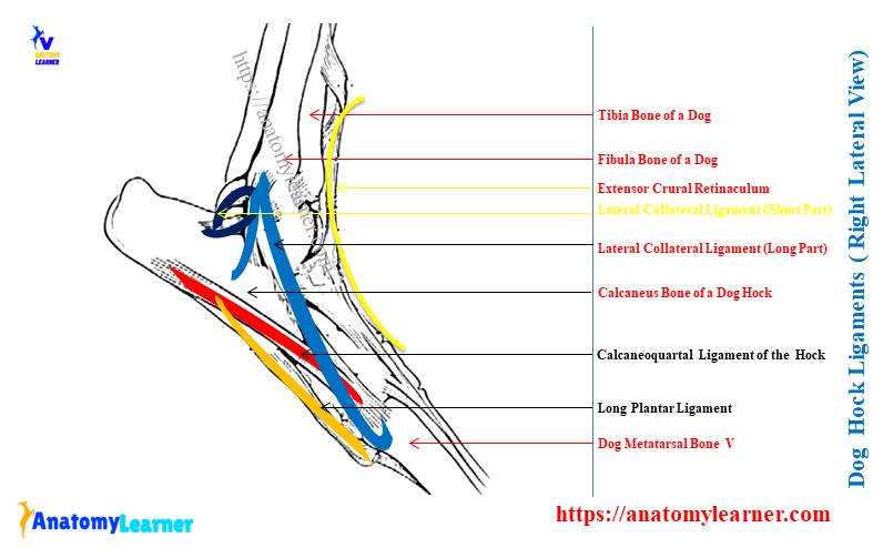
Collateral ligaments in dog hock
There are mainly two collateral ligaments in the canine hock structure – medial and lateral. The medial collateral ligament of the hock shows long and short parts.
Here, the larger long part is more superficial that runs from the medial malleolus to the first tarsal, and finally to the metatarsal I and II. You will find the strong attachment of this long superficial collateral ligament in between the medial malleolus and first tarsal.
Again, there is a weak attachment of the long superficial medial collateral ligament between the tarsal and metatarsal I and II. You will also see the attachment of the long part of the medial collateral ligament with the free surface of the talus and central tarsal bone of the hock.
Again, the short segment of the medial collateral ligament extends from the craniodistal to the medial malleolus to the talus bone. Finally, this part of the medial collateral ligament joins with the long segment.
The lateral collateral ligament of the hock joint also divides into short and long segments. Here, the long segment of the lateral collateral ligament passes from the lateral malleolus to the base of the metatarsal V. It also attaches to the calcaneus and fourth tarsal bones of the hock.
Now, the short part of the lateral collateral ligament lies deep in the long segment and shows two bands. One band of the short collateral ligament extends from the lateral malleolus to the calcaneus tubercle.
Again, the other small band extends from the lateral malleolus to the talus. Now, both band runs near the long part of the lateral collateral ligament.
Other ligaments of canine hock
There are various dorsal ligaments on the dorsal surface of the tarsus bone of a dog hock. One of the ligaments attaches to the distal extension of the crural extensor retinaculum. Here, you will see the tendons from various extensor muscles of the dog leg.
In between the central and second tarsals and also between the central and third tarsals, you will find the oblique dorsal centrodistal ligament.
There are various thick special plantar ligaments on the plantar surface of the (tarsus) dog’s hock. Most of these ligaments fuse with the thickened part of the capsular ligament (joint capsule).
A long plantar ligament passes from the calcaneus body and crosses the fourth tarsal bone. Finally, this long plantar ligament continues to the fourth and fifth metatarsal bones.
Dog hock muscles anatomy
Most muscles or their tendon from the dog leg (crus) have a direct or indirect connection with the dog’s hock anatomy. There are two (2) groups of muscles in the dog leg –
- Craniolateral muscle of the dog’s crus (extensor muscle of hock), and
- Caudal muscles of the crus – flexor muscle of leg and hock,
Most of these muscles or their tendons provide stability to the hock anatomy. First, let’s see the list of the craniolateral and caudal groups of muscles from the dog leg. Then I will describe these important muscles that greatly contribute to the canine hock structure.
Craniolateral muscles of the dog leg –
- Tibialis cranialis muscle,
- Fibularis longus of the leg,
- Extensor digitorum longus,
- Extensor digitroum lateralis muscle of the dog leg, and
- Digit I longus extensor muscle,
Now, let’s see the below-mentioned caudal group of muscles from the dog’s leg –
- Gastrocnemius muscle in the dog leg,
- Flexor digitorum superficialis and profundus muscles,
- Flexor digitorum lateralis and medialis muscles of the dog leg,
- Tibialis caudalis muscle of the canine leg, and
- Popliteus muscle in the dog leg,
Most of these craniolateral and caudal groups muscles from the dog crus are identified in the labeled diagram. This diagram might help you to understand the contribution of these muscles in the hock anatomy.
From the diagram, you will understand that tendons from most of these muscles pass over the hock structure and provide stability. Now, let’s know a little about these craniolateral and caudal groups of muscles of the dog leg. Or you may read the below-mentioned article to get a basic idea of the dog leg muscles –
- Dog leg anatomy with a labeled diagram (with bone, muscles, and vessels),
Tibialis cranialis and dog’s hock
The most superficial and flattened muscle lies cranial to the tibia bone. This tibialis cranialis muscle arises from the cranial part of the articular margin of the lateral tibial condyle.
It passes over the craniodistal surface of the leg and forms a thin and flat tendon. This tendon from the tibialis cranialis muscle extends obliquely over the tarsus to the medial aspect.
Finally, it attaches to the plantar surface of the rudiment of metatarsal I and metatarsal II (but rear). This muscle helps to flex the tarsus and rotate the paw laterally.
Extensor digitorum longus muscle of hock
The extensor digitorum longus is a spindle-shaped muscle between the tibialis cranialis and fibularis longus. Proximally, this muscle is covered by the tibialis cranialis muscle. Again, distally, the extensor digitorum longus runs free lateral and caudal to the tibialis cranialis.
Do you know from where the extensor digitorum longus muscle arises in a dog? It arises from the lateral aspect of the articular surface of the lateral condyle of the femur. Above the hock joint, it forms a terminal tendon that divides into four branches.
All these tendons from the extensor digitorum longus are held in place by the crural extensor retinaculum. Finally, these tendons extend distally along the dorsal surface of the metatarsals and reach digits II, III, IV, and V.
How does this muscle contribute to the canine hock structure? This muscle helps to flex the tarsus and extend the digits. Again, they provide great stability in the hock.
Fibularis longus and canine hock anatomy
The fibularis longus is a short muscle on the lateral surface of the proximal half of the leg. But its tendon has an active contribution in forming a dog hock anatomy.
This muscle runs between the tibialis cranialis and flexor digitorum lateralis muscle. Again, the fibularis longus muscle of a dog leg covers the proximal part of the extensor digitroum lateralis and fibularis brevis muscle.
From the lateral condyle of the dog tibia, this fibularis longus muscle originates and becomes an elliptical tendon in the middle of the tibia. The tendon of the fibularis longus mass with the tendons of extensor digitorum lateralis and fibularis brevis muscle.
Then the tendon of the fibularis longus runs through the sulcus of the lateral malleolus and passes over the tarsus bone. It runs cranial to the distal tibiofibular ligament and crosses the tendons of the extensor digitorum longus and fibularis brevis.
Finally, the tendon passes over the groove on the lateral side of the fourth tarsal bone of the hock. The main action of the fibularis longus muscle is to flex the tarsus and also rotate the pelvic limb medially.
Fibularis brevis of the hock
The fibularis brevis is a deep muscle in the dog leg that passes between the fibularis longus and flexor digitorum lateralis muscles. This extensor muscle of the dog leg arises from the lateral surface of the distal two-thirds of the tibia and fibula bones.
The fibularis brevis become tendinous after the origin and crosses the long lateral collateral ligament of the hock. It again runs between the tendons of the fibularis longus and lateralis digital extensor and attaches to the metatarsal V.
This muscle and its tendon provide great stability in the hock joint structure. Practically, this muscle contributes to flexing the tarsal joint.
Other muscles from the craniolateral group
You will also find the extensor digitorum I longus and extensor digitorum lateralis muscles in the dog leg (on its craniolateral aspect). They also have little contribution to the hock structure.
The extensor digit I longus is a small muscle band covered by the extensor digitorum longus and fibularis longus muscle. It directly lies on the tibia and arises from the cranial border of the fibula bone.
It extends obliquely distomedial deep to the long digital extensor. The tendon from the extensor digit I longus passes over the tarsus and metatarsus II and finally reach the metatarsophalangeal joint of the digit I and II.
This muscle greatly contributes to the flexion of digits I and II. Now, let’s talk about the extensor digitorum lateralis muscle from the dog leg that has a small contribution to the stability of a canine hock.
The extensor digitorum lateralis is another small muscle that runs between the fibularis longus and flexor digitorum lateralis muscles. It arises from the proximal third (above) of the tibia, passes over the hock joint, and is finally inserted on digit V.
Extension and abduction of the digit V are the main functions of the extensor digitorum lateralis muscle of the dog leg.
Tibialis caudalis and dog hock
This is the flexor group of muscle in the dog leg that lies deep and the medial aspect. It is a completely separated muscle in the dog leg from the two heads of the flexor digitorum profundus muscle.
This muscle contributes little to the structure of a canine hock joint anatomy. It is covered by the flexor digitorum medialis muscle and arises from the medial part of the proximal end of the fibula bone.
It forms a delicate tendon that extends distally cranial to the larger tendon of flexor digitorum medialis muscle. Now, the tendon of the tibialis caudalis inserts on the medial ligamentum tissue of the tarsus bone or hock.
Gastrocnemius muscle of the dog leg
This is a large muscle in the caudal group in the dog leg that has a potential contribution to the dog hock anatomy. It forms a common calcaneus tendon along with some other muscles of the dog leg that insert into the tubercle of the calcaneus bone.
You already know two heads in the gastrocnemius muscle – lateral and medial. Here, the lateral head of this muscle arises from the large tendon of the lateral supracondylar tuberosity of the femur bone. Again, the medial head of the dog’s gastrocnemius muscle arises from the medial supracondylar tuberosity.
The biceps femoris muscle covers the proximal lateral part of the gastrocnemius muscle. Again, in the medial to this muscle, you will find the semimembranosus, semitendinosus, and gracilis muscles.
As mentioned earlier, the tendon from the muscles forms the common calcaneus tendon that attaches to the hock joint (tubercle of the calcaneus). Thus, this muscle provides great stability in the hock structure.
The primary action of the dog’s gastrocnemius muscle is to extend the hock joint. It also contributes to slight flexion of the stifle joint of a dog.
Flexor digotroum muscles and hock
The superficial, deep, medial, and lateral flexor digital muscles of the dog leg have also a great contribution to providing stability in the canine hock. A tendon from these muscles passes over the hock joint and attaches to it.
The flexor digitorum superficialis muscle lies on the flexor digitorum profundus, tibialis caudalis, and popliteus muscle. A great part of the proximal extremity of the flexor digitorum superficialis muscle is covered by the head of the gastrocnemius muscle.
This muscle arises from the lateral head of the femur along with the lateral head of the gastrocnemius muscle. The tendon from this muscle attaches to the tendon of the gastrocnemius muscle in the middle of the tibia bone.
They form a broad ligament that inserts on the calcaneus tubercle and divides into four branches at the plantar surface. These four divisions of the tendon finally pass over the metatarsal II – V and insert into the corresponding digits.
The flexor digitorum profundus of the dog leg lies on the caudal surface of the tibia and is covered by the gastrocnemius and superficial digital flexor muscles. From the proximal caudolateral border of the tibia, the flexor digitorum lateralis originates.
It forms a long tendon that passes over the medial aspect of the calcaneus bone of the hock. This tendon divides into four branches in the middle of the metatarsus.
Finally, the flexor digitorum medialis is a short, flat muscle in the dog leg just medial to the flexor digitorum lateralis and lateral to the popliteus muscle. It arises from the head of a fibula and also forms the popliteal lines.
You will also find the four different divisions in the tendon of a flexor digitorum medialis that inserts the digits.
Vessels and nerves in the dog hock
The blood and nerve supply in the dog’s hock is also very complicated. But, I will try (sure) to make it simple as I will only show you the main arterial and nerve supply from the hock area.
But, if you want to know more about the different arterial and nerve supplies from the hind limb of a dog, I would like to suggest you read the below-mentioned article –
- Dog leg and its arterial supply with the diagram,
This article will provide deep knowledge of the different branches of the internal and external iliac arteries of the dog. You may also see the overview of the arterial supply in the hind limb of the dog from the below-mentioned diagram.
Here, in the diagram, you will find the main branches of the external iliac artery in the different regions of the pelvic or hind limb of the dog. So, you can understand and identify the main arteries that directly supply to the hock joint structures and their surrounding muscles.
So, the main arterial supply in the hock anatomy is –
- Dorsal pedal artery,
- Medial and lateral tarsal arteries,
- Dorsal common digital artery I,
- Cranial branches of a saphenous artery, and
- Arcuate artery,
Now, let’s learn a little about the courses of these arteries from the dog’s hind limb. But, keep in mind, I will only show you these arteries that pass over or have a direct connection with the hock anatomy.
Arteries in hock
You know, the popliteal artery is the continuation of the femoral artery through the popliteal fossa. Again, this popliteal artery divides into small caudal tibial and longer cranial tibia arteries.
Now, the cranial tibial artery of the dog continues to the tibiotarsal joint as the dorsal pedal artery. You will see different small branches that arise from the dorsal pedal artery and supply to the hind paw of the dog.
Medial and lateral tarsal arteries in the hock area run deep to the side of the tarsus. These two arteries of the dog terminate in the collateral ligament (lateral and medial).
Again, the dorsal pedal artery terminates in the proximal metatarsus and forms the arcuate artery. This arcuate artery runs transversely and disappears in the ligamentum tissue of the lateral and medial side of the proximal end of the metatarsals.
You will also see the cranial branches of the saphenous artery at the dorsal aspect of the hock. This artery joins with the superficial branch of the cranial tibial artery and forms the dorsal common digital arteries (II, III, IV, and V).
Nerves in canine hock
Again, the nerve supply of the hock region is also complicated. But, most of the nerves of the dog hock anatomy come from the tibia and fibular nerve. And you know, the tibia and fibular nerve are the branches of the main sciatic nerve in the dog’s hind limb.
So, you will find the below-mentioned main nerves that are associated with the canine hock structure –
- Branch of the caudal tibial nerve,
- Small branches of the saphenous nerve,
- Caudal cutaneous sural nerve, and
- Branches of deep and superficial fibular nerves,
Again, the muscles associated with the hock anatomy are supplied by different branches of the tibial and fibular nerve. For treatment or surgery, you might know the courses of the main nerve (branches of the ischiatic or sciatic nerve) from the hind limb of a dog.
There is a details guide on the dog’s sciatic nerve (branches, courses, and major supply) here in anatomy learner. You may read the following article to get the basic idea of the course of the sciatic nerve in dogs –
- Dog sciatic nerve anatomy – branches, courses, and distribution
I hope the dog hind limb nerve labeled diagram might help you to identify the different branches of the tibial, fibular, and saphenous nerves that are related to the canine hock structure. The branches, courses, and distribution of the canine sciatic nerve on the hind paw are more complicated than the ruminant.
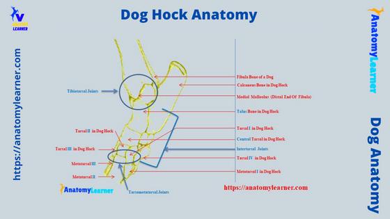
Dog hock anatomy diagram
Now, let’s memorize the different features from the dog hock anatomy with the help of the labeled diagram. Here, in the diagram, I tried to show you the different bones (tibia, fibula, 7 tarsal bones, and 5 metatarsal bones) and different extensor and flexor muscles from the hock joint.
Again, I tried to show you the different blood vessels (focus on arteries) that pass over the hock anatomy. Even the diagram shows different branches of the femoral (branch of an external) artery that supply to the different extensor and flexor muscles of the dog leg.
The dog leg Achilles tendon diagram also shows the formation of this tendon along with its insertion. Here, I also tried to show the different branches of the tibial and fibular nerves in the hock region of the dogs.
You may find more diagrams on the canine hock on social media of anatomy learners.
Dog hock injury symptoms
You already know the canine hock anatomy, consisting of the bones (tibia, fibula, tarsal, and metatarsal), muscles, and ligaments. The dog will show different symptoms if any deviation occurs in the normal structure.
Here, I will enlist (Important) some of the most common injuries in the hock joint. You will find the details guide (short) on every single injury of the canine hock here in anatomy learner.
So, first, let’s see the common injuries in the hock joint –
- Fracture in the dog’s hock – malleolar fracture, calcaneus fracture,
- Joint instability – can cause in any joints of the hock anatomy, but more commonly, stability occurs in the intertarsal and tarsometatarsal joints,
- Ligament rupture – lateral and medial collateral ligaments rupture are very common in the canine hock,
- Hygroma in the hock or ankle joint of the dogs – may occur occasionally,
- Tarsal hyperextension in the canine hock joint,
- Injuries in the Achilles tendon (common calcaneus tendon), and
- Osteochondritis dissecans in the dog hock,
You will also find other different injuries in the dog’s hock anatomy.
Fractures in the dog’s hock and instability
Fractures may occur in any bones of the dog’s hock. Most commonly, you may find fractures in the malleolus bones (both lateral and medial) and also in the calcaneus bone (fibular tarsal). If the dog has a malleolus fracture, it will show tarsocrural instability.
Thus, tarsocrural luxation or subluxation may develop, followed by this instability. So, you will find the lameness in the affected dog first; then, it will show swelling and pain in the tarsocrural joint.
If there is a fracture in the calcaneus bone, you will find instability in the common calcaneus tendon of the dog leg. Again, the specific area of the calcaneus will show swelling with severe pain.
If any fracture occurs in the talus (tibial tarsal bone), you will find the tibiotarsal joint instability, resulting in the hock luxation (complete). Again, any fracture in the middle or distal row of tarsal bones will also lead the intertarsal joint instability.
So, for any kind of fractures in the dog’s hock joint anatomy, you need emergency veterinary care for your pet dog.
Collateral ligaments rupture in the hock
The lateral and medial collateral ligaments from the dog hock joint are very much susceptible to rupturing. Any rupture in the medial and lateral collateral ligaments may cause the subluxation or luxation of the hock joint.
Rupture may occur in any position of the medial and collateral ligaments. But, the mid-body of these ligaments is very susceptible to become rupture. Again, the attachment part of these ligaments to the tibia and fibular bone is also susceptible to being ruptured.
In this case, surgical approaches are required to repair and recover this situation. Diagnostic imaging and determination of the level of injuries might help the veterinarian to confirm the collateral ligament rupture. So, it is very important to know the basic structure of the canine hock joint anatomy.
Hygroma dog hock and hyperextension
A fluid-filled false bursa may occur in the bony prominence and in the presser point of any bone. But, this will not create a problem for the dog’s leg.
In the case of a dog, this fluid-filled, painless swelling may generally occur in the carpus and tarsus joints. But, this condition may commonly occur in the larger dog breed compared to the small breed of dogs.
Externally, you will find a fluid-filled swelling at the hock region (tarsus), which surface is very hard to touch. If you confirm that is hygroma in the hock, then some home management can remedy this condition.
But, I am not interested in suggesting home remedies; rather, it will be better if you bring your dog to the veterinarian. They will confirm whether the structure is hygroma or not.
The dog hock hyperextension is the serious conformation fault where you will find a straight tarsal joint. In this condition, the distance between the common calcaneus tendon and the hock joint reduces.
Fortunately, this condition (hock hyperextension) is rare and breed-specific. You may find this hock hyperextension in the boxer dog’s breed.
How do I know if my dog has a hock injury?
You may easily identify the hock injury in your dog. However, the dog shows unique symptoms in different types of injuries in their hocks. But, you will commonly find the lameness for various kinds of injuries in the hock.
I have already enlisted different common injuries previously from the canine hock joint. You will commonly find hock instability due to fracture, rupture of the collateral ligaments, and damage to the tendons.
Again, you may also find hygroma, hyperextension, and Achilles tendon injuries in the canine hock joint. So, if there is any deformity in the tarsal bones of the dog, it will show complete or partial luxation.
That means you will find the lameness with severe pain in the dog’s hock region. Again, they will remain cool rather they jump or play. Furthermore, if there is a rupture of the collateral ligament, it will show complete luxation and sudden lameness.
In addition, the dog will show swelling in the tarsus region in hygroma. You will find a fluid-filled cavity (hard from the surface) in the tarsus or hock region of the dogs.
Thus, you may identify the different injuries in your dog’s hock.
How do you know if your (pet) dog’s Achilles tendon is ruptured?
The Achilles tendon of a dog may be ruptured completely or partially. You may identify the Achilles tendon rupture of the dog by the degree of lameness and physical condition of the hock.
In the complete Achilles tendon rupture in a dog, it will show non-weight bearing lameness, and the dog will stand on its ankle. You may also find the skin wound in the area of the dog’s calcaneus. But, the dog may not show pain in the complete rupture of the Achilles tendon.
Again, you may easily palpate the defect of the Achilles tendon in the completely ruptured condition.
In the partial Achilles tendon rupture, the dog shows swelling in the tarsus region. Again, there may occur tendon insertion avulsion. You may also find hyperflexion in the tibiotarsal articulation and even in the digits.
In some traumatic injuries, the complete tendon rupture may occur commonly in the dog. Again, low-grade strain, gastrocnemius muscle avulsion may cause a dog’s partial rupture of the Achilles tendon.
So, according to the degree of lameness and other specific symptoms in the hock region, you may easily identify the Achilles tendon rupture in your dogs.
How do you treat a dog’s hock?
As you see, the injuries in the dog’s hock are numerous; so according to the injuries, your veterinarian will provide the orthopedic brace or other different management. In some cases of dog hock injuries, your veterinarian will provide only some physical therapy.
Again, your veterinarian will perform the surgeries in some cases, like fractures and rupture of the collateral ligaments. In the lower grade strain and sprains, swelling on the canine hock may occur. General management and some medicinal care may recover this condition so quickly.
How long does it take for a dog hock to heal?
Normally, it depends on the type of injuries in the dog’s hock. If there is a fracture or rupture injury in the canine hock (performed surgery), it will take 1 – 2 months on average.
Again, in subluxation of the hock, or incomplete rupture of the collateral ligament, it will take 3 – 5 weeks (if you provide the proper management).
What causes dropped hocks in dogs?
The complete or partial rupture of the Achilles tendon, rupture of the medial and lateral collateral ligaments, and instability of the hock cause dropped conditions in the dogs. Again, the fracture of any hock bones also causes the dropped leg condition in the dog.
Enough these conditions, the complete rupture of the dog’s Achilles tendon is more serious to the dog. First, the dog shows lameness, and in severe conditions, foot drops occur. Then the dog stand on the ankle or hock instates of paws.
How many articulations are in the dog’s hock anatomy?
The dog’s hock anatomy shows three important articulations among the tibia, fibula, tarsal (7 bones), and metatarsal (5 bones) – tibiotarsal, intertarsal, and tarsometatarsal. Here, the tibiotarsal or tarsocrural articulation permits the greatest degree of hock movement.
You will find different types of intertarsal articulations in the hock joint anatomy. The most important intertarsal articulation of the hock is – the talocalcaneal central and calcaneoquartal joints. These are also known as the proximal intertarsal articulation of the canine hock.
You will also see the centrodistal or distal intertarsal articulations between the central and tarsal I, II, and III. Finally, the distal row tarsal forms the tarsometatarsal articulations with the 5 metatarsal bones of the dog leg.
Conclusion
The dog hock joint anatomy components are mainly the distal articular surface of the tibia, fibula, tarsals, and proximal end of the metatarsal. Again, the different ligaments like capsular, collateral and plantar provide the conformity and stability of the hock joint.
The dorsal and medial tarsal arteries pass over the dorsal surface of the hock. Again, there is a branch of a saphenous artery that also passes over the canine hock anatomy. The different branches of the tibial and fibular nerves pass over the structure of the dog’s hock.
Now, you should practice all these structures from the dog hock joint anatomy with the real sample. I hope all the provided labeled diagrams on the dog’s hock might help you to learn these structures perfectly.