
Esophagus is a long hollow muscular tube extending from pharynx to stomach in animal. In esophagus histology, you will find all the layers of typical tubular organs of animal’s body.
Hi dear anatomy learner, are you tired to find out the best guide to learn esophagus histology with slide images and labeled diagram? Don’t worry, I have a solution for it and going to provide you a best guide to learn esophagus histology with slide images.
In today’s article you will find the different layers of normal esophagus histology images with description. You will also find the identification points of esophagus histology slide so that you may easily identify esophagus slide under light microscope.
I will show you the general histological features of different layers of esophagus and also tell you the differences among different animals. So, if you want to learn histological features of esophagus from different animal then this article is for you.
Okay, let’s get into the article; but make sure you know the general organizational pattern of a tubular organ from animal’s body.
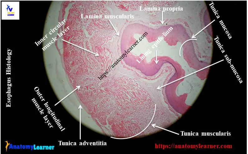
Esophagus histology
In this part of the article I am going to show you the most important histological features from a esophagus histology slide image. You might find out the following list of histological structures from the esophagus slide images carefully.
#1. Stratified squamous epithelium on mucosa (keratinized or nonkeratinized; vary with animals)
#2. Lamina propria of mucosa layers of esophagus
#3. Lymhatic nodules (not in all amimals) oin lamina propria layer
#4. Connective tissue papillae of esophagus
#5. Lamina muscularis layer of esophagus
#6. Mucosa acini of esophageal gland in sumucosa area of esophagus
#7. Adipose tissue and blood vessels at submucosa area of esophagus structure
#8. Tunica muscularis (two layers; skeletal muscles or smooth muscles; vary with animals)
#9. Tunica adventitia or serosa layer of esophagus (vary with different parts of esophagus)
Identification points of esophagus slide
Do you want to identify the esophagus histology slide under light microscope with proper identification points? Well, here in this section I am going to enlist the proper identification points of esophagus slide under light microscope.
#1. The wall of the provided sample tissue show four different layers of tubular organ structure – tunica mucosa, tunica submucosa, tunica muscularis and tunica adventitia or serosa.
#2. Presence of three different parts (lamina epithelium, lamina propria and lamina muscularis) in mucosa of this tissue sample
#3. The mucosa is folded and lined by stratified squamous epithelium (keratinized or non keratinized; you will find details guide at description part of esophagus).
#4. Presence of mucous acini glands and adipose tissue in submucosa of the sample tissue
#5. The tunica muscularis layer consists of inner circular and outer longitudinal muscle layers (skeletal or smooth; vary with animals)
#6. Presence of tunica adventitia or tunica serosa (vary with different parts of esophagus)
So, this smaple tissue is a esophagus slide. You may add other different identification points for esophagus slide.
Layers of esophagus histology with labeled diagram
So, are you interest to learn details histological characteristics of esophagus? You know esophagus is the best organ that have all the layers of a tubular organ. I am going to describe the different layers from esophagus histology slide.
Mucosa of esophagus structure
The mucosa of esophagus consists of lamina epithelium, lamina propria and lamina muscularis. The lamina epithelium of esophagus mucosa is stratified squamous and the keratinization may vary with different animals.
You will find the ideal keratinization in the esophagus of ruminant, slightly in pig and horse. But in the mucosa of carnivores, there is no keratinization.
In the lamina propria of esophagus in different animals, you will find atypical characteristics; this part consists of more dense collagen fibers with numerous elastic fibers. So, the connective tissue is more dense in this part than the connective tissue tissue of submucosa layer of esophagus histology.
The lamina muscularis part of esophagus mucosa contains only longitudinally oriented smooth muscle bundles. You will also find the difference in the apperence of smooth muscle in different animals. In ruminant, horse and cat, there are isolated smooth mucles bundles near to the pharynx and increase it’s number towards stomach.
In the cranial part of pig and dog esophagus, there is no lamina muscularis layer but in caudal part you will find the well developed lamina muscularis.
Submucosa of esophagus structure
In the submucosa of esophagus histology, you will find the loose connective tissue that conatis large longitudinally oriented arteries, vein, lymph vessels and nerves. Submucosa contains mixed acini with serous demilune only ar pharyngoesophageal junction in ruminant, horse and cat.
But in pig, you will find the seromucous glands in the cranial half of esophagus and throughout the esophagus of dog.
Tunica muscularis layer of esophagus slide
The tunica muscularis layer of esophagus histology consists of two layers of smooth or skeletal muscles. Generally in ruminant and dog, the tunica muscularis consists entirely skeletal mucles whereas in horse you will find the skeletal muscle in the cranial two third of tunica muscularis. Then it will change to smooth muscle in the caudal third tunica muscularis of horse esophagus.
You will find the interdigitation and spiraling of the two layers of muscle at the cranial end of esophagus structure. But these layers change orientation into inner circular and outer longitudinal at caudally.
A thin layer of connective tissue lines between the two layers of muscle in esophagus.
Tunica serosa or adventitia of esophagus
The tunica muscularis is surrounded by an adventitia that consists of loose connective tissue along with blood vessels, lymph vessels and nerves at the cervical part of esophagus. But in the thoracic and abdominal part of esophagus are invested by serosa (mesothelium lining). In some animal, mesothelium lining may lack at esophagus stomach junction.
Esophagus histology slide drawing
Do you want to get esophagus histology slide drawing tutorial? Here in this section I am going to share esophagus slide image drawing with you. You may follow the same but must try to draw better than this esophagus drawing.
Do you need more images related to esophagus histological structure? You may follow anatomy learner at social media for more updated picture and video related to esophagus histology.
You might read other different article from anatomy learner blog (both anatomy and histology of different organs from animal body) –
#1. Identification of tongue histology slide under light microscope – core connective tissue and different types of papillae
#2. Identification of salivary galnds under light microscope
Conclusion
Hope you got the best guide to learn different layers of esophagus histology with proper slide images and labeled diagram. If you think this article is really helpful to learn histological features of esophagus then you may share it with your friend who wants to learn esophagus structure.
Are these identification points helpful to identify the esophagus histology slide? If you need more esophagus slide image then let me inform.
Don’t forget to join with anatomy learner’s social media to get more updates on articles, pictures and videos of different organ