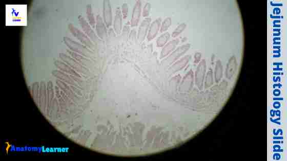
The jejunum is the second part of the small intestine of an animal. In the jejunum histology, you will find the four different layers like a tubular organ with some other specific identifying features. Here, I will show you the different features of a jejunum histology slide with a labeled diagram.
I will also describe the jejunum anatomy from an animal in a little. That might help you to get a clear concept of the histological features of a jejunum. Again, I will help you to make the difference among duodenum, jejunum, and ileum histology slides at the end.
So, after finishing this article, you will know the normal jejunum wall histology and other parts of the small intestine of an animal.
Jejunum histology
The wall of the jejunum consists of four layers – the mucosa, submucosa, muscularis mucosa, and serosa. From the jejunum histology slide, you might identify some important features under the light microscope. So, first, let’s know these histological features that you should know and identify from the jejunum.
In the tunica mucosa of the jejunum, you will find some permanent fold (plica circularis) and villi (modification of mucosal surface). Again, in the core of the plica circularis, submucosa contains numerous arteries and veins. The club-shaped villi cover the plica circularis of the jejunum.
“ Please read the species difference of jejunum structure at the end of this article.”
The villus of the jejunum fold lines with the simple columnar epithelium. Within each villus of the jejunum, you will find the lamina propria. At the bottom of the villi, there presence intestinal glands.
Between the villi, you will find the intervillous spaces. The intestinal glands of the jejunum open into these intervillous spaces.
Sometimes, you may find the lymphatic nodule with the germinal center in the lamina propria of the jejunum histology slide. The tunica muscularis consists of two layers (inner circular and outer longitudinal) of smooth muscle.
There is also some nerve plexus present between the two layers of smooth muscles of the jejunum. Again, you will find a loose connective tissue layer with blood vessels, lymphatics, and adipose cells in the jejunum serosa.
Okay, let’s summarize the histological features from the jejunum –
- The plica circularis, villi, intervillous spaces, and intestinal glands
- The lining epithelium of mucosa, muscularis mucosa, few glands on submucosa layer
- The lymphatic nodule in the lamina propria (optional), and
- An inner circular, the outer longitudinal muscle layer, and tunica serosa layer
Fine, try to identify these structures from the jejunum.
Jejunum histology slide identification points
Sometimes, you may be asked to identify the jejunum slide under the light microscope at the laboratory. Now, I will provide the jejunum histology slide identification points with the actual sample.
“Here, I will enlist only the main identifying features of the jejunum slide. If you want, you may add more identifying features of jejunum.”
- The sample tissue section shows four different layers (tunica mucosa, submucosa, musculris, and serosa) like a tubular organ
- Presence of prominent and permanent mucosal fold (known as plica circularis) in the mucosa of the provided tissue sample
- There are club-shaped villi present on the mucosal fold of the jejunum that lines by the simple columnar epithelium.
- Presence of intervillous spaces and intestinal glands in the lamina propria of the sample tissue
- There are an inner circular and an outer longitudinal smooth muscle layer in the tunica muscular layer of the provided sample tissue
- Presence of loose connective tissue, blood vessels, lymphatics, and adipose cells in the serosa of the tissue sample.
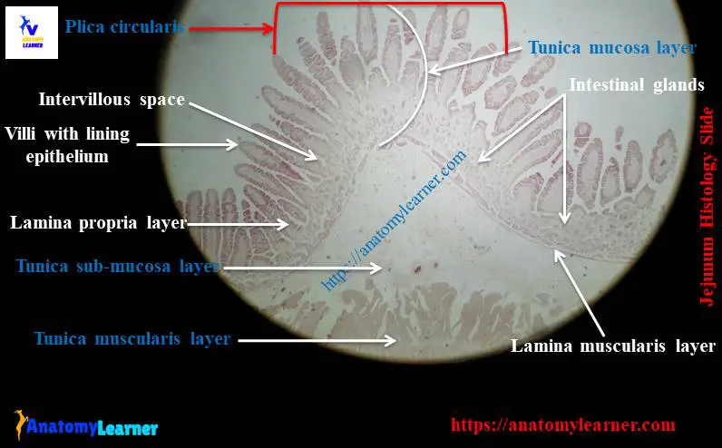
So, this is the slide of a jejunum. In the labeled diagram, I tried to show you all these above-mentioned histological features from the jejunum.
Normal jejunum wall histology with diagram
If you want to learn the detailed histological features of the jejunum, you may continue this part of the article. I will describe the single layer from the normal jejunum wall histology slide with a labeled diagram.
You know, the small intestine (duodenum, jejunum, and ileum) of an animal is the main site for the absorption of products of digestion. Again, these digestive and absorptive functions are facilitated by several specialized structures.
In the jejunum slide, you will find the following three important features that help to increase the efficiency of absorption –
Numerous circularly disposed mucosal folds present in the jejunum extend approximately two-thirds of the way around the lumen. Another name for these circular folds of the jejunum is plica circularis.
The surface of the mucosa covers with club-shaped mucosal projections (villi).
Again, the villi lines with the simple columnar epithelium that contains microvilli.
Species differences of these three structures
In the ruminant like cow, sheep, and goat, you will find prominent and permanent circular folds. But, in other mammals, these circular folds of the jejunum mucosal wall will disappear when the organ distend.
Again, the length of the jejunum villi varies with different species. You will find a short but wide villus in the jejunum of ruminant. But, in a carnivore, you will find the long and slender villi in the jejunum.
Again, the lining epithelium of the villi will remain almost the same in the jejunum of all domestic animals.
Now, let’s know the detailed structures of four layers (tunica mucosa, submucosa, muscularis, and serosa) of the jejunum histology slide with diagrams.
Tunica mucosa of jejunum
The mucosa of the jejunum histology slide includes lining epithelium, lamina propria, and a lamina muscularis. As I told you before, there are numerous plica circularis with villi present in the mucosal surface of the jejunum.
There are also mucosal glands (intestinal glands) that open between the base of the villi. These mucosal glands are the simple tubular gland and remain in the lamina propria of the jejunum slide.
The lining epithelium of jejunum mucosa
The epithelium covering the villi and the mucosal surface area is predominately simple columnar cells with absorptive functions. Numerous mucous secreting goblet cells scatter distributed within the simple columnar epithelium.
The columnar absorptive cells have ovoid nuclei located near the base of the cell. They have prominent microvilli that form a striated border.
In the electron microscope, you will find mitochondrial near the absorptive columnar epithelium’s nucleus and in the basal region. The apical cytoplasm contains the terminal web and extensive smooth endoplasmic reticulum.
Other cells of the jejunal mucosa
You will find numerous goblet cells that disperse among the columnar absorptive cells in jejunum mucosa. The apical part of the goblet cells become distends, accumulates mucinogen droplets. And the nuclei and remaining cytoplasm push into the narrow cell base that rests on the basement membrane.
The number of goblet cells in the jejunum slide will decrease at the tip of the villi. Again, you will find more goblet cells in the ileum than the jejunum part.
The crypts (at intervillous spaces) are the tubular invegination of the epithelium into the lamina propria. They are simple tubular intestinal gland that lines with a variety of cell types. The main cell type of the intestinal gland is the undifferentiated columnar cells.
These cells multiply, differentiate, and migrate onto the villus, giving rise to the columnar absorptive and goblet cells. They are pushed towards the tip of the villus by succeeding cells, where they slough off into the lumen.
Near the base of the jejunum intestinal glands, you will find some acidophilic granular cells (paneth) in horses and ruminants. These are the pyramidal-shaped cells that possess prominent spherical, acidophilic granules in between the nucleus and apex of the cell. The acidophilic granular cells have all the characteristics of the enzyme-producing cells.
You will also find enteroendocrine cells in the intestinal glands of the jejunum that produce various paracrine and endocrine hormones. Again, you will find some specialized microfold cells covering the lymphatic nodules in the lamina propria of the jejunum histology slide.
I have described every cell from the small intestine in my previous article (duodenum histology slide) in detail. You may read that guide to know a good piece of knowledge on the intestinal cells if you want.
Lamina propria of the jejunum slide
The lamina propria of the jejunum slide forms the core of the villi and surrounds the intestinal glands. You will find loose connective tissue with a prominent reticular framework in the lamina propria of the jejunum histology slide.
You will also find the following structure within the extensive fiber network of the jejunum –
More blood and lymph vessels
The leukocytes, fibrocytes, smooth muscle cells, plasma cells, and mast cells.
Sometimes, you may find globule leukocytes in the jejunum mucosa of some domestic species. They contain large eosinophilic globular material surrounding a small nucleus.
Some diffuse lymphatic tissue and solitary lymphatic nodules may present in the lamina propria of the jejunum slide. But, this phenomenon in the jejunum slide is not common in the animal. These diffuse lymphatic tissue and solitary lymphatic nodules commonly occur in the ileum than that of the jejunum.
A single lymphatic capillary is present in the center of the lamina propria within the villi of the jejunum. The longitudinally oriented smooth muscle cells that originated from the lamina muscularis extend to the tip of the villus.
The Contraction of these smooth muscle cells causes the villus to shorten. Again, this contraction is also responsible for the lateral movement of the jejunum.
Lamina muscularis of the jejunum
The lamina muscularis of the jejunum slide arranges into two layers. You will find the inner circular and outer longitudinal layers of smooth muscle in the lamina muscularis layer of the jejunum. But, these two layers of smooth muscles are thin and incomplete.
The evidence of the two layers of smooth muscles is not common in the lamina muscularis of the jejunum. They (lamina muscularis of the jejunum) may vary with the species, the individual animal, and the specific region.
Tunica submucosa of the jejunum histology slide
The tunica submucosa of the jejunum histology slide contains more dense connective tissue than the lamina propria. You will find numerous blood vessels (arteries and veins) in the submucosa of the jejunum slide.
The tunica submucosa of the jejunum also contains submucosal nerve plexus. In addition, nerve fibers from this submucosal nerve plexus extend into the villi.
Generally, you will not find any submucosal glands in the jejunum of a small ruminant like sheep, goat. They are mainly found in the duodenum part of most of the domestic species.
But, in the jejunum of the horse, cattle, and pig, you may find some submucosal gland in the tunica submucosa.
The diffuse lymphatic tissue and solitary lymphatic nodules are not common in the tunica submucosa of the jejunum slide. You will find these structures in the other part of the small intestine, especially in the ileum part.
There are larger lymphatic nodules present in the ileum of the cattle. Again, you will find numerous lymphatic nodules in the ileum of a horse.
So, this might be a good point to differentiate the jejunum slide from the ileum.
Tunica muscularis of the jejunum
The tunica muscular of the jejunum slide consists of two layers of smooth muscle cells. So, you will find an inner layer of circularly arranged smooth muscle cells and an outer layer of longitudinally arranged smooth muscle cells.
The thickness of the tunica muscularis is more in the jejunum of a horse. Both the inner and outer layers of the smooth muscles are nearly equal in thickness. The connective tissue between the two layers contains the myenteric plexus.
The tunica muscularis of the jejunum shows two types of muscle contraction – segmentation and peristalsis.
Segmentation contraction – local contraction displace intestinal contents both proximally and distally. These contractions primarily involve the circular muscle layer. They help to circulate the contents locally, mixing it with the digestive juices and moving it into contact with the mucosa for absorption.
Peristalsis contraction – is the second type of contraction that involves the coordinated action of both circular and longitudinal muscle layers. This contraction helps to move the intestinal contents distally.
Tunica serosa of the jejunum histology slide
A thin layer of serosa covers the entire jejunum. In the jejunum histology slide, you will find a thin layer of loose connective tissue covering mesothelium in the tunica serosa layer.
You may also find the adipose cells in the tunica serosa layer of the jejunum part of the small intestine.
Jejunum histology slide labeled diagram
Now, it is better to review all the histological features from the jejunum slide. I will try to show you all the features from the jejunum histology slide with the labeled diagram.
Here, I tried to show you all the histological features from the four different layers of the jejunum wall. You may join anatomy learner on social media if you want to get a more updated labeled diagram on the jejunum slide.
Jejunum anatomy
I will not describe the detailed anatomical facts of the jejunum. Rather, I prefer to summarize all the anatomical features from the jejunum in short. Let’s know the main features of the jejunum anatomy of the animal.
The jejunum constitutes most of the length of the small intestine and comprises a good number of colis. These close coils are constricted and dilated and form the U-shaped tubular loops that attach at the border of the mesentery.
The jejunum is the most mobile part of the small intestine of an animal. It occupies the space between the right surface of the rumen and the right abdominal wall below the large intestine.
The blood vessels of the jejunum are arranged in a few layers of arches in the mesentery. The straight vessels connect the loops of the jejunum with the convexity of vascular arches.
You will find some jejunal or mesenteric lymph nodes within the double layer of the mesentery. The advanced pregnancy and fullness of the rumen influence the movement of the jejunum.
Frequently asked questions on jejunum slide.
Now, I will try to solve the common inquiries on the jejunum slide. If you have any inquiries on the jejunum slide, please let me know.
What type of tissue is in the jejunum?
You will find the basic tissue in the jejunum slide. The fold of tunica mucosa lines with the simple columnar epithelium with goblet cells. Again, in the tunica submucosa, you will find dense connective tissue. There is muscular tissue (two layers of smooth muscle layers) present in the tunica muscularis layer of the jejunum.
In addition, the serosa of the jejunum slide contains the loose connective tissue with mesothelium covering. You will also find adipose cells in the serosa of the jejunum slide.
How do you differentiate duodenum, jejunum, and ileum histology?
The most common inquiries are how you may differentiate the duodenum, jejunum, and ileum histology slide. Fine, I will try to make this simple for you to identify these slides under a light microscope.
For duodenum histology slide
Presence of short leaf-like intestinal villi that lines with the simple columnar epithelium and goblet cells
You will find the Brunner’s gland in the tunica submucosa of the duodenum histology slide.
Presence of the crypt of Liberkuhn in the mucosal surface of the duodenum
For the jejunum histology slide
You will find finger or club-shaped mucosal villi that also lines with the simple columnar epithelium and goblet cells.
There are no submucosal gland and Peyer’s patches in the tunica submucosa of the jejunum slide.
Sometimes, you may find fewer lymphatic nodules in the lamina propria of the jejunum slide.
The lamina muscularis are thin but arranged in two incomplete layers in the jejunum.
Presence of a thin serosa that contains loose connective tissue and adipose cells
For the ileum histology slide
The villi of the ileum mucosal surface are thin and slender
In the tunica submucosa, you will find the aggregated lymphoid follicles (Peyer’s patches) that is more characteristics feature of the ileum
Are these histological features enough to identify the duodenum, jejunum, and ileum slide? You may also find the histological differentiation of this three-part of the small intestine here in tabular form.
What is the feature of the jejunum?
I have already described the features of the jejunum in detail. Would you please read the full article and try to identify the important features of the jejunum slide? You might know and identify the histological features from the four different layers of the jejunum slide.
The mucosa of the jejunum slide contains the plica circularis, villi, and simple columnar epithelium lining. You will find numerous intestinal glands in the lamina propria that open in the intervillous spaces (at the base of the villi).
There are no submucosal glands or Peyer’s patches in the tunica submucosa of the jejunum slide. The histological features of the other two layers (tunica muscularis and serosa) are similar to the common tubular organs.
What is the histological difference between the small and large intestines?
It is very important to know the histological features of the small and large intestine for identification and differentiation. Here, I will try to show you the main identifying characteristic of the small and large intestine.
In the small intestine histology slide, you will find some special features in the tunica mucosa. You will find the plica circularis, longer villi with microvilli in the tunica mucosa of the small intestine. This plica circularis of the small intestine is the prominent and permanent structure of the mucosa.
The lining epithelium of the tunica mucosa is a simple columnar epithelium with goblet cells. You may also find other different cells in the mucosa of the small intestine like penath cells, enteroendocrine cells, and others.
The number of the plica circularis and villi mary varies in the different parts of an animal’s small intestine. In the submucosa of the small intestine, you will find Brunner’s glands in the duodenal part, Peyer’s patches in the ileum part, and more.
In the large intestine, you will not find any well-defined plica circularis and villi. But the crypts are well-developed and lines with numerous goblet cells. The number of the goblet cells of the crypts is increasing towards the large intestine.
You will not find any paneth cells in the crypt of the large intestine. In addition, you will find a well-defined tunica muscrularis layer in the histology slide of a large intestine.
There is also a significant difference in the tunica serosa and adventitia of the large intestine compared to that of the small intestine.
If you want to get the short notes on identifying the jejunum histology slide with a more labeled diagram, please visit this page.
Conclusion
I hope you got the basic idea on the jejunum histology slide with the labeled diagram. The larger and more numerous plica circularis (circular mucosal folds) with numerous villi are the most identifying histological features of the jejunum slide. Generally, you will not find submucosal glands in the tunica submucosa of the domestic species. This might be another important identifying feature of the jejunum slide.
Again, the tunica submucosa of the jejunum histology slide lacks diffuse lymphatic nodules and Peyer’s patches. These are other identifying histological features that might help you differentiate the jejunum histology slide from the duodenum and ileum. At last, I would like to suggest you read this article again to understand the normal jejunum wall histology slide.