The ileum histology slide consists of the four layers like tunica mucosa, submucosa, muscular, and serosa. Here, I will show you the detailed histological features of the wall of the ileum slide with a labeled diagram. I will also provide you with the important identification points for the ileum histology slide so that you may identify it so quickly.
The histological features of the ileum wall are almost similar to the tubular organs. But, you will find the most characteristic features of the ileum slide in the mucosa and submucosa layers. The mucosa of the ileum histology slide possesses some modified structures (villi). Again, the submucosa of the ileum slide consists of Peyer’s patches.
In addition, in the end, I will provide some points that might help you distinguish among duodenum, jejunum, and ileum slide under the light microscope.
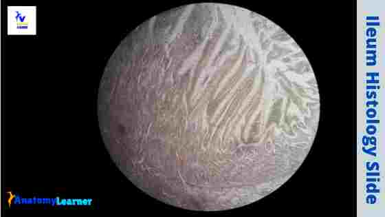
Ileum histology
The wall of the ileum histology consists of four distinguished layers – tunica mucosa, submucosa, muscular, and serosa. You will find thin and slender villi on the tunica mucosa layer of the ileum slide. The simple columnar epithelium lines these villi of the ileum.
So, you will find the intervillous spaces between the villi of the ileum structure. Within the villi, there presence the lamina propria that contains loose connective tissue. At the bottom of these villi, you will find numerous intestinal glands.
These intestinal glands of the ileum open into the intervillous spaces. The same characteristic features were found in the jejunum histology slide. Sometimes, you may find the diffuse lymphatic tissue in the lamina propria of the ileum histology slide.
This diffuse lymphatic tissue forms the lymphatic nodules that extend into the submucosa of the ileum. As the lymphatic nodules extend from lamina muscularis to tunica submucosa, the disruption of the lamina muscularis occurs. For that, you will find the disrupted lamina muscularis in the ileum tissue section.
The tunica submucosa of the ileum slide possesses numerous aggregated lymphatic nodules (known as Peyer’s patches). These are the most interesting and identifying features of the ileum histology slide. Here in most of the lymphatic nodules, you will find the germinal center.
The tunica muscularis and serosa of the ileum slide did not possess any exceptional features. Fine, you will find two layers of smooth muscle (an inner circular and an outer longitudinal layer) in the tunica muscularis of the ileum slide.
Again, the tunica serosa of the ileum contains the connective tissue cells, blood vessels, and adipose tissue.
Ileum histology slide identification points
I want to provide you with the most important identifying features of the ileum histology slide. You may add more identification points for the ileum slide if you want.
Let’s try to find out the below-mentioned identification points from the ileum slide –
- The wall of the provided tissue sample shows four distinguished layers – tunica mucosa, tunica submucosa, tunica muscularis, and tunica serosa.
- Presence of thin and slender villi on the tunica mucosa of the sample tissue that is lining with the simple columnar epithelium
- There are numerous intestinal glands at the bottom of the thin and slender villi of the provided sample tissue.
- Presence of the Peyer’s patches (aggregated lymphoid follicles or nodules) in the tunica submucosa of the sample tissue
- The tunica muscularis of the tissue sample consists of inner circular and outer longitudinal layers of smooth muscles.
So, the provided sample is the ilium histology slide.
I think you learn the basic histological features of the ileum slide. If you want to learn the details of every single layer of the ileum slide’s wall, you may continue this article.
Ileum structure
I will describe everything about the ileum structure practically. You will find the detailed histological characteristics of every single layer of the ileum wall. You know there are four different layers present in the wall of the ileum slide.
- The tunica mucosa layer of the ileum
- Tunica submucosa layer of a ileum
- The tunica muscularis layer and
- Tunica serosa layer of the ileum slide
But, it will be better if you have a good piece of knowledge on the below-mentioned topics earlier. These might help you to understand easily and quickly the histological features of the ileum structure.
General organizational features of a tubular organ (hollow organ histology)
The histological features of duodenum or jejunum slides
That’s fine; let’s move into the histology of the four different layers of the ileum structure.
The tunica mucosa of the ileum slide
As you know, the tunica mucosa of any tubular organ contains three basic layers – lining epithelium, lamina propria, and lamina muscularis. In the ileum histology slide, you will also find the same structures in the tunica mucosa.
You know, the surface area of the mucous membrane of three parts of the small intestine are extensive and possess the following features in common –
- The presence of numerous circular folds in the tunica mucosa of different parts of the small intestine (duodenum, jejunum, and ileum parts)
- Numerous finger-like processes project from the mucosal surface to the lumen (known as villi)
- In addition, the presence of microvilli on the luminal surface of the cells that line the tunica mucosa
- Some numerous crypts or depressions invade the lamina propria of the small intestine (duodenum, jejunum, and ileum)
You will also find numerous intervillous spaces in the duodenum, jejunum, and ileum structure where the intestinal glands open.
The circular of the ileum is made up of all layers of the tunica mucosa. But these circular folds are fewer and less marked in the ileum structure than that of the other part of the small intestine. Again, in the terminal part of the ileum structure, you will not find such circular folds.
The villi of the ileum slide are thin and slender that contain a connective tissue core with numerous blood vessels. Each villus of the ileum also contains a distinguished central lymphatic channel that is known as lacteal.
The crypts of the ileum are numerous as the intestinal glands open in these crypts.
Lining epithelium and other cells of the ileum
The villi of the ileum slide lines with the simple columnar epithelium (with microvilli). These are the absorptive columnar cells in the ileum structure. You will also find some other cells in the ileum structure.
You may find the following cells in the ileum structure –
- The goblet cells, undifferentiated cells of the ileum
- Numerous cells in the intestinal glands of the ileum
- The paneth cells (but less in number in the ileum part)
- And other cells in the lamina propria of the ileum structure like fibroblast, lymphocytes, and other
Nice, if you want to know more about the ileum structure cells, as mentioned earlier, you may read my previous article (duodenum histology slide). In that article, you will get basic information about the cells of the small intestine.
The lamina propria of the ileum histology slide
The basic structure of the lamina propria of the ileum histology slide is similar to that of a tubular organ. But, you will find some more interesting and characteristic features in the lamina propria of the ileum slide.
You will find diffuse lymphatic tissue and aggregated lymphoid follicles in the lamina propria of the ileum structure. These diffuse lymphatic tissues become numerous and aggregated to form numerous follicles (the Peyer’s patches).
These Peyer’s patches are numerous at the terminal part of the ileum slide. Again, these lymphoid follicles extend up to the tunica submucosa of the ileum.
The epithelium covering the lymphatic follicles contains special types of cells (micro folds or membrane cells or M cells). In addition, you will find numerous intestinal glands in the lamina propria of the ileum slide that opens in the intervillous spaces.
The lamina muscularis of the ileum slide
The lamina muscularis of any tubular organ contains a thin layer of smooth muscles (sometimes, two layers of smooth muscles). But, in the ileum structure, you will find a disrupted lamina muscularis layer. I have shown these disrupted lamina muscularis layers in the ileum slide diagram.
Tunica submucosa of ileum histology slide and Peyer’s patches
In the tunica mucosa layer, you will find the more identifying characteristics features of the ileum histology slide. There are solitary or isolated lymphatic nodules present in the submucosa throughout the ileum structure.
You will also find the large aggregated lymphatic nodules (Peyer’s patches) in the tunica submucosa of the ileum structure. These large aggregated lymphatic nodules are more numerous in the terminal part of the ileum slide.
The lymphatic nodules are largest in the tunica submucosa of the cow’s ileum. In addition, you will find more numerous lymphatic nodules in the ileum of a horse.
The intestinal glands sometimes may extend into the tunica submucosa of the ileum structure. Again, the tunica submucosa of the ileum slide also contains the submucosal nerve plexus. The nerve fibers from this submucosal plexus extend into the villi of the ileum.
Tunica muscular and serosa layers of the ileum slide
You will not find any exceptional histological features in the tunica muscular layer of the ileum slide. The tunica muscularis of the ileum slide consists of inner circular and outer longitudinal smooth muscle layers.
Again, the thickness of the tunica muscularis of the ileum may vary with the animal species. You will find more thicken tunica muscularis layers in the ileum of a horse.
The serosa of the ileum consists of a loose connective tissue layer that covers the mesothelium. You will also find the blood vessels, adipose cells in the tunica serosa of the ileum slide.
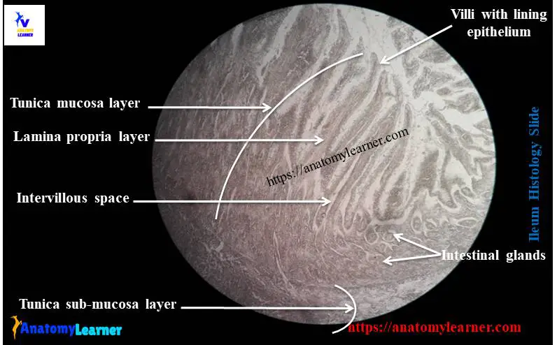
Ileum histology slide labeled diagram
I think the features mentioned earlier in the ileum histology slide labeled diagrams might help you a lot. Again, I will show you the ileum histology diagram where you might identify the following important features.
Okay, let’s try to identify the following histological features from the ileum slide diagram –
- The villi with lamina propria and the lining epithelium (simple columnar epithelium)
- Intervillous spaces and intestinal glands of the ileum slide
- The submucosa with the Peyer’s patches (germinal centers of the lymphatic nodules)
- Disrupted muscularis mucosa of the ileum slide
- The tunica muscularis layers (inner circular and outer longitudinal muscle layers) and
- A tunica serosa (containing simple squamous lining, loose connective tissue, and adipose tissue)
If I update the ileum slide diagram in the future, you will get notified on the social media of the anatomy learner. In addition, you will find more histology slide diagrams here.
Ileum anatomy
The ileum is a terminal part of the small intestine of an animal. It is a tube-like structure that attaches to the cecum at its cranial part. The ileum terminates at the medial surface of the cecum at the cecocolic junction.
Frequently asked questions on ileum slides.
Now, I will try to solve the common inquiries on the ileum histology slide and others. If you have any inquiries on ileum slides, let me know.
What is found in the ileum?
Great, this is a nice question on the ileum slide. I think it will cover all the important histological features of the ileum slide. You will find four basic layers in the ileum histology structure – tunica mucosa, submucosa, muscular, and serosa.
Again, in the tunica mucosa, you will find a different important structure like lining epithelium with other cells, modification of the mucosal surface (villi), lamina propria, and lamina muscularis.
The lining epithelium of the ileum slide is mainly the absorptive simple columnar with microvilli. You will not find any distinguished circular folds in the terminal part of the ileum structure compared to a jejunum.
But, you will find numerous villi and intervillous spaces in the mucosal surface of the ileum slide. In the ileum’s vill core, you will find the connective tissue, blood vessels, and lacteals. Again, there presence numerous intestinal glands at the bottom of the villi of the ileum structure.
These intestinal glands of the ileum slide open into the intervillous spaces. In addition, these intestinal glands of the ileum may extend into the tunica submucosa layer.
The most characteristic feature of the lamina propria of the ileum slide is the presence of diffuse lymphatic nodules. These nodules aggregated to form the larger lymphatic nodules extending up to the ileum structure’s tunica submucosa.
The histological features of the tunica muscularis and tunica serosa of the ileum histology slide are similar to a hollow organ. To get a list of exceptional features of the ileum slide, you may read the ileum slide identification points (listed earlier).
What is the ileum in anatomy?
The ileum in anatomy is a tube slike structure of the small intestine. It is the terminal part of the small intestine of any animal species. The length of the ileum may vary with the different animal species.
The ileum is the continuation of the jejunum and attaches with the cecum at its cranial part. It joins with the medial surface of the cecum and colon at the cecocolic junction.
Grossly, you may easily identify the ileum part from the small intestine of any animal. You know the jejunum part of the small intestine is a coiled structure. Then this part becomes straight and continues with the cecum and colon of the large intestine. This straight part of the small intestine may consider as the ileum part.
What cells make up the ileum?
You will find different types of cells that make up the ileum. First, you will find the absorptive columnar epithelium in the mucosal surface of the ileum structure. Again, you will find the glandular epithelium in the intestinal glands of the ileum slide.
The surface epithelium (simple columnar epithelium) contains microvilli on their surfaces. You may also find the goblet cells, fewer panteth cells with the mucosal lining epithelium of the ileum slide.
Again, in the lamina propria and the tunica submucosa of the ileum histology slide, you will find the lymphocytes, fibroblast, micro folds cells. The Peyer’s patches of the tunica submucosa and lamina propria consist of lymphocytes and micro folds cells.
You may know the detailed histology of different types of cells in the ileum (or small intestine) from the previous article (duodenum slide).
What are the three parts of the ileum?
There are no defined parts of the ileum. But, you may divide the ileum into three parts: the proximal, middle, and terminal parts.
Grossly, it is very difficult to identify the part of the ileum. But, you may identify the proximal and terminal parts of the intestine histologically.
You may find a few circular folds in the proximal part of the ileum slide. In addition, you will find fewer diffuse lymphatic nodules and larger lymphatic nodules in the starting part of the ileum slide.
But, in the terminal part of the ileum slide, you will not find any distinguished circular folds compared to the proximal part. Again, you will find numerous diffuse lymphatic tissue and larger lymphatic nodules in the terminal part of the ileum histology slide.
In addition, the microvilli of the ileum are more at the end part.
What is the histology of the colon?
Fine, I will provide a short description of the histology of a colon. You know the colon is also a tubular organ that consists of four defined layers. But, the four layers of a colon histology slide possess prominent histological features.
The lining epithelium of the mucosal surface of a colon is simple columnar cells. But, you will not find any intestinal villi in the colon histology slide.
There presence of more crypts of Liberkuhn with a large number of goblet cells in the colon structure. You will find well-defined lamina propria and lamina muscularis in the histology of a colon slide.
The tunica submucosa of a colon slide contains dense connective tissue with numerous blood vessels. Again, the outer longitudinal layer of the tunica muscularis layer shows ribbon-like bands (known as taenia coli).
The tunica serosa of the colon slide shows a more fat-filled peritoneal pocket.
How can you distinguish between duodenum jejunum and ileum in histology?
The very common question on the duodenum, jejunum, and ileum histology slide is how to differentiate these three parts histologically? I have already provided a short guide on how you may differentiate among the duodenum, jejunum, and ileum histology slide practically. You may read that guide to know the main identifying features for the different parts of a small intestine or may continue the following part.
You may identify the duodenum slide by the following identifying points –
The presence of short leaf-like villi on the mucosal surface of the duodenum slide that lines with the simple columnar (with microvilli) epithelium with goblet cells.
There are numerous crypts of Liberkuhn in the mucosal surface that invade into the lamina propria of the duodenum.
Presence of numerous Brunner’s glands (submucosal glands) in the tunica submucosa of the duodenum slide
You may identify the jejunum histology slide by the following identifying points –
The presence of long club-shaped intestinal villi that lines with the simple columnar epithelium with goblet cells
There are well-developed plica circularis (circular mucosal folds) present in the structure of a jejunum.
Absence of the Brunner’s glands and Peyer’s patches in the jejunum slide structure
In addition, you may identify the ileum histology slide by the following identifying points –
The presence of short, thin finger-like slender villi on the mucosal surface that lines with the simple columnar cells and with goblet cells
There is numerous diffuse lymphatic tissue in the lamina propira of the ileum slide.
Presence of numerous solitary and larger lymphatic nodules in the tunica submucosa of the ileum slide structure
I think these identifying features might help you to identify the duodenum, jejunum, and ileum slide under the light microscope.
Ileum slide picture drawing
Now, you should draw the ileum slide picture with the help of the provided sample diagram. You might draw every single part of the four different layers of the ileum so carefully.
Let’s draw the tunica mucosa, submucosa, muscular, and serosa layers of the ileum slide. You might care about the lamina propria and tunica submucosa layers as solitary and larger lymphatic nodules are present.
In this ileum diagram, I tried to show you all the histological features from the four different layers. You will get a more ileum slide diagram on social media of anatomy learners.
Suggested reading from anatomylearner.com –
The simple and compound stomach histology slide with labeled diagrams
Conclusion
I think you got a basic idea of the ileum histology slide with the necessarily labeled diagram. The most important characteristic of the ileum histology slide is the presence of larger lymphatic nodules (Peyer’s patches) in the tunica submucosa layer.
In the tunica mucosa of the ileum slide, you will find thin and slender villi that also identify features from the other part of the small intestine (duodenum and jejunum). The disrupted lamina muscularis is another important identifying feature of the ileum histology slide under the light microscope.