While studying the gross animal anatomy, you will find different body cavities. These body cavities are the fluid-fill spaces or compartments that hold and protect the animal’s internal organs. Here, in this article, I will discuss the boundary of different body cavities and organs from the animal.
You will also find the body cavities and organs labeled diagram so that you may identify them so quickly from the actual sample. First, I will provide short information on the cavities; then, I will go with anatomical facts.
Body cavities of the animal
There are almost seven major body cavities in the animal. So, I would like to introduce these cavities to you first. You will find the following major cavities in the body of an animal.
- The cranial cavity of the animal
- A nasal cavity of an animal
- The orbital cavity of the animal
- A thoracic cavity of an animal
- The abdominal cavity of the animal
- A pelvic cavity of an animal
- The vertebral cavity of an animal
You will also find some minor cavities within the cavities as mentioned above of an animal. Under the thoracic cavity, you will find the pericardial, pleural, peritoneum cavities. So, I will also describe these minor cavities with the labelled diagram.
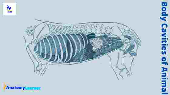
All the cavities are well-separated from each other and possess a distinct boundary. Here in the diagram, I tried to show you all the cavities from the animal body as a whole.
But, don’t worry; you will get detailed information about the individual cavity with their boundary and contents.
Body cavities and organs
Now, I will show you the boundary of all the major body cavities and organs from an animal. Make sure you know the anatomical facts of the animal skeleton and some other structures like a diaphragm, peritoneum, and others.
In each animal cavity, you will find the roof, floor, rostral wall, caudal wall, and lateral wall. You may also find the rostral opening and posterior opening in each cavity.
The cranial cavity of an animal is an oval elongated irregular structure that contains the brain. Again, the nasal cavity of an animal is an elongated structure bounded by some facial bones.
The thoracic cavity of an animal is cone-shaped and laterally compressed. In addition, the abdominal cavity of the animal is the largest cavity that extends from the diaphragm to the pelvic inlet.
The vertebral cavity contains the spinal cord and the roots of the spinal nerves of an animal. Most of the visceral organs of the animal body are covered by the peritoneum. You will find two layers of the peritoneum – a parietal and one visceral layer. The cavity in between the parietal and visceral layers of the peritoneum is known as the peritoneal cavity.
Again, the heart is covered by the pericardial covering, where you will also find two layers. These two layers of the pericardium also form the pericardial cavity. You will also find a pleural cavity in the covering of the animal lung.
All these body cavities of an animal contain important organs and structures that I will enlist at the end of the individual cavity’s description.
The cranial cavity of the animal
The cranial cavity of the animal contains the brain, with its covering and vessels. Its capacity varies more with the body size than the head shape. This is the most oval elongated irregular shape space among all body cavities of the animal.
The boundaries of an animal’s cranial cavity may be considered a base, roof, rostral wall, caudal wall, and lateral wall. Let’s see the bones that involve in the different walls of the cranial cavity.
- Roof – bounded by the frontal bone (parietal and occipital bone; partly)
- Floor – bounds by the basilar part of the occipital and sphenoid bones
- The rostral wall – bounds by the ethmoid and sphenoid bones,
- Caudal wall – bounded by the occipital bone,
- The lateral wall – bounds by the temporal, orbital, and wing of sphenoid bones (parietal and frontal bones; partly)
So, the roof of the cranial cavity forms the frontal and parietal bones. But the interparietal process of the occipital bone also contributes to the roof of the cranial cavity in the animal.
The base of the animal cranial cavity divides into rostral, middle, and caudal cranial fossae. It forms by the basilar part of the occipital and sphenoid bones.
The rostral two-third of the base of the cranium forms by the sphenoid bone. Again, the caudal third of the cranium forms by the basioccipital bone. You will also find the contribution of the ethmoid bone (cribriform plate) in the formation of the rostral wall of the cranial cavity.
In addition, the caudal wall of the cranial cavity forms by the occipital bone. Both lateral walls of the cranial cavity form by the temporal, parietal and frontal bones.
Contents of the cranial cavity: Brain, it’s covering, and vessels.
The base of the animal cranial cavity
Here, I will provide a little information on the different divisions of the base of the animal cranial cavity. You know there is three-division (rostral, middle, and caudal cranial fossae) present in the base of the cranial cavity.
The rostral cranial fossa supports the olfactory bulbs, tracts, and remaining part of the brain’s frontal lobe. It continues rostrally by the concave cribriform plate. Several cribriform foramina perforate this cribriform plate.
The transverse body of the presphenoid bone forms most of the floor of the rostral cranial fossa.
The middle cranial fossa locates at a more ventral level than that of the rostral fossa. In addition, the floor of the middle cranial fossa forms by the body of a basisphenoid bone. Caudally, it is limited by the rostrodorsal surface of the petrosal parts of the temporal bone.
The middle cranial fossa of the cranial cavity gives accommodation to the cerebral hemispheres.
Again, the caudal cranial fossa forms by the dorsal surface of the basisphenoid bone and the petrous part of the temporal bone. It locates caudal to the middle cranial fossa. In addition, it is bounded rostrally by the dorsum sellae and ends at the foramen magnum.
In this part of the animal cranial cavity, you will find the cerebellum, pons, and medulla oblongata.
The nasal cavity of an animal
The nasal cavity is an elongated space and the facial part of the respiratory tract of an animal. It consists of two symmetric halves (facial bones) separated by the nasal septum. You will find the septal cartilage in the rostral part of the nasal septum of an animal.
Again, in the caudal part of the animal nasal septum, you will find the septal process of the frontal and nasal bones. First, let’s see the bone involvement in the structure of the nasal cavity of an animal.
Roof – forms by the nasal and frontal bones
Floor – forms by the horizontal palatine, maxilla, incisive, and premaxilla bones
Lateral wall – forms by the maxilla, nasal process of the incisive bone; partly by the palatine and pterygoid bones.
Nasal septum – forms by the vomer and ethmoid bones (partly)
Rostral opening – bounded by the premaxilla and the nasal bones
Posterior opening – bounded by the palatine, sphenoid and pterygoid bones
The bony nasal opening is formerly known as the piriform aperture. I hope you can understand the boundary of the nasal cavity of an animal. Now, you might know the different structures of the animal nasal cavity.
Hey, don’t forget to check the frequently asked question and labelled diagram sections on animal body cavities with their contents.
Structure of the animal nasal cavity
Each nasal cavity of an animal is filled largely by the ventral nasal conchae rostrally and ethmoturbinates caudally. You will find three nasal conchae in the nasal cavity of an animal.
- A dorsal nasal concha
- The ventral nasal concha, and
- A middle nasal concha
Another name of the dorsal nasal concha is nasoturbinate. It is a curved shelf of bone that protrudes from the ethmoidal crest into the dorsal part of the nasal cavity.
Again, another name of the ventral nasal concha is maxilloturbinate that protrudes from the choncal crest into the nasal cavity. The basal lamina of the ventral nasal concha curves medially and ventrally into the cavity.
The conchae divide the nasal cavity into four primary passages (known as meatuses). The dorsal nasal meatus locates between the dorsal nasal chocha and the nasal bone. Again, the middle nasal meatus locates between the dorsal nasal concha and ventral nasal concha.
In addition, the ventral nasal meatus locates between the ventral nasal concha and dorsum of the hard plate. You will also find the common nasal meatus in the longitudinal space between the conchae and nasal septum.
There is another nasopharyngeal meatus present in the nasal cavity that is responsible for air passage. This meatus extends from the caudal end of the ventral and common nasal meatus to the concha.
The thoracic cavities of the animal’s body
The thoracic cavity is one of the important spaces among all animal body cavities. It is a cone-shaped structure and bounded by subserous endothoracic fascia. It is formed by the thoracic vertebrae, ribs, sternum, muscles (including the diaphragm).
Let’s see the structures (bones and muscle) that involve in the formation of the thoracic cavity in an animal –
Dorsally – bounded by a series of thoracic vertebrae
Ventrally – forms by the sternum
Laterally – you will find thirteen pairs of ribs, cartilage of the sternal ribs (in ruminant) and some intercostal muscles; eighteen pairs of ribs in the horse.
Caudally – there is a diaphragm at the caudal part of the thoracic cavity
Cranially – bounded by the thoracic inlet
The thoracic inlet is the roughly oval opening into the cranial part of the thoracic cavity. It is bounded bilaterally by the first pair of ribs with their cranially extended costal cartilage.
Again, dorsally you will find the body of the first thoracic vertebrae. In the ventral part of the thoracic inlet, you will find the manubrium of the sternum.
The endothoracic fascia is the areolar tissue that attaches the costal and diaphragmatic pleurae to the underlying muscles, ligaments, and bones. You will find a scanty endothoracic fascia where it closely attaches to the costal pleura to the ribs.
In the thoracic wall, you will find the ribs and intercostal muscles on both sides. Again, dorsally there are bodies of the thoracic vertebrae and intervening fibrocartilages.
Contents of the thoracic cavity
In the thoracic cavity of the animal, you will find the following important organs –
The heart, lung, oesophagus, trachea, bronchi, aorta, brachiocephalic trunk, cranial and caudal vena cava, and vagus nerve.
Minor body cavities (in thoracic region)
In the thoracic region of an animal body, you will find some minor cavities like the pericardial and pleural cavity. Let’s know a little about these minor cavities of the animal body.
You know, the pleura is the serous membrane that covers the lung and lines the thoracic cavity wall. There are two surfaces of the pleura (parietal and visceral) that forms the pleural cavities.
The parietal pleura forms the wall of the pleural cavities. It is further designated as costal, mediastinal, and diaphragmatic pleura.
The costal pleura is the part of the parietal pleura that attaches to the medial surface of the lateral wall of the thoracic cavity. It is thin and firmly adheres to the medial surface of the ribs.
The mediastinal pleura forms the wall of the mediastinal space in the animal thoracic cavity. In addition, the diaphragmatic pleura is the pleural covering of the diaphragm.
The pericardium (heart sac) is the serous fibro envelope of the animal heart. You will find two parts in the pericardium of the heart – fibrous and serous parts.
The serous pericardium possesses two distinct layers (parietal and visceral) that form the pericardial cavity. It is the smallest serous body cavities containing the liquor pericardii (clear and light yellow fluid).
The parietal layer of the serous pericardium firmly attaches with the fibrous pericardium. Whereas the visceral layer of the serous pericardium firmly attaches to the heart muscle.
The abdominal cavity of an animal body
This abdominal cavity is the largest among all the body cavities of an animal. It extends from the diaphragm to the pelvic inlet of an animal. Here, I will show you the structured involvement in forming the abdominal cavity with its contents.
So, let’s see the structures (bones and muscles) that are involved to form the abdominal cavity in an animal –
Roof – the roof forms by the lumbar vertebrae, lumbar muscles and part of the diaphragm
Floor – the floor of the abdominal cavity forms by the two recti muscles, aponeurosis of the muscles of the lateral abdominal walls and xiphoid cartilage.
Cranial wall – forms by the concavity of the diaphragm
Caudal extremity – continue with the pelvic cavity
Lateral wall – form by the external and internal oblique muscles, transverse abdominal muscle, and some part of the few caudal ribs
You will find the parietal peritoneum that attaches to the abdominal wall with the help of subserous tissue. Again, the muscles are covered by a superficial and deep fascia.
The deep fascia is a sheet of elastic tissue and intimately attached to the extraneous oblique muscle. This is also known as an abdominal tunic that support the muscles to bear the weight of the abdominal viscera.
The three openings at the diaphragm pierce the abdominal cavity. These three openings are –aortic aperture, oesophagal aperture, and vena cava aperture.
Contents of the abdominal cavity
You will find lots of organs in the animal abdominal cavity. Let’s see the organs or structures that are present in the abdominal cavity of an animal.
You will find different parts of the stomach, spleen, kidney, large and small intestine parts, part of the ureter, part of the urinary bladder, and part of the uterus.
Different regions in the abdominal part
There are nine different regions in the abdominal part of an animal. The nine different regions of the abdomen are –
The right hypochondriac region contains the part of the right kidney, the caudate and main lobe of the liver, gall-bladder, and a part of the pylorus and beginning of the duodenum.
A left hypochondriac region contains the spleen, fundus of the stomach, reticulum and cranial end of the dorsal sac of the rumen. You will also find the part of the left kidney in the left hypochondriac region of the abdomen.
The xiphoid region contains the liver, stomach, pancreas, parts of the aorta, vena cava. You will also find the part of the reticulum, omasum, part of the abomasum, and cranial end of the dorsal sac of the rumen in the xiphoid region.
The left lateral abdominal region contains the caudal part of the kidney, part of the colon.
A right lateral abdominal region contains the caudal part of the right kidney, parts of the duodenum, and cecum.
Again, the umbilical region contains the part of the small intestine, mesentery, and duodenum. You will also find the ventral sac of the rumen and part of the small intestine.
The left inguinal region contains the left ureter, part of the colon, and a small portion of the female organs. In the right inguinal region, you will find the right ureter cecum, caudal flexure of the duodenum, and other parts of the intestine.
The prepubic region of the animal contains the urinary bladder, coils of the small intestine and the part of the uterus.
Minor body cavities in the abdominal region
You will also find some minor body cavities (oral and peritoneal) in the abdominal region of an animal. Let’s know how these cavities form in the animal.
The peritoneum is a serous membrane that lines the abdominal wall and is reflected over the viscera. You will find a surface mesothelium covering (simple squamous lining) and connective tissue framework in the composition of a peritoneum.
You will have two layers in the peritoneum – the parietal and a visceral layer. The parietal peritoneum cover a large part of the inner surface of the organ from abdominal, pelvic, and scrotal cavities. Again, the visceral peritoneum covers the organs of the abdominal, pelvic, and scrotal cavities wholly or partly.
Sometimes you may hear about two terms – retroperitoneal and intraperitoneal.
Retroperitoneal – organs that lie against the wall of the abdominal or pelvic cavities and cover only one surface by peritoneum.
Intraperitoneal – most organs project freely into the abdominal, pelvic, and scrotal cavities and receive nearly complete coverage of the peritoneum.
The oral cavity is an opening or space between the lips to the hard plate. The oral cavity of an animal divides into the vestibule and oral cavity proper.
The vestibule of the oral cavity is a space external to the teeth and gums and internal to the lips and cheeks. In addition, the oral cavity is proper bound dorsally by a hard plate and a small part of the adjacent soft palate.
Dorsally and rostrally, you will find the dental arches and teeth in the boundary of the oral cavity proper. Again, the floor of the oral cavity is properly formed by the tongue.
The pelvic cavity of an animal
The pelvic cavity is another important space in the animal body. Let’s see the structures involved in forming the animal pelvic cavity.
The floor of the pelvic cavity – forms by the ventral part of the ischium and pubis bones
The roof of the pelvic cavity – forms by the bodies of the sacrum and a few coccygeal or caudal vertebrae
The pelvic inlet – is a cranial opening of the pelvic cavity. Dorsally you will find the bodies of the first sacrum vertebrae. The two lateral walls of the pelvic inlet form by the body of the ilium and ischium bones. Again, you will find the iliopubic eminence and cranial part of the pubic symphysis ventrally.
The pelvic outlet – is the caudal opening of the pelvic cavity. Dorsally, you will find the body of the last sacrum vertebrae and ischial arch ventrally.
The most important organs of the animal pelvic cavity are – urinary bladder, parts of the uterus, parts of the large intestine, parts of the ureter.
Body cavities diagram from an animal
You already got a different diagram of the body cavity from an animal. Here, I will show you again some of the cavities in one diagram. But, if you need more updated diagrams on the body cavity, you may join anatomy learner on social media.
I hope you will find a lot of diagrams on social media of anatomy learners that might help you understand the gross anatomy of an animal.
Frequently asked questions on body cavities of an animal
Now, I will try to solve some inquiries on the body cavities of an animal that the learners commonly ask. If you have any questions related to the body cavity, please let me know.
What are the 7 major body cavities?
As I told you before, there are seven major body cavities in an animal. You will find cranial, thoracic, abdominal, pelvic, vertebral, orbital cavities in an animal. If you read the whole article, you will find an informative description about all these cavities from an animal with their contents.
So, I would like to request you to read the full article on the body cavity to get a basic idea.
What are the 4 major body cavities?
There are different major and minor cavities in the animal. Here, I will tell you the most important four cavities from an animal. The most important four body cavities of animals are – cranial cavity, thoracic cavity, abdominal cavity, and pelvic cavity.
What is the body cavity?
The body cavity is a space or compartment in the animal’s body that hold and protect the visceral or soft organs. There are several minor and major cavities in an animal that hold and protect the important organs.
How many body cavities are there?
In an animal, you will find major cavities like cranial, thoracic, abdominal, pelvic, vertebral, orbital. Again, you will find some minor cavities like – peritoneal, oral, pleural, scrotal, pericardial, and others.
What body are cavities located superior to the diaphragm?
The cavity that locates superior to the diagram is the thoracic cavity. Some important organs like the heart, lung, parts of the aorta, cranial and caudal vena cava are present within this thoracic body cavity of an animal.
What are the major body cavities and their organs?
Are organs inside body cavities?
What are the main body cavities?
Conclusion
I think this article might help you get an idea of the different body cavities of an animal with their contents. As a beginner of anatomy learning, you might learn the boundary of all these body cavities and organs perfectly with the labelled diagrams.
You might also practice and identify these body cavities with their contents from the actual sample of an animal.