Lymphocytes under a microscope show a round to slightly indented nucleus with clumped heterochromatin. You know these lymphocytes are agranulocytes and form the second-largest population of white blood cells.
This article will show you the structure (morphology) of different types of lymphocytes (small, large, B, and T) under light and electron microscopes. So that after completing this article, you can identify the lymphocytes from the microscope slide.
Again, you may easily differentiate lymphocytes from other white blood cells like monocyte and basophil. So, if you want to know the histological features of lymphocytes, let’s continue this article until the end.
Lymphocytes under microscope
Lymphocytes are numerous and constitute about 20 – 25% of all leucocytes in blood. But, the number of lymphocytes in peripheral circulation varies among species.
Lymphocytes under a light microscope are variable in their size. You may find smaller lymphocytes (6 -9 micrometers in diameter) larger than the red blood cells. Again, the larger lymphocytes measure up to 15 micrometers.
You will find numerous small lymphocytes in the blood, lymph circulation, and lymphatic tissue. A large number of lymphocytes are also present in the bone marrow.
The smaller lymphocytes are more common in the blood of dogs and cats. Again, you will find both large and small lymphocytes in the blood of cows, goats, and sheep.
You will find the lymphocytes as round cells in the normal blood smear. But, they may be pleomorphic when they migrate through connective tissue.
The rounded lymphocytes possess a round to slightly indented nucleus with clumped heterochromatin. You know lymphocytes are agranulocytes (have no granules), but sometimes, they may have a few azurophilic granules in their cytoplasm.
These azurophilic granules of lymphocytes may find in the periphery of the cytoplasm. The small lymphocyte possesses a large nucleus and a modest pale blue cytoplasm.
In addition, the large lymphocytes also have similar features. You will find 2 types of large lymphocytes – lymphoblast and ideal lymphocyte.
Here, the lymphoblasts are capable of dividing to form smaller lymphocytes. In contrast, the ideal lymphocytes have no nucleoli in their nucleus and present antigens.
Lymphocytes microscope slide identification
You may be asked to identify the lymphocytes from the microscope slide at your histology learning laboratory. Here, I will enlist some of the identification points of the lymphocyte microscope slide so that you may easily identify them.
Let’s see the identification points for lymphocytes and identify them from the microscope slide –
- The sample microscope slide shows a large round and smooth cell with a rounded nucleus (somewhat eccentrically located),
- Here, the rounded nucleus occupies most of the cell and possesses a dense, heterochromatin,
- The peripherally located sparse cytoplasm shows a light blue color which contains a few azurophilic granules,
So, this is the lymphocytes microscope slide. It is very hard to differentiate the small and large lymphocytes from a histology slide with the help of a light microscope.
They both possess similar histological features, only the large lymphocyte lack nucleoli.
You may easily differentiate lymphocytes from other white blood cells with the help of their histological structure. If you want to know their (white blood cells) basic structural difference, let’s continue the article.
I will show you the important differences in their histological features, which may help you differentiate these cells.
Lymphocytes in peripheral circulation in different species
As I told you before, the lymphocytes in peripheral blood circulation vary in animal species. Let’s see how the number of lymphocytes varies among different species –
- In dog, cats, and horses – lymphocytes account for 20 – 40% of the total white blood cells (leukocytes),
- The cow, mice, and pigs – contain 50 – 60% lymphocytes of the total leukocytes,
Lymphocytes have a key component of adaptive immune response in animals. These lymphocytes play an important role in cell mediate (T – lymphocytes) and antibody mediates (B – lymphocytes) immunity.
Now, I will discuss the different types of lymphocytes from the circulating blood, lymph circulation, bone marrow, and lymphoid tissue.
Types of lymphocytes in animal
The lymphocytes in the circulating blood and lymphoid tissue may divide in 2 ways. According to the morphology (size), lymphocytes may be divided as follow –
- Small lymphocytes – have 6 – 9 micrometer diameter,
- Medium lymphocytes – diameter is about 10 – 13 micrometers, and
- Larger lymphocytes – have more than 15-micrometer diameter,
The medium and larger lymphocytes in the circulating blood are less in number compared to small lymphocytes. Earlier in this article, you already have a few microscopic features of the small and larger lymphocytes. But, I will later describe the detailed features of these lymphocytes under a microscope (small, medium, and large) in this article with the labeled diagram.
Again, functionally the lymphocytes are subdivided into 3 major types –
- B – lymphocyte ( B cells),
- T – lymphocytes ( T cells), and
- Null cells (NK cells),
It is very hard to distinguish the B – lymphocyte, T – lymphocyte, and Null cells under the light microscope with the help of their morphology. But, you may easily understand the types of these cells immunohistochemically by the differences in their surface marker.
In animal circulating lymphocytes, you may find 80% T – lymphocytes. In addition, 15% are the B –lymphocytes, and the remaining are the Null cells.
Again, the lifespan of these T and B lymphocytes may widely differ. Here, the T – lymphocytes may live for years, whereas the B – lymphocytes last for a few months.
Now, let’s see the microscopic features of functionally divided three lymphocytes.
T – lymphocytes under a microscope
Under the immunohistochemical process, you may easily understand the T – lymphocytes with their surface marker. Here, I will discuss the microscopic and general features of T – lymphocytes from animal leukocytes.
Here, the T- lymphocytes are formed in the bone marrow and migrate to the cortex of the thymus. Now, they become immunocompetent cells.
These T – lymphocytes can recognize the epitopes displayed by the major histocompatibility complex molecule (MHC molecule). The only function of these T –lymphocytes (T cells) is in cellular mediate the immune response.
Again, they also (T cells) are responsible for forming cytokines. These cytokines may facilitate the initiation of the humoral-mediated immune response.
There are various types of T – lymphocytes in the lymphoid tissue. Each of the T cell subtypes possesses –
- A T cell receptor surface determinant and
- The cluster of differentiation determines,
Note: Generally, lymphocytes have no function in circulating blood, but these cells are responsible for the proper functioning the immune response when they are in connective tissue.
To become immunocomponent cells, lymphocytes should migrate to specific tissue from the bloodstream and represent the specific surface marker and receptors. Both the B and T cells can proliferate and differentiate into two subtypes after stimulation by the specific antigen –
Memory cells (maybe T or B memory cells) – these memory cells do not participate in the immune response. But, they remain as the immunological memory cells which can undergo cell division and response against a specific antigen,
Effector cells – are the immunocompetent cells that perform their immune functions. They divide into major B and T cells with their different subtypes.
B – lymphocyte and NK cell under a microscope
Again, the electron microscope can reveal the presence of B – lymphocyte and NK cells with the help of their surface marker. Here, the B – lymphocytes are formed and become immunocompetent within the bone marrow of the animals.
These B – lymphocytes are responsible for the humoral-mediated immune response. So, they differentiate into plasma cells, and these cells produce antibodies against the antigens.
In the structure of the B – lymphocytes, you will find the major histocompatibility molecule surface marker. Again, these B cells also possess surface immunoglobulin (Ig) on their plasmalemma.
Here, the B – lymphocytes express IgM and IgD on their surface. Again, B cells possess the specific markers – CD9, CD19, CD20, and CD24.
Null cells comprise 2 distinct populations – circulating stem cells and Natural k-cells. The circulating stem cells give rise to all of the formed (1) elements of blood.
Natural k-cells fight against foreign and virally altered cells. They (nk) never influence the thymus or T cells.
The natural k-cells are larger than these of the T and B lymphocytes. You will find several large cytoplasmic granules in the cytoplasm of natural k-cells.
For this reason, this cell (type of B – lymphocytes) can be identified under a light microscope. But, you need to perform the immunohistochemical analysis of that cell to be confirmed.
The natural K-cells possess a kidney-shaped nucleus. Several larger granules exist in their cytoplasm, so this cell is also known as the larger granular lymphocyte.
Their cell surface (plasmalemma) shows the specific markers – CD16, CD56, and CD94.
Subtypes of T – lymphocytes
There are 3 basic types of T –lymphocytes – cytotoxic, helper, and suppressor. Identifying these specific types of T – lymphocytes with an immunohistochemical staining procedure is possible.
Here, the cytotoxic T –lymphocytes act as the main effector cell in the cell-mediated immune response. You know CD8 cells specifically synthesis T – lymphocytes which can recognize the antigen on the host cell.
When the antigen bind to the major histocompatibility molecule, cytotoxic T – lymphocyte can only recognize it at that time. Now, the T – lymphocyte secretes lymphokines and perforins, which can produce the ion channel in the membrane of infected cells.
Helper CD4 T –lymphocytes induce the immune response in the case of foreign antigens. Any antigen bind to the major histocompatibility molecule can be presented by antigen-presenting cells (macrophage) to the Helper CD4 T – lymphocytes.
Now, the T – lymphocytes produce interleukin II, which acts in the autocrine mode. This condition stimulates the proliferation and differentiation of more helper CD4 T – lymphocytes.
These newly differentiated cells release lymphokines, affect function, and differentiate T and B cells. Now, the T cells differentiate into plasma cells and synthesize antibodies.
Suppressor T – lymphocytes suppress antibody formation by the B cells. These cells also downregulate the ability of the T lymphocyte.
Now, they initiate the cellular immune response. The suppressor cells also regulate the erythroid cell maturation in the bone marrow of animals.
Lymphocyte under the electron microscope
You may easily identify and understand the organelles and other different segments of lymphocytes under the electron microscope. The general features, like the shape of the lymphocyte and its nucleus, are similar under the electron microscope.
That means you will see the rounded cell with an eccentrically placed nucleus under the electron microscope. The normal light microscope shows a smooth outer surface (plasmalemma of lymphocytes). But, under the electron microscope, you will find little different features in the outer surface of the lymphocyte (plasmalemma).
There may be short microvilli present on the outer surface of lymphocytes under the electron microscope. Again, the rounded eccentrical nucleus of the lymphocyte with clumped heterochromatin may easily understand under the electron microscope.
The scanty amount of peripheral cytoplasm of lymphocytes shows few mitochondria, small Golgi bodies, and few profiles of rough endoplasmic reticulum (under electron microscopic view). A small amount of the lysosome, azurophilic granules, and an abundant supply of ribosomes are also present in the lymphocyte.
On the other hand, T cells and B cells (lymphocytes) are indistinguishable under the light microscope if you perform a normal blood smear. You may easily understand their structure with the help of immunohistochemical staining.
This staining aims to identify the different types of markers and receptors on their cell surfaces. I have already enlisted the markers from the T and B – lymphocytes in the previous section of this article.
Finally, null cells (type of lymphocyte) can be distinguished under the light microscope. With the help of their shape, size,
nucleus size, and the presence of larger cytoplasmic granules, you may easily identify these null cells from the normal blood smear.
But, it is highly recommended to identify these null cells with the immunohistochemical staining for their specific markers.
How do lymphocytes form? (Lymphopoiesis)
The term lymphopoiesis means the production of new lymphocytes, including the T cells, B cells, and null cells. In very short, I would like to describe the process of lymphocyte formation with a labeled diagram.
So, first, let’s see the diagram of lymphopoiesis (a process of lymphocyte formation). This diagram might help you to clear the basic concept of lymphocyte formation.
Like the other hematopoietic cells, lymphocytes also originate from the pluripotential stem cells. You will find the earliest progenitor cells (lymphoblast) in the development process.
The number of lymphoblasts is very low in the bone marrow of any animal. Again, it is very difficult to distinguish the earlier lymphoblasts from rubriblasts and early myeloblasts.
The lymphoblast cells possess a spherical nucleus, fine chromatin, and a variable number of nucleoli. In lymphoblast cells, you will also find a thin rim of deeply basophilic agranular cytoplasm.
Now, the lymphoblast cells of this process undergo division and differentiate to produce prolymphocytes. Under certain conditions, these prolymphocytes subsequently differentiate into the ideal lymphocytes.
But, the number of mitotic divisions between the progenitor stem cell and the last stage of this process is unknown. In most of the bone marrow of animals, lymphocytes are small with clumped chromatin. You will not find any distinct nucleoli in these lymphocytes (bone marrow).
You will find 20% active lymphocytes in the dog bone marrow, whereas 10% active lymphocytes are found in the other species’ bone marrow.
T and B cells production
In the process of lymphopoiesis, the cloning-forming unit of lymphocyte divides and differentiates into cloning-forming B and T lymphocytes. B lymphocyte B cell is formed from the cloning forming, and subsequently, it divides to form the plasma cells and memory cells.
Again, the cloning forms unit T lymphocytes from the T cells. From these, T cells immediately divide into 3 main subtypes – cytotoxic cells, T helper cells, and T suppressor cells.
Sometimes, some lymphoid precursor is believed to leave the marrow cavity and go to the thymus. Within the thymus, these precursors differentiate into the thymic or T lymphocytes.
The bone marrow contains a small number of lymphocytes which contain an abundant amount of light basophilic cytoplasm. Again, these lymphocytes in the bone marrow also contain larger azurophilic granules.
So, these lymphocytes are sometimes known as the larger granular lymphocytes. They may be natural K-cells or cytotoxic T lymphocytes.
These two types of cells play a major role in tumor resistance and host immunity. So if there is any viral or microbial infection in the host, these two types of cells may play a major role in host immunity.
T and B cells in lymphopoiesis
There are 2 types of main cells that are produced in the process of lymphopoiesis – T and B lymphocytes. These 2 major cells play an important role in cell-mediate and humoral-mediated immunity response (system).
You can not distinguish the T and B lymphocytes under the light microscope with their morphological features. They need to special staining method (like immunophenotyping) to understand their actual structure.
The bone marrow and Peyer’s patches are the site for B – lymphocyte maturation in most mammals. And you know this B – lymphocytes express the cell surface immunoglobulins.
Again, B lymphocytes undergo further differentiation in the bone marrow and become plasma cells. In this process, a small number of B cells (memory cells) are also produced.
You may easily distinguish the plasma cells from the resting lymphocytes. Here, in the plasma cells contain more abundant, deeply basophilic cytoplasm, prominent light–stained Golgi complex, and an eccentrically placed nucleus.
In the case of a bird, the maturation of B – lymphocytes occurs in the cloacal bursa or bursa of Fabricius.
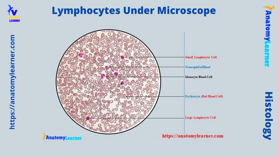
Lymphocyte under microscope labeled
Now, in this section, I will show you both the small and large lymphocytes under the light microscope with the labeled diagram. You know the lymphocytes of an animal’s blood vary in size and are larger or smaller than the erythrocyte.
Let’s see the below-mentioned labeled diagram, where I tried to show both the small and large lymphocytes. The red–staining erythrocytes surround these small and large lymphocytes.
The small lymphocytes constitute more percentage than these of the large lymphocytes in the blood leukocytes. Of most of the lymphocytes in the animal’s blood, about 90% are small lymphocytes.
You will only find 3% of larger lymphocytes in most animals’ circulating blood. So, the labeled diagram shows more small lymphocytes which are surrounded by the erythrocytes.
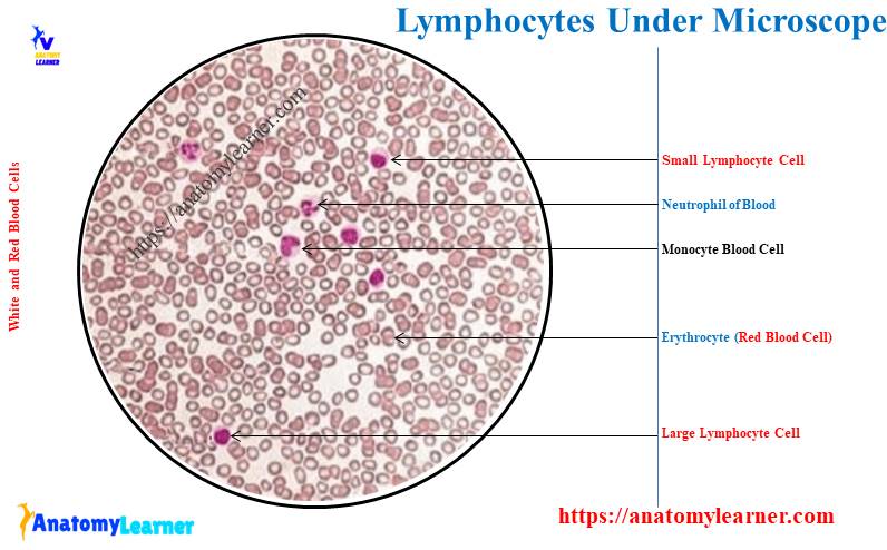
Small lymphocytes under a microscope
Though it is very difficult to distinguish between the small and larger lymphocytes from the microscopic slide. But, I will try to show you the basic difference between these 2 lymphocytes with the labeled diagrams.
First, let’s see the structure of both the small and large lymphocytes. Here, the most important feature of the small lymphocyte is that it possesses a relatively larger nucleus.
Again, this larger nucleus is surrounded by a thin rim of cytoplasm. The nucleus of the small lymphocytes appears as spherical and generally shows a small indentation to one side.
In addition, the nucleus of the small lymphocytes is intensely stained with routine slide preparation. The cytoplasm of the small lymphocytes is slightly basophilic and has a thin rim around the nucleus.
Normally, the granules (azurophilic) can not see from the small lymphocytes under the routine stain. But, in some special stains for the lymphocytes, you may see a few granules in the cytoplasm of the small lymphocytes.
I hope you can identify the small lymphocytes from the microscopic slide with the help of these labeled diagrams and features. Now, let’s see what the large lymphocytes look like in the microscope.
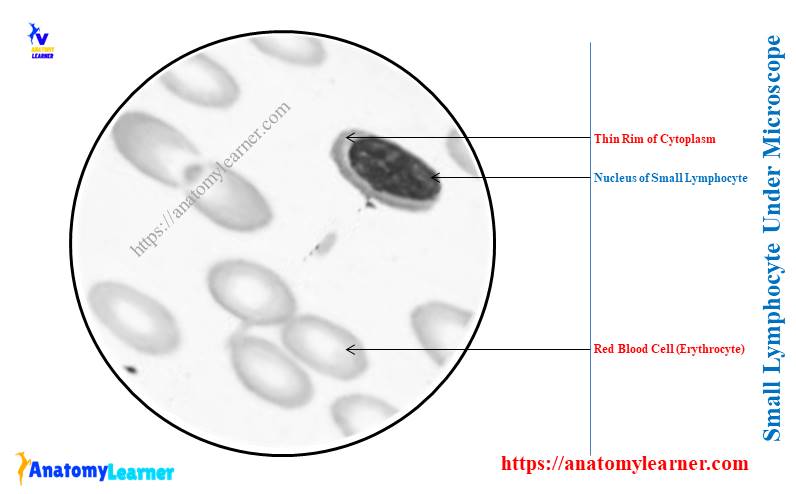
Large lymphocytes under a microscope
As the number of large lymphocytes is few in total leukocytes, you may hardly find one or two cells in the microscope focus. Here, I tried to show you the large lymphocyte under the microscope with the labeled diagrams.
Again, I will provide the important features of large lymphocytes so that you can easily distinguish them from the small ones.
The labeled diagram shows a larger nucleus in the center position of the cell (lymphocyte). But, the nucleus of the larger lymphocyte is paler compared to the nucleus of the small lymphocyte.
Again, this nucleus of the smaller lymphocytes doesn’t show any distinct nucleoli. Now, let’s see the amount and state of the cytoplasm of the larger lymphocytes. Have you found any difference between the cytoplasm of large and small lymphocytes?
The amount of cytoplasm of the large lymphocytes is abundant compared to the small lymphocyte. Again, the cytoplasm of the large lymphocyte is more basophilic compared to the small lymphocyte.
So, the basic difference between the large and small lymphocytes is found in their position in the nucleus, the structure of nucleoli, and the amount of cytoplasm. I hope you can also identify the larger lymphocytes from the blood smear slide and distinguish it from the small lymphocytes.
You may see more labeled diagrams on different types of lymphocytes here on the social media of anatomy learners.
Difference between monocyte and lymphocyte under a microscope
Sometimes you may be asked to differentiate between monocyte and lymphocyte under the microscope. As you know the all-important histological features of blood lymphocytes, you may compare this cell with the monocyte.
Let’s know some of the basic features of blood monocyte with the labeled diagram. Here, I will only enlist some of these important histological features by which you may easily identify this cell under the light microscope.
Monocytes are the largest active leukocyte in the peripheral circulating blood of animals. The diameter of the blood monocyte may vary from 12 – 20 micrometers.
The nucleus of the blood monocyte possesses some distinct and peculiar features. Here, the monocyte possesses a large, centered, notched or indented (horse-shoe-shaped) nucleus.
If you see this monocyte with the help of an electron microscope, you will find the fine reticulated chromatin network. The cytoplasm of the blood monocyte is more than that of large or small lymphocytes.
Again, the cytoplasm also presents some other special features compared to the cytoplasm of lymphocytes. You will see the numerous dust-like fine granules and also the vacuoles in the cytoplasm of the blood monocytes.
So, these are the basic (primary) difference between the blood monocyte and lymphocyte under the light microscope. Now, let’s see the summary of the different microscopic features between the blood monocyte and lymphocyte in Table 1 –
| Features | Monocytes | Lymphocytes |
| Cell | Rounded larger | Rounder larger |
| Nucleus | Horse shoe shaped | Rounded |
| Cytoplasm | Abundant | Thin rim surround the nucleus |
| Granules | Fine dust like | Few (occiassionally) |
| Vacuoles | Present | No vacuoles |
I hope you can easily differentiate the monocyte from the lymphocyte under the light microscope.
Frequently asked questions on lymphocytes under a microscope
Now, I will try to concisely answer the frequently asked question on lymphocytes under a light microscope. But, it is highly recommended to read the full guide on the lymphocytes from start to end.
Let’s see the commonly asked questions on the lymphocytes by the learners.
What do lymphocytes look like under a microscope?
Lymphocytes look like rounded cells with a rounded nucleus under a light microscope. As the circulating peripheral blood of animals contains numerous small lymphocytes, you will see a densely stained nucleus.
There is a thin rim of cytoplasm (basophilic) that surround the larger rounded nucleus of the small lymphocytes. The nucleus is somewhat eccentric in the case of small lymphocytes.
If you notice the structure of the nucleus, you will find distinct nucleoli and clumped chromatin under the light microscope. All these features of small lymphocytes (microscopic features) are described (enlisted) in detail in the previous section (1) of this article.
Again, the large lymphocyte shows similar features under the light microscope. But, their rounded nucleus locates at the center of the cell.
You will see more cytoplasm that surrounds the nucleus of the larger lymphocyte under the light microscope.
How do you identify lymphocytes?
Identifying the lymphocytes from a normal blood smear is a straightforward task. You may easily identify the lymphocyte’s basic structure (morphology) under the light microscope (especially the small and large lymphocytes).
But, you might perform immunohistochemical staining to visualize the organelles from the small and large lymphocytes. This immunohistochemical staining also helps you to identify the T and B lymphocytes.
Again, you may identify the null cells (natural k-cells) under the light microscope with the normal blood smear. But, to confirm these natural k-cells, you might perform the immunohistotyping.
All these identification processes are described (enlist) in detail in the previous section (2) of this article. So, let’s get the full concept on lymphocyte identification from that part of this article.
What are the characteristics of lymphocytes?
Morphologically, lymphocytes are 3 types – smaller, medium, and larger. The smaller lymphocytes are more numerous in the peripheral circulating blood. You may also see numerous small lymphocytes in the lymphatic circulation and lymphatic tissue.
The lymphocyte cell profile is rounded and smooth under the light microscope. But, this same cell (lymphocyte) may show smaller microvilli on their surface or plasmalemma.
There is a great variation in the cytoplasm between the small and larger lymphocytes. Again, the nucleus of both small and large lymphocytes shows a little difference.
How to differentiate lymphocytes from neutrophils and basophils?
Under the light microscope, neutrophils show 3 – a lobed nucleus with a rounded cell. The cells’ size of the neutrophil is smaller compared to the lymphocyte.
Again, the cytoplasm of the neutrophil contains fine granules which can easily be visible under the light microscope. The cytoplasm is more abundant than the cytoplasm of lymphocytes.
Again, the basophil contains a bilobed nucleus (which seems like as rounded structure) with a small rounded cell. You will find coarse granules in the cytoplasm of a basophil cell, whereas a few granules may occasionally be found in a lymphocyte’s cytoplasm.
What is the basic difference between erythrocytes and leukocytes?
You may differentiate the erythrocyte from different types of leukocytes in different ways. Here, I will provide some essential features that help you distinguish the erythrocyte from other leukocytes.
Leukocytes are the cells that possess a nucleus, mitochondria, Golgi bodies, and other organelles. In the erythrocyte, you will find hemoglobulin, whereas the leukocytes don’t possess any hemoglobulin.
Thus, they become colorless in unstained slide preparation. The erythrocyte don’t have any mobility of their own, whereas leukocytes can move actively.
Erythrocytes can’t move from the vascular system, but the leukocytes can move from the vessels to the surrounding or particular tissue.
Conclusion
Both small and large lymphocytes under a microscope will show different distinct features. With the help of labeled diagrams and identification points, you may easily distinguish these lymphocytes from the microscope slide.
Functionally classified lymphocytes – T, B, and Null cells required the special stain to visualize their structure. The amount of cytoplasm, the size of the nucleus, and their position are key features to distinguish the small and large lymphocytes under the light microscope.