The adipose tissue under a microscope shows an aggregation of fat cells or adipocytes. You will find this adipose tissue subcutaneously throughout the body except over the eyelid, auricle, and other parts of the animal’s body.
The adipose tissue is considered the specialized connective tissue which plays an essential role in energy homeostasis. Microscopically, you will see 2 types of adipose tissue in the animal body – white adipose tissue and brown adipose tissue.
This article will help you know the histology of the adipose tissue under a light microscope. So that you will identify and differentiate the white and brown adipose tissue from microscope slides.
Again, I will provide adipose tissue labeled diagrams at the end of this article which might also help to understand their structure perfectly. Finally, at the end of the article, you will find the concise answer to some of the common inquiries on the adipose tissue microscope slide.
So, let’s continue this article to learn the microscopic features of adipose tissue.
Adipose tissue under a microscope
First, I would like to point out the most important features of adipose tissue under a microscope. The microscopic features of adipose tissue show 2 types of appearances –
- Yellow (white) or unilocular adipose tissue (adult type), and
- Brown or multilocular adipose tissue (embryonic type),
When the individual fat cell (adipocyte), and a group of adipocytes aggregate throughout the loose connective tissue and form the adipose tissue. So, first, you should know the structure of the adipocyte and loose connective tissue.
You may learn the details of histological fats of loose connective tissue from the below-mentioned article of anatomy learner –
- Loose connective tissue histology with labeled diagram,
The cells of white fat (adipocyte) are separated by loose connective tissue. Each adipose cell is surrounded by a delicate collagen and reticular fibers network.
These delicate networks of collagen and reticular fibers of white adipose cells support a dense capillary plexus and nerve fibers. You will also find a narrow intercellular space (in between white adipose cells) that may contain few fibrocytes, mast cells, and sparse amorphous ground substances.
On the other hand, brown adipose tissue composes of aggregates of multilocular adipocytes. The intercellular connective tissue (between brow adipose cells) contains fibrocytes, collagen, and reticular fibers.
Here, the capillaries from a dense plexus and the brown adipocytes are directly innervated by adrenergic axons.
Now, I will show you the different microscopic features of 2 types of adipocytes (white and brown). So that you can understand the basic structure of the white and brown adipocytes structure perfectly and identify them from the light microscope easily.
Yellow adipocyte vs brown adipocyte under a microscope
If you see the diagrams of yellow and brown adipocytes structure, you may point out the following important features –
- Yellow adipocytes – a large rounded cell is containing a thin cytoplasm rim and a single unilocular lipid droplet. The nucleus of the yellow adipocyte locates at the periphery (flat nucleus).
- Brown adipocytes – is a small polygonal cell that possesses many multilocular lipid droplets and contains a spherical central nucleus,
Now, let’s see the main histological features of the white (yellow) and brown adipocytes under the microscope from table 1 –
| Features | White adipocytes | Brown adipocytes |
| Adipocyte | Large rounded | Small polygonal |
| Lipid droplets | Single – unilocular | More – multilocular |
| Nucleus | Flat, peripheral | Spherical, central |
| Endoplasmic reticulum | More | Less |
So, from table 1, you can understand the white or yellow adipocyte is the larger cell compared to the brow adipocyte. Again, the lipid contents of these adipocytes may vary, and this is an important feature to differentiate them under the light microscope.
You will find only a single unilocular lipid droplet in the white or yellow adipocyte. In contrast, the brown adipocyte shows many multilocular lipid droplets under the light microscope.
Again, table 1 also shows another important microscopic feature between the white and brown adipocytes. In the white or yellow adipocyte, you will see the flat nucleus located at the cell’s periphery.
On the other hand, the brown adipocyte contains a spherical nucleus at the center of the cell. So, now you may easily differentiate the yellow or white adipocytes from the brown adipocyte under a light microscope.
Adipose tissue under microscope identification
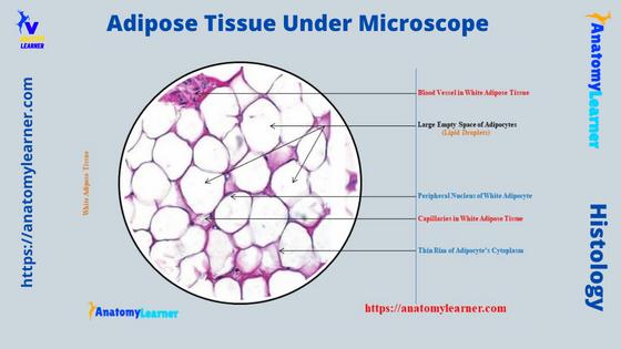
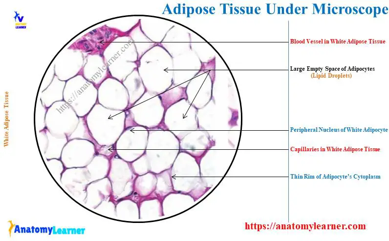
Sometimes you may be asked to identify white or brown adipose tissue under a microscope. The below-mentioned identifying features might help you to identify the adipose tissue from the microscope slide –
- The sample tissue section shows aggregates of white adipocytes,
- The center of each adipocyte appears empty as the fat in them gets dissolved during the preparation of the tissue section (and gives them a honeycomb appearance),
- Again, the cytoplasm of each white adipocyte shows a thin pink-colored rim,
- There is a flat nucleus in each white adipocyte that lies to one side or eccentrically,
- The sample tissue section also shows some capillaries (in between white adipocytes) that line with endothelium,
- There is also the presence of a delicate network of collagen and reticular fibers that surrounds each adipocyte or cell,
So, this is a white or yellow adipose tissue histology slide.
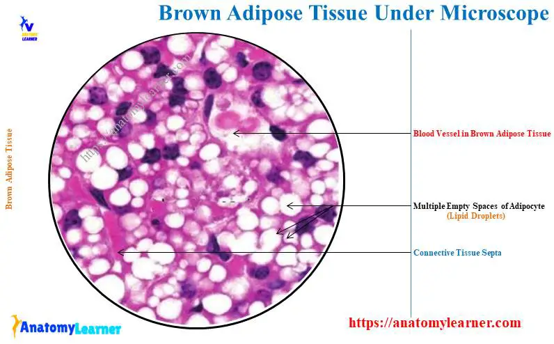
In this histology slide, the adipose tissue shows the larger rounded cell with a central empty space. But, if you see multilocular empty spaces (as there is the presence of lipid droplets dissolved during slide preparation) in the sample tissue section with a centrally placed nucleus, you may identify it as brown adipose tissue.
Sometimes you may be asked to identify the white and brown adipocytes under the light microscope. At that time, you might write only the microscopic features of the specific adipocyte.
You may easily write the identification points for individual white (yellow) and brown adipocytes if you read the previous section of this article. Again, I would like to point out the identifying characteristics of white and brown adipocytes below.
White or yellow and brown adipocytes identification
You will find white or yellow adipocytes in most parts of the animal’s body (generally throughout the loose connective tissue). These white or yellow adipocytes are also known as ordinary adipocytes.
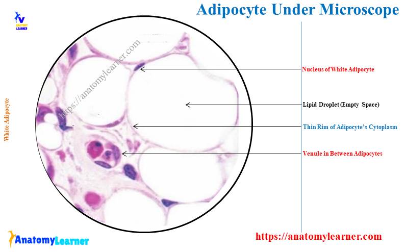
The white or yellow adipocytes show the below-mentioned microscopic features (identification points) –
- The sample tissue section shows larger rounded cell (compared to the brown adipocyte),
- Here, the cytoplasm and nucleus of this cell push to the periphery,
- The sample cell shows the cytoplasm as a thin rim (pink),
- Again, the nucleus is flat and located at the periphery of this adipocyte,
- The center of the cytoplasm shows a large single unilocular lipid droplet (empty space),
So, this is the white (yellow) adipocyte under the light microscope. Now, let’s try to identify the brown adipocytes from the microscope with the help of followings identifying characteristics –
- The tissue section shows smaller and polygonal adipocytes (compare to the ordinary adipocytes),
- Here, the cytoplasm of the sample cell shows mitochondria and many small empty spaces (especially lipid droplets),
- This adipocyte also shows a centrally placed spherical nucleus,
So, this is a brown adipocyte under a light microscope. The big difference between the white and brown adipocytes is in their nucleus and lipid droplets.
The fat in the cytoplasm of brown adipocytes occurs in the form of several small droplets. Thus the brown fat cell is also known as the multilocular adipose tissue.
I hope you learn all the basics of the adipose tissue microscope feature. Now, it’s time to learn the details of the adipose tissue (description) with the different labeled diagrams.
Adipose connective tissue under microscope description
In this article section, I will describe adipose connective tissue under a microscope with different labeled diagrams. You will find separate descriptions of white (yellow) and brown adipose tissue with their location, functions, and specific structure with diagrams.
So, this section contains the following topics on adipocytes and adipose tissue –
- Location or distribution of adipose tissue or adipocytes in an animal’s body,
- Specific functions of white and brown adipose tissue in the body, and
- Details microscopic features of the white and brown adipose tissue,
Again, I will show the development process of adipose tissue cells (differentiation of white and brown adipocytes) with the diagram. So, if you want to know these topics from adipose tissue with the diagram, let’s continue till the end.
Adipose tissue location
In very short, white adipose tissue distribution is widespread in animals. In contrast, the distribution of brown adipose tissue is limited and found in fetuses and newborns. Again, you will find brown adipose tissue in some parts of the animal’s body (described later in detail).
In a normal healthy individual, the white adipose tissue represents at least 10% of their body weight. This white adipose tissue form a fatty layer of subcutaneous fascia (known as the panniculus adipose). You will find this white adipose tissue in the connective tissue just beneath the skin.
Here, the subcutaneous fascia (made of white adipose tissue) provides significant thermal insulation against the cold by reducing the rate of heat loss.
In the mammary fat pads, you will find white adipose tissue. The mammary fat pad is essential in supporting the mammary gland’s functions.
The white adipose tissue also fills the several hollow spaces of the animal’s body. These hollow spaces of the animal body include orbits, axillae, and ischial fossae.
Internally, the white adipose tissue finds in the greater omentum, mesentery, and retroperitoneal spaces. You will find abundant white adipose tissue around the abdominal organs, especially the kidneys.
The white adipose tissue also fills in the bone marrow and in other tissue. Again, much adipose tissue is present in the synovial folds of many joints in the animal’s body.
You will also find the white adipose tissue in the paws, sole, beneath the visceral pericardium, and in orbit around the eyeball.
Brown adipose tissue location
Generally, you will find brown adipose tissue in the fetus and a newborn. But, you may also find brown adipose tissue in some parts of the animal’s body (different animal).
The brown adipose tissue is widespread and abundant in rodents and hibernating mammals. You will also find the brown adipose tissue in the following locations in the rodents and hibernating mammals –
- Axillary and neck regions (interscapular adipose tissue),
- Along the thoracic aorta and in the mediastinum, and
- In the mesenteries, around the abdominal aorta, and vena cava near the kidneys,
But other mammals (except hibernating mammals) also possess a small amount of brown adipose tissue in the exact locations.
Structure of white adipocyte and adipose tissue
When you view the isolated white adipocyte from the adipose tissue under a light microscope, you will see them as a spherical or oval structure. But, sometimes, they may become polyhedral when crowded in the adipose tissue.
The size of the white adipocyte depends on the accumulation of lipid droplets on its cytoplasm. Typically, you will see the larger white adipocyte due to many lipid droplets on their cytoplasm.
In the routine histologic section of white adipose tissue, the lipid droplet is lost through extraction by organic solvents like xylene. Thus, the adipocyte appears as the delicate meshwork of a polygonal profile.
You will see a thin strand of meshwork that separate adjacent adipocytes. And they also represent the cytoplasm of both cells (adipocytes) and the extracellular matrix.
This thin rim or strand of white adipocyte cytoplasm may easily identify under the light microscope as pink colored structure. Now, let’s see the shape and location of the white adipocyte nucleus under the light microscope.
The nucleus of the white adipocyte is flat and placed on one side of the lipid droplet. That means you will find the flattened nucleus in the periphery of the white adipocyte.
The white adipocyte surrounds by a delicate network of collagen and reticular fibers. They are richly supplied with blood vessels and capillaries.
You will find these vessels and capillaries at the angle of a network where adjacent white adipocytes meet. The narrow intercellular spaces between 2 adjacent white adipocytes show fibrocytes, mast cells, and little amorphous ground substance.
Adipose tissue structure
The microscopic figure shows the aggregation of many white adipocytes, which form the adipose tissue. So, in the adipose tissue histology slide, you will see the empty spaces with a thin rim of cytoplasm along with a flattened peripheral nucleus.
Except for the routine stain, you may use some other stain to visualize the structure of white adipose tissue. In the silver stain, the reticular fibers (type III collagen fibers) surround the individual white adipocyte.
Do you know who secretes these reticular fibers around the white adipocytes? The white adipocytes secrete these reticular fibers surrounding the individual white adipocyte. The special stain of the white adipose tissue shows some unmyelinated nerve fibers and numerous mast cells.
In the electron microscope, you will find a thick layer of parallel vimentin filament in between the lipid and cytoplasm of individual white adipocytes. This filamentous layer separates the hydrophobic contents of the lipid droplets from the hydrophilic cytoplasmic matrix.
Again, the perinuclear cytoplasm of white adipose tissue contains small Golgi bodies, free ribosomes, rough endoplasmic reticulum, microfilaments, and intermediate filaments. The thin rim of the adipocyte’s cytoplasm also shows the mitochondria and multiple smooth endoplasmic reticula.
Describe the appearance of brown adipose tissue under the microscope
The brown adipose (less) tissue is abundant in newborns and growing animals (stated before). They (brown adipose tissue) gradually reduce in the animal with age.
This brown adipose tissue is a key thermogenic tissue that helps to offset excessive heat loss. Thus it results from the newborn’s high surface-to-mass ratio and to avoid hypothermia.
In newborn animals, you will find 5% brown adipose tissue in their body weight. Usually, a newborn’s brown adipose tissue may be found on the back, along the upper half (arrow) of the spine, and towards the shoulder.
The amount of brown adipose tissue gradually reduces as the body grows. But they remain widely distributed in the cervical, axillary, paravertebral, mediastinal (sternum), sternal, and abdominal regions of the individual animal.
Again, you will find the brown adipose tissue in the following region of the animals – around the kidneys, adrenal glands, larger vessels, supraclavicular region, interscapular, paravertebral, and thorax region.
Microscopic appearance of brown adipose tissue
The cells of the brown adipose tissue are smaller than these of the white adipose tissue. Again, the shape of the brown adipose tissue (actually the brown adipocytes) is polygonal compared to the white adipocyte.
Here, the cytoplasm of the brown adipocyte contains many lipid droplets; hence the name is multilocular. But, the white adipocyte is unilocular as it contains only a single lipid droplet.
Now, let’s see the nucleus of the brown adipocytes (adipose tissue) from the diagram. Normally, the nucleus of the growing brown adipose tissue is spherical and eccentric in position (sometime in the center).
So, the nucleus is not flattened in the brown adipocyte like the white adipocyte (making a huge difference between 2 types of adipocytes or tissue).
In the routine stain, the cytoplasm of a brown adipose tissue shows large empty spaces. This is because the lipid droplets of the brown adipose tissue that occupies the spaces are lost during slide preparation.
You will not find numerous connective tissue fibers between brown adipocytes as in white adipose tissue. The connective tissue fibers are sparse in the brown adipose tissue structure.
A small amount of connective tissue forms septa that divide the brown adipose tissue into different lobules. You will find numerous blood vessels and capillaries in the structure of the brown adipose tissue.
Electron microscopic view of brown adipose tissue
You will find numerous large spherical mitochondria with numerous cristae in the structure of the brown adipose tissue under the electron microscope. Again, a small Golgi complex and only a small amount of rough endoplasmic reticulum and smooth endoplasmic reticulum are also found in the structure of the brown adipocyte’s cytoplasm.
The numerous mitochondria of the brown adipocytes possess a large amount of cytochrome oxidase, which imparts the brown color into the cells (brown adipocytes).
In the silver and special stain of brown adipose tissue, you may find numerous unmyelinated noradrenergic sympathetic nerve fibers. They (nerve fibers) locates among the fat cells.
Metabolism in brown adipose tissue generates heat in a process known as thermogenesis. Hibernating animals have a large amount of brown adipose tissue (enlisted previously).
This brown tissue serves as a ready source of lipids. After oxidization, this tissue produces heat to warm (heat production) the blood flowing through the brown adipocytes.
Thus, the brown adipose tissue maintains the body temperature in cold conditions. This type of heat production by the brown adipose tissue is known as nonshivering thermogenesis.
The brown adipose tissue is also found in non-hibernating animals and again serves as a heat source. Usually, the heat is produced by the brown adipose tissue when the sympathetic nervous system stimulates them.
So, the metabolic activity of brown adipose tissue (more) is regulated by the sympathetic nervous system and is related to the ambient outdoor temperature. Here, the metabolic activity of brown adipose tissue is largely (more) regulated by the norepinephrine released from the sympathetic nerve terminals.
They stimulate lipolysis and hydrolysis of triglycerides as well as increase mitochondrial expression.
Difference between white and brown adipose tissue
I hope you can understand the basic difference between white (yellow) and brown adipose tissue. Here, I will show you the basic difference between these 2 types of adipose tissue in table 2 –
| Features | White adipose tissue | Brown adipose tissue |
| Adipocytes | Unilocular, spherical, flattened nucleus, thin rim of cytoplasm | Multilocular, polygonal, round eccentric nucleus, |
| Precursor cells | Mesenchymal stem cells | Skeletal myogenic progenitor cells |
| Mitochondria | Few, elongated, few cristae | Many, large, round, more cristae |
| Innervation | Few sympathetic nerve | Noradrenergic sympathetic nerve |
| Vasculization | Few blood vessels | Numerous vessels and capillaries |
| Locations | Subcutaneous layer, Mammary gland, Omentum, mesenteries, Retroperitoneal space, Bone marrow, orbits, | Newborn, retroperitoneal space, Deep cervical, supraclavicular, Intrascapular, mediastinum regions, |
| Functions | Metabolic energy storage, Insulation, cushion, Hormone production, | Heat production (thermoregulation) |
| Growth | Throughout inter life | During fetal period |
| Differentiation | May undergo to brown adipose | Decrease with animal’s age |
Table 2 shows some essential differences between the white and brown adipose tissue. You may learn more about their metabolic activities, regulation of these adipose tissues, their growth, and differentiation from another article of anatomy learner.
Development of adipose tissue cells (differentiation)
The development process of white and brown adipose tissue is complex and divides into different stages. But, I will try to describe the development process of white and brow adipose tissue straightforwardly.
So, I will describe the development process of the following adipose tissue with a labeled diagram –
- Differentiation of white or yellow adipose tissue, and
- Differentiation of brown adipose tissue,
First, let’s summarize the differentiation of white and brown adipose tissue from the diagram below.
Here, the diagram shows the white adipose tissue differentiates from the perivascular mesenchymal stem cell. Under certain transcription factors (which will describe in the full development process), it becomes an early lipoblast or preadipocyte.
Again, the preadipocyte becomes midstage lipoblast under certain conditions. Finally, the midstage lipoblast forms the later lipoblast, followed by the white adipocyte.
Again, the differentiation of brown adipose tissue occurs in 2 ways – transdifferentiation of white adipose to brown and also from skeletal myogenic progenitor cells.
Now, let’s discuss the development process of white and brown adipose tissue. First, start with the differentiation process of white adipose tissue.
Differentiation of white adipocyte
First, let’s see the important features or stages of the white adipocyte differentiation process –
- White adipocytes differentiate from the perivascular mesenchymal stem cell under the control of retinoid Xe-receptor transcription factors,
- This adipose tissue begins to form midway through fetal development,
- The early lipoblast of this process looks like the fibroblast but develops small lipid inclusion and a thin external lamina,
- Now, the midstage lipoblasts (stage 2) become ovoid as lipid accumulation changes the cell dimension, and
- The white adipocyte is characterized by a single, large lipid droplet (arrow) surrounded by a thin rim of cytoplasm,
Now, let’s try to understand these developmental processes for white adipose tissue with details information.
Mesenchymal cells of white adipocyte
You know that white adipose tissue derives from the undifferentiated perivascular mesenchymal stem cell during embryonic development. These stem cells are associated with the small venules of tunica adventitia.
Here, the major transcription factors are – peroxisome proliferator-activated receptor gamma and retinoid Xe-receptor. These 2 receptors (factors) play an important role in white adipocyte differentiation and initiation of lipid metabolism.
Early lipoblast or preadipocytes are formed in this white adipocyte differentiation stage. Here, the 2 factors for white adipocyte differentiation are regarded as the master switch.
The lipoblasts initially develop within the fetus from the stromal vascular cell and the small blood vessels. In this stage, these lipoblasts remain free from lipid droplets.
Now, they become adipocytes at the early stage by expressing retinoid receptors transcription factor. The early adipocytes aggregated to form primitive fat organs.
Here in the primitive fat organ, you will see the proliferation of lipoblasts and capillaries. Now, the lipid droplets become to accumulate in the early adipocytes.
Early adipocyte of adipose tissue
The early adipocyte of adipose tissue under the elector microscope reveals some important features. These early lipoblasts have an elongated configuration with multiple cytoplasmic processes.
For these multiple cytoplasmic processes of the early lipoblasts, they look like fibroblasts. But, you will find small lipid droplets in the cytoplasm (arrow) of the early lipoblasts.
You will also find the numerous endoplasmic reticulum and Golgi bodies in the cytoplasm of the early lipobalsts of adipose tissue. The external lamina and numerous pinocytotic vesicles are also found in the structure of early lipoblasts.
These external laminae of the early lipobalst will help to distinguish them from the connective tissue cell. At the lipoblastic differentiation, the vesicles and rough endoplasmic reticulum increase.
Midstage lipoblast to white adipocyte
Under the electron microscope, the midstage lipoblast shows the oval configuration. The midstage lipoblast cell shows the eccentrically placed nucleus.
You will find excessive vesicles and small lipid droplets (arrow) around the nucleus of the lipoblast. These vesicles and lipid droplets may extend toward both poles of the midstage lipoblasts.
You may also find glycogen deposition at the periphery of the lipid droplets. More pinocytic vesicles and a distinct basal lamina appear in the early lipoblast cell.
Now, the midstage lipoblast differentiates from the later-stage lipoblast. In contrast, you will find lipid droplet accumulation in a single adipocyte structure. And the nucleus of the lipoblast pushes into the periphery of the cell.
In the final differentiation stage, the lipoblast increases in size and becomes more spherical. Many lipid droplets coalesce (identified) to form a single large lipid droplet that remains in the cell’s center.
You will find more smooth endoplasmic reticulum and less rough endoplasmic reticulum in the structure of this lipoblast. Now, the single large lipid droplet creates pressure and pushes the nucleus more peripherally.
Thus the nucleus of the white adipocyte goes to an eccentric position and produces a signet-ring appearance in routine stain (H&E preparation). As there is a single large lipid droplet in this cell, so it is known as the unilocular white adipocyte.
Differentiation of brown adipocyte
The brown adipocyte is derived from the common skeletal myogenic stem cell. These skeletal myogenic stem cell for brown adipocyte differentiation is found in the dermatomyotomes of developing embryo.
The transcription factors for brown adipocyte differentiation are different than the white adipocyte. Here, you will find the PG-coactivator transcription factor to differentiate the brown adipocyte.
These transcription factors regulate gene expression essential for brown adipocyte metabolism. This is a very complex process of brown adipocyte development, but typically, you may describe this process as follow.
Under the control of PG-coactivator transcription factors, skeletal progenitor myogenic cells become early lipoblast. The configuration of the early lipoblast shows the oval structure.
You will also find a small eccentrically placed nucleus in the early lipoblast. The cytoplasm of the early lipoblast also shows the diffuse deposition of lipid droplets.
Under certain conditions, the early lipoblasts differentiated into multiple brown adipocytes. They contain a polygonal shape with an eccentric nucleus and more lipid droplets.
Transdifferentiation of adipose tissue
Adipocytes can undergo white-to-brown and brown-to-white transformations. This transformation is a response to the thermogenic needs of an organism.
If the animal exposure to chronic cold temperatures, they need to increase its thermogenic condition. After prolonged exposure to cold an animal, the accumulation of white adipose tissue undergoes a brown phenomenon.
Again, the brown adipose tissue can also transform into white adipose tissue. This condition occurs when the energy balance (nutrition) is positive, and the body requires increased triglyceride storage capacity.
These transformation phenomena (white to brown and brown to white adipose tissue) are known as the transdifferentiation of adipose tissue.
Adipose tissue under microscope labeled diagram
Now, I will show you the 2 types of adipocytes (white and brown) and adipose tissue with the labeled diagram. This might help you understand the basic structure and identify the adipose tissue under a microscope.
First, let’s see the labeled diagram of white and brown adipocytes. Then I will provide the labeled diagrams of white and brown adipose tissue.
In the first labeled diagram, I tried to show you the structure of a white adipocyte. Here, the diagram shows a larger rounded cell with a central empty space.
The nucleus of the white adipocyte is identified from the labeled diagram (located at the periphery of the cell and appears as a signet ring). Here, the labeled diagram shows a thin rim (pink color) of the cytoplasm of the white adipose tissue.
Again, in the same diagram, I tried to show the structure of the brown adipocyte. Here, the shape of the cell is polygonal (spherical) and possesses an eccentric rounded nucleus.
The cytoplasm of the brown adipocyte labeled diagram shows multiple lipid droplets (you will see multiple empty spaces). In both diagrams of adipocytes (white and brown), I tried to show the connective tissue between the cells and different vessels.
Adipose tissue labeled
Now, I will show you the higher-magnified white adipose tissue labeled diagram. In the provided diagram of white adipose tissue, you will see several lobules of adipose cells.
Dense irregular connective tissue septa separate the individual adipose tissue lobules from other structures. Here, the aggregated adipocytes of white adipose tissue show a very thin rim of cytoplasm that surrounds large, single, fat-containing empty spaces.
The diagram shows a delicate, thin layer of connective tissue stroma in the white adipocytes. This thin, delicate connective tissue stroma shows small blood vessels (primarily capillaries and venules), and different types of connective tissue cells.
The diagram shows the clear difference between the fibroblast nucleus and white adipocytes. A thin cytoplasm with a peripheral nucleus gives the white adipose tissue the signet ring appearance.
Let’s see the brown adipose tissue labeled diagram, which shows the small adipocytes. These adipocytes are closely packed with minimal intercellular spaces.
Because of this type of arrangement of brown adipose tissue, it is very hard to define the individual brown adipocyte. But, the brown adipocyte and adipose tissue diagrams might help you understand their structure perfectly.
Each cell from the brown adipose tissue shows numerous fat-containing empty spaces that surround the cytoplasm. The diagram shows numerous blood vessels in the brown adipose tissue labeled diagram.
You will get more labeled diagrams on the white (yellow) and brown adipose tissue here on social media of anatomy learners.
Adipose tissue function
You will find many adipose tissue functions together (both white and brown). In short, let’s see the main functions (act) of white and brown adipose tissue (at a glance) –
- White adipose tissue – storage of metabolic energy, insulation, cushioning, production of the hormone, and source of metabolic water, and
- Brown adipose tissue – regulates thermogenesis (heat production),
So, you see, the adipose tissue has many functions. Now, let’s enlist some of the major functions of adipose tissue from them –
- The subcutaneous adipose tissue works as an insulator against heat loss (this condition occurs when the adipose tissue layer is thick in that specific region),
- This is a storehouse of nutrition (fat can deposit when excess and remove when deficient in the diet),
- The brown adipose tissue can generate heat which can pass rapidly on to the neighboring tissue (due to the presence of numerous blood vessels),
- Adipose tissue perform a mechanical function in different condition of the body (especially for kidneys, eyeball, paws, and soles),
The adipose tissue around the kidneys keeps them in their position. Again, the adipose tissue around the eyeball performs an important function, allowing them to move smoothly.
At the paws and soles, adipose tissue acts as a cushion and protects them from external pressure. In these adipose tissues, you will find many elastic fibers microscopically.
More inquiries on adipose tissue under a microscope
Many histology learners have more inquiries on adipose tissue under a light microscope. Here, I will try to make concise answers to their inquiries on both white and brown adipose tissue.
But, it is highly recommended to read the full article from start to end to get a basic idea of the adipose tissue structure. Okay, let’s see the common inquiries on the white and brown adipose tissue by the learners.
What is the importance of adipose tissue?
You will find the adipose tissue (commonly the white or yellow adipose tissue) throughout the body just under the connective tissue have a great role. Here, the white adipose tissue of an animal’s body has insulating and cushioning functions.
Again, the white adipose tissue helps to store metabolic energy and production of different hormones. In the case of different organs of an animal’s body, the white adipose tissue performs mechanical functions.
On the other hand, brown adipose (less) tissue is less in the animal body and has a great role in thermoregulation.
What does adipose tissue look like under a microscope?
The white (yellow) and brown adipose tissue look different under the light microscope. You may easily understand the structure of white and brown adipose tissue from the microscope slide.
More commonly, you will find white adipose tissue in the animal body. So, let’s find the appearance of the white adipose tissue from a microscopic slide.
The white adipose tissue looks like the signet ring (a thin rim of cytoplasm with a flattened peripheral nucleus). Again, the brown adipose tissue shows different empty spaces in each cell.
You may see this video where the adipose tissue looks from the microscope.
How do you identify adipose tissue under a microscope?
You might know the identifying features to identify the adipose tissue under a microscope (provided earlier). Here, the white adipose tissue shows the aggregation of numerous white adipocytes and forms the lobules.
The thin connective tissue septa separate these lobules of the white adipocytes. You may easily identify the white adipose tissue with its adipocytes which possess larger central empty spaces and a flattened peripheral nucleus.
Again, this diagram and video might help you to identify the adipose tissue from the microscopic slide.
Conclusion
The adipose tissue under a microscope shows 2 types of appearance – white (unilocular), and brown (multilocular). All these 2 types of adipose tissue are specialized connective tissue that plays an essential role (active) in energy metabolism and hormone production.
The provided labeled diagrams on the white (yellow) and brown adipocytes and adipose tissue might help you to identify them under the light microscope. Again, the development process of white and brown adipose tissue might provide a clear concept of their specific structure.