The reticulum is a non-glandular part of the ruminant stomach. In the ruminant reticulum histology, you will find some permanent interconnecting mucosal folds known as reticular crests. I will show you every essential histological feature from the ruminant reticulum histology slide with labeled images.
I will also show you some differences among the three different parts of the ruminant forestomach (rumen, reticulum, and omasum). So that you may differentiate and identify all the three parts of the ruminant non-glandular stomach under the light microscope so quickly.
In the first part of this article, I will summarize the basic histological features of the ruminant reticulum. Then I will provide the identification points for identifying the reticulum histology slide under the light microscope. Finally, you will get a detailed description of the different layers of the ruminant reticulum histology and a drawing guide.
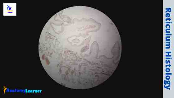
Ruminant reticulum histology
The wall of the ruminant reticulum histology slide also follows the general pattern of a tubular organ. You will find two types of reticular folds (tall and short) in the tunica mucosa of the ruminant reticulum. The reticular or mucosal folds line with the keratinized stratified squamous epithelium like ruminant rumen.
Some reticular papillae remain in between the reticular crest of the tunica mucosa surface. The lamina muscularis is not ideally found in all the reticular crests (mucosal folds). Generally, the taller reticular crests possess a condensed lamina muscularis in their upper part.
In other mucosal folds (reticular crests), you will find the propria-submucosa. The propria submucosa of the ruminant reticulum slide does not possess any special features. You will find a thin layer of loose connective tissue in the propria submucosa of the ruminant reticulum.
The tunica muscular layer of the ruminant reticulum shows some differences from that of the general pattern of a tubular organ. You will find two defined layers of smooth muscle fibers (inner oblique and outer circles-cross) in the tunica muscular layer of the ruminant reticulum slide.
Again, the serosa of the ruminant reticulum shows a weak layer of loose connective tissue. These layers also contain numerous blood vessels, nerves, and an outer mesothelium lining.
Identification of ruminant reticulum histology slide
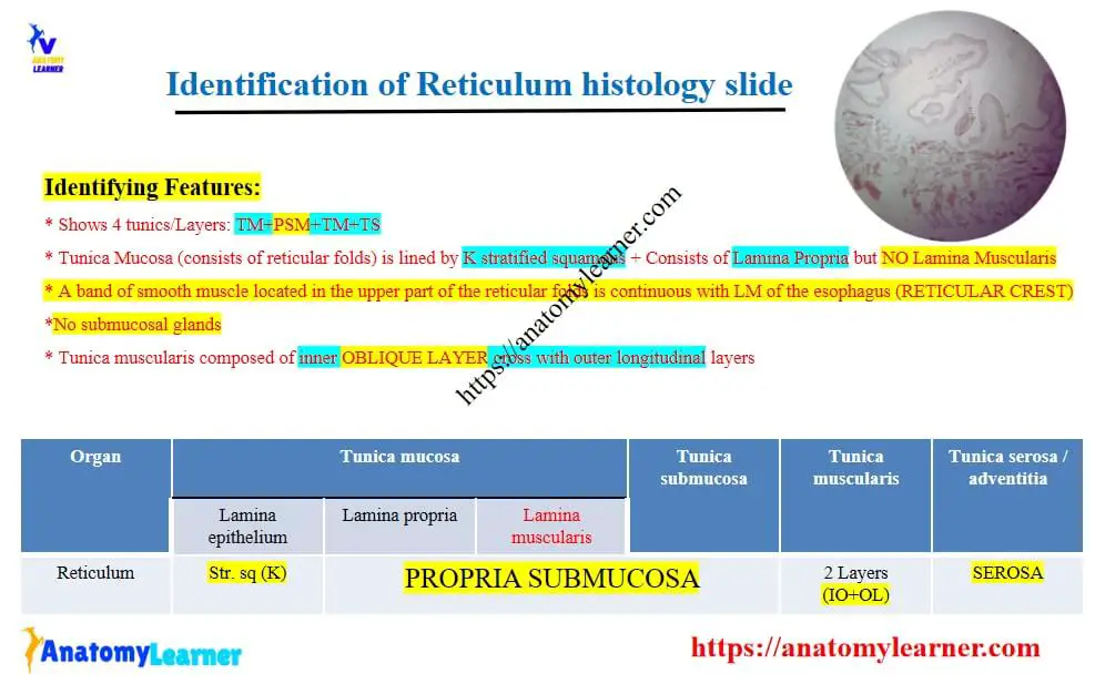
Fine, now let’s identify the ruminant reticulum under the light microscope. Here, you will find some perfect identification points for identifying the ruminant reticulum microscope slide under the light microscope.
All the identification features are listed here based on a realistic view. But, if you find any more remarkable identifying characteristics of the ruminant reticulum slide, please add them.
- The sample tissue section shows the anastomosing and interconnecting permanent mucosal folds (reticular folds or crests)
- There are two types of mucosal folds (tall and short) present in the same focus under the light microscope.
- The whole mucosal surface of the provided sample is lined with the keratinized stratified squamous epithelium.
- A condensed lamina muscularis present in the upper part of the larger (taller) mucosal folds.
- The tunica muscular layer shows smooth muscle’s inner oblique and outer cross pattern.
So, this is a ruminant reticulum histology slide. It would be best to try to identify the reticulum microscope slide with the help of the above-mentioned identifying features. I am sure that you will ideally locate all the essential structures from the different layers of the ruminant reticulum.
It will be better to enlist the structures you might focus on when identifying a ruminant reticulum slide under the light microscope.
- The tunica mucosa with lining epithelium (reticular crests or mucosal folds – tall and short)
- A condensed lamina muscular in the upper part of the tall reticular crest
- The propria submucosa of the ruminant reticulum
- A thick tunica muscularis layer (inner oblique and outer cross pattern of smooth muscle)
- The thin serosa of the ruminant reticulum
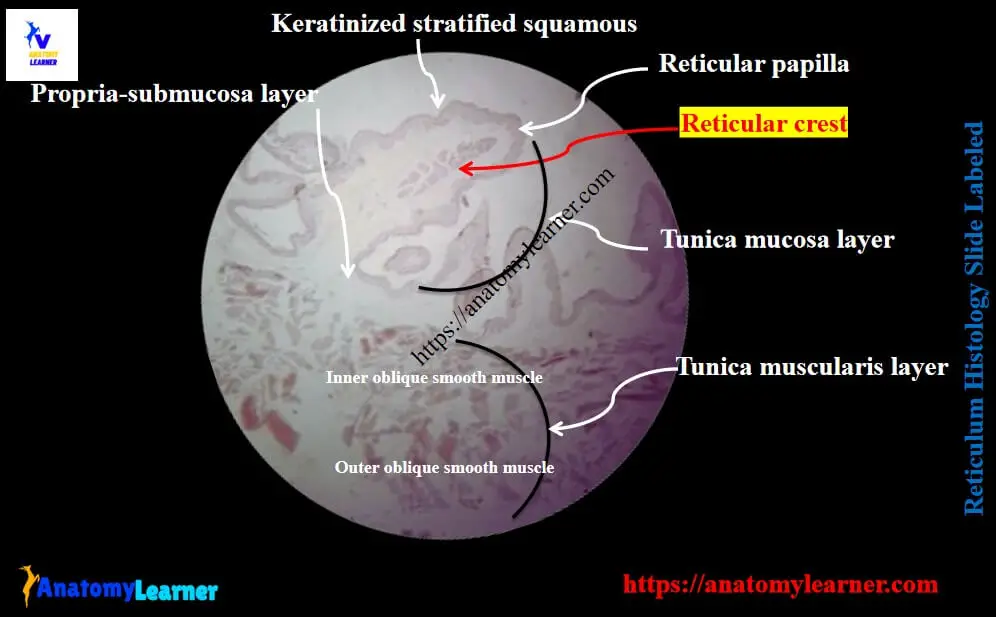
All the structures from different layers of the ruminant reticulum are shown in the labeled image and diagram.
Layers of ruminant reticulum slide
You may skip this part of the article as you have a good piece of knowledge on the basics of a ruminant reticulum histology slide. Moreover, you have a basic understanding of the different layers of a tubular organ. Here, I will describe all the structures from the different layers of the ruminant reticulum slide.
- The tunica mucosa layer of the ruminant reticulum
- A tunica submucosa (propria submucosa) layer of reticulum
- The tunica muscular layer of the ruminant reticulum, and
- A thin tunica serosa layer of the reticulum
Fine, let’s know the details of every single layer from the ruminant reticulum microscope slide with the labeled images and diagrams.
The tunica mucosa of the reticulum
The reticulum shows a well-defined tunica mucosa layer with some permanent interconnecting and sometimes anastomosing folds. These interconnecting folds of the reticulum mucosa are known as the reticular crests. There are two different types of reticular folds present in the tunica mucosa of the ruminant reticulum microscope slide.
According to the height of the crest, they are two types – tall and short. The tall reticular crests separate the mucosa surface into the shallow compartment. These compartments are further divided into small areas by the shorter reticular crests.
You will find some vertical crest on both sides of the reticulum’s reticular crest. Again, there are reticular papillae present in between the reticular crests. So, these reticular papillae of the reticular mucosa project into the lumen.
You will find a well-defined keratinized stratified squamous epithelium lining over the tunica mucosa surface of the ruminant reticulum. The most characteristics histological feature of the ruminant reticulum is the presence of condensed lamina muscularis. It is present in the upper part of the taller reticular crest.
But, this condensed lamina muscularis is not commonly occurring in the short reticular crests. So, the lamina propria of the reticulum blend with the tunica submucosa and form the propira submucosa layer.
The lamina muscularis of the tall reticular crest of the ruminant reticulum is the continuation of the lamina muscular layer of the esophagus.
A propria submucosa layer of the ruminant reticulum
No more salient histological features are present in the propria submucosa layer (of the ruminant reticulum). The propria submucosa of the ruminant reticulum slide consists predominantly of a feltwork of collagen and elastic fibers. You will find some blood vessels and nerve plexus in the propria submucosa of the reticulum slide.
The tunica muscularis layer of a reticulum histology slide
You will find a variation in the tunica muscular layers of the ruminant reticulum compared to the tubular organ. Most tubular organs possess inner circular and outer longitudinal smooth muscle layers in their tunica muscularis.
Here, you will find two defined smooth muscle layers in the ruminant reticulum microscope slide. The inner layer of the tunica muscularis shows the oblique pattern. Again, the outer layer of the tunica muscularis shows the cross pattern.
But, you may find some variation in the muscular layer in large ruminants.
A thin tunica serosa layer of reticulum
The ruminant reticulum histology slide possesses a thin layer of tunica serosae like that of the rumen or omasum. You will find a thin layer of loose connective tissue layer that consists of connective tissue cells, fibers, numerous blood vessels, and nerves.
Again, the outer part of the thin tunica serosa consists a single layer of mesothelium lining (simple squamous epithelium lining).
The reticular sulcus histology
The reticular sulcus or groove begins at the cardiac orifice and passes ventrally on the medial wall of the reticulum. This reticular sulcus ends at the reticulo-omasal orifice. You will find two thick folds (known as lips) that surround the reticular sulcus.
The reticular sulcus is lined with the keratinized stratified squamous epithelium. You will not find the continuous lamina muscular layer in the reticular sulcus. The lamina muscular layer may discover in the lip of the reticular sulcus. But, it is the extension of the esophageal lamina muscularis layer.
You will find a complete layer of lamina muscularis near the omasum. In the area where the lamina muscularis is absent, you will find the propria submucosa. Again, the propria-submucosa of the reticular sulcus predominantly contains collagen and elastic fibers.
The tunica muscularis of the reticular sulcus consists of two layers of smooth muscles. Sometimes, you may find the skeletal muscle layer near the cardiac orifice. This skeletal muscle layer extends the lamina muscularis of the esophagus. But, you will not find any skeletal muscle layer in the reticular sulcus.
On the floor of the reticular sulcus, you will find both longitudinal and transverse oriented muscle fibers layers. Again, at the ventral end of the reticular sulcus, the muscle fiber enters into the reticulo-omasal orifice.
You will find the longitudinally oriented smooth muscle fibers in the tunica muscular layer of the lips of the sulcus. Again, a longitudinal muscle fiber of the lip forms a loop around the cardiac orifice.
At the side of the reticular sulcus, the muscle fibers spread out into the inner layer of the reticulum. In addition, the tunica serosa of the reticular sulcus contains loose connective tissue and mesothelium.
Ruminant reticulum histology slide labeled diagram
Now, I will show you all the ruminant reticulum histology slide structures with the labeled diagram. The following reticulum labeled diagram shows the different layers with their essential structures.
I tried to show you some longer, and shorter reticular creates in the reticulum microscope labeled image. The longer reticular crests of the labeled diagram show the condensed lamina muscularis layer. But, the shorter reticular ridges of the reticulum slide image do not display any lamina muscular layer.
Again, the reticulum microscope labeled image shows the distinguished keratinized stratified squamous epithelium. The propira submucosa of the diagram shows numerous collagen and elastic fibers.
Again, the reticulum labeled diagram shows the inner oblique pattern of the smooth muscle layer. The outer smooth muscle layer of the labeled diagram shows the cross pattern.
In addition, the tunica serosa layer of the reticulum microscope labeled image shows a thin layer of loose connective tissue with numerous blood vessels. The outer part of this delicate, flexible connective tissue layer contains a single layer of simple squamous epithelium (mesothelium).
Ruminant reticulum microscope image drawing
This part of the article might help you learn the drawing of ruminant reticulum microscope images. You might draw the four different layers of the reticulum. First, you should draw the more prominent and shorter reticular crest, as shown in the figures.
You might provide a condensed lamina muscular layer in the more prominent reticular crests. Again, it would be best to give a keratinized stratified squamous epithelium on the mucosa of the reticulum microscope image.
In the propira-submucosa, let’s draw numerous collagen fibers and some elastic fibers. Again, in the tunica muscular layer of the reticulum microscope image, you should draw the inner oblique pattern of the smooth muscle layer. In addition, you should try to draw a cross pattern of smooth muscle layer at the outer layer of the tunica muscularis.
Finally, let’s draw the thin connective tissue layer with a single layer of the simple squamous epithelium of the reticulum microscope image.
Frequently asked questions on a reticulum microscope slide
I will solve the common inquiries on the ruminant reticulum histology slide. If you have any questions on the reticular microscope slide, please let me know.
What are the salient features of a ruminant reticulum microscope slide?
If you read the full article, you will get the salient features of the ruminant reticulum microscope slide. The most salient features of the ruminant reticulum are – the presence of the taller and shorter reticular crests in the tunica mucosa of the reticulum slide.
A lamina muscular layer is present in the larger or taller reticular crest. At the same time, no lamina muscularis present in the shorter reticular crest. You will find a significant variation in the tunica muscular layer of the reticulum microscope slide (inner oblique and outer cross pattern of smooth muscle layers).
What is the reticulum in histology?
In the histology of the reticulum, you will find four different layers like the tubular organ. I have already described all the essential features of the ruminant reticulum with the microscope labeled images and diagrams.
Conclusion
I think you got the basic idea on the ruminant reticulum histology with the actual image and labeled diagram. The condensed lamina muscularis was one of the significant salient microscopic features in the ruminant reticulum.
Again, the tunica muscularis layers show another salient microscopic feature in the reticulum slide. It would be best to identify the reticular microscope slide under the light microscope and differentiate it from the other different parts of the ruminant stomach.