The cow stomach anatomy comprises four compartments – rumen, reticulum, omasum, and abomasum. Here, I will focus on the anatomical facts of these four compartments of cow compound stomachs with a diagram.
Quick overview: rumen is the larger and more capacious compartment than the reticulum, omasum, and abomasum of a cow’s stomach. Except for the omasum, the other 3 compartments lie left of the median plane of the cow’s body. Here, the reticulum is the smaller among these 4 compartments and lies cranially.
I will focus on the surface and topographic anatomy of the different parts of a cow’s stomach. Again, you will find the external and internal identifiable features from all compartments of the cow compound stomach.
This might help you to understand the basic features of another ruminant stomach anatomy (sheep, goat). You will see (find) very little difference between the large and small ruminant stomach anatomy.
First, let’s learn the anatomical facts of the cow compound stomach.
Cow stomach anatomy
The cow compound stomach is very capacious and occupies nearly 3/4th of the abdominal cavity. This compound stomach fills the left half of the abdominal cavity except a small space occupied by the spleen and intestine.
In cow stomach anatomy, you might describe the features of the four different compartments with their blood and nerve supply. You might also describe the composition or layers of the stomach wall.
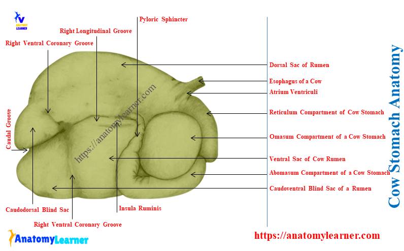
First, let’s identify the 4 compartments of the cow compound stomach from the diagram –
- Rumen – a big muscular elongated sac that compressed side to side,
- Reticulum – a pyriform smallest sac of the stomach that lies between the diaphragm and liver in front,
- Omasum – roughly oval and laterally compressed structure that lies right of the median plane, and
- Abomasum – an elongated saccular structure that is also known as the true stomach and lies ventral to the rumen and abdominal floor,
The provided labeled diagram identifies the four compartments of the cow compound stomach. From the right side of the body (abdomen), you will clearly identify the different parts of the compound stomach.
The rumen, reticulum, and omasum of the cow stomach are known as the forestomach or proventriculus. They possess the non-glandular mucous membrane in their internal wall.
Again, the last part, or abomasum of the compound stomach, is the true glandular compartment. You will find the glandular mucous membrane in the internal structure of the abomasum.
The size of the compartments of a cow’s stomach changes with age. Thus, the capacity of the stomach also changes.
A medium-sized cow has a 115 – 150 liters capacity in their compound stomach. Again, the sheep and goats have only 15 – 18 liters capacity in their stomach.
Summary of the cow compound stomach
Here, I will summarize the anatomical facts of every single compartment of a cow compound stomach. Table 1 shows the number of surfaces, curvatures, extremities, and other special features from the rumen, reticulum, omasum, and abomasum –
| Bovine stomach anatomy | Features (external) | Internal features |
| Rumen | Surfaces – parietal, visceral Curvatures – dorsal, ventral Ends – cranial, caudal Grooves – Cranial transverse Right and left longitudinal Right accessory Caudal transverse (2) Sac of rumen – Dorsal larger sac Ventral larger sac Cado-dorsal blind sac Caudo-ventral blind sac | Carpet like appearance |
| Reticulum | Surfaces – parietal, visceral Curvatures – lesser, greater Ends – neck, fundus | Honeycomb appearance |
| Omasum | Surfaces – right and left Curvatures – dorsal, ventral Ends – base and neck | Possess laminae omasi |
| Abomasum | Have three parts – Cranial blind fundus Longer body, and Caudal pyloric part Surfaces – parietal, visceral Curvatures – lesser, greater | Possess larger permanent folds (plica abomasa) |
What is rumen in bovine stomach anatomy?
Rumen is the larger muscular elongated sac of bovine stomach anatomy. It occupies most of the left half of the cow’s abdominal cavity.
The rumen of a cow extends considerably to the right of the median plane ventrally and caudally.
You may get the basic idea of the animal’s abdominal cavity with its contents from the below-mentioned article –
Where is the rumen of a cow located?
Quick answer: the rumen of a cow extends from the seventh or eighth intercostal space to the pelvic inlet. Its left surface contacts with the left lateral abdominal wall.
Again, the ventral sac of the rumen encloses by a greater omentum (modification of the peritoneum). It lies on the ventral abdominal wall just caudal to the transverse plane of nine costochondral junctions.
You may also learn the anatomical facts of the cow abdomen from the below-mentioned article – Cow abdomen anatomy – boundary, muscles, and organs with diagram,
Cow rumen anatomy
If you open the abdominal cavity of the cow, you will see the rumen almost cover the left side. This compartment of the cow stomach possesses the followings –
- Two surfaces – parietal (left) and visceral (right),
- Two curvatures – larger dorsal and smaller ventral, and
- Two extremities – cranial extremity and caudal extremity,
Let’s discuss the surfaces, curvatures, and extremities of the cow rumen.
Surfaces of cow rumen
The parietal or left surface of the cow rumen is convex and related to the diaphragm. Again, the visceral surface faces right and is irregular.
This visceral surface of the rumen is related to the variety of organs. You will find the relationship of the visceral surface of the rumen with the omasum, intestine, liver, pancreas, kidney, vena cana, and aorta.
The surfaces of the cow rumen represent two grooves –
- Right longitudinal groove, and
- Left longitudinal groove,
These longitudinal grooves externally divide the rumen into dorsal and ventral sacs. The right and left longitudinal groove joins caudally and form the caudal groove.
You will see the dorsal coronary groove at the left of the longitudinal groove. This groove extends caudodorsally and fades out later.
The right surface of the cow rumen shows another longitudinal groove just above the main groove. This is the dorsal accessory longitudinal groove on the right side of the cow rumen.
Thus, you will find two (2) longitudinal grooves on the right surface of the cow rumen –
- Ventral main longitudinal groove, and
- Dorsal accessory longitudinal groove,
Now the dorsal accessory longitudinal groove is convex dorsally. It joins the main ventral longitudinal groove on both ends.
Thus, these dorsal and ventral longitudinal grooves on the right surface form the elliptical area. This elliptical area of the right surface is insula ruminis.
Here, the cow rumen labeled diagram shows the various groove on parietal and visceral surfaces.
Curvatures of cattle rumen anatomy
The cattle rumen shows two distinct curvatures – dorsal and ventral. Here, the dorsal curvature of the rumen is larger compared to the ventral curvature.
The dorsal curvature is convex and attaches dorsally to the crura of the diaphragm and sublumbar muscles. Again, these curvatures are also firmly attached to the left aspect of the abdomen by the peritoneum and connective tissue.
The ventral curvature of the rumen is also convex and related to the abomasum. This ventral curvature lies on the floor of the abdomen.
The dorsal and ventral curvatures of the cow rumen are also identified in the labeled diagram.
Extremities of cow rumen
The rumen of a cow’s stomach shows two distinct extremities – cranial and caudal. Let’s discuss the features of the cranial and caudal extremities of the cow rumen.
The cranial extremity of the cow rumen divides by the transverse cranial groove into two sac –
- A cranial sac or atrium ruminis, and
- A dorsal sac of the rumen,
Here, the cranial sac continues caudally as the dorsal sac. Again, the reticulum is the cranial continuation of the atrium ruminis or cranial sac.
But, you will find an external line of demarcation between the rumen and reticulum. This external demarcation is known as the rumenoreticular groove.
There is no natural dorsal separation between the rumen and reticulum. Now, the rumen and reticulum form the atrium ventriculi, where the esophagus opens.
This atrium ventriculus is the dome-like structure between the rumen and reticulum. You may know the course and termination of the cow esophagus on this atrium ventriculi from the below-mentioned article –
The caudal extremity of the cow rumen extends nearly to the pubis bone. This extremity is also related to the intestine and urinary bladder of the cows.
You may learned he features of pubic bones from the below-mentioned article of anatomy learners –
You will see the deep, transverse caudal groove at the cow rumen’s caudal extremity. This caudal transverse groove contact with the longitudinal grooves and form two sacs –
- Caudo-drosal blind sac, and
- Caudo-ventral blind sac,
These two blind sacs are marked off from the remainder of the rumen on each side by the dorsal and ventral coronary grooves.
How to differentiate the cow rumen from goats or sheep?
Answer: you may differentiate the sheep or goat rumens from the cows with the appearance of dorsal and caudoventral blind sacs. The ventral sac of goat or sheep rumen is larger compared to the cows.
Again, the ventral sac or rumen extends more right off the median plane of the body compared to the cows. The blind sac of the goat rumen extends further caudally than the dorsal sac.
Again, the parietal attachment of the dorsal sac of goat or sheep rumen extends caudally to the second lumbar vertebra. The dorsal coronary groove on the sheep or goat rumen is short compared to the cows.
You may learn the features of the lumbar vertebrae of a cow from the below-mentioned article of anatomy learner –
Reticulum of bovine stomach anatomy
Reticulum is the most cranial and smallest part of the cow stomach anatomy. It is somewhat pyriform in shape and compressed craniocaudally.
The reticulum of the cow lies on the left side of the median plane. You will see the extension of this compartment from the sixth to seventh or eighth (6th – 7th /8th ) ribs of the thoracic cavity.
The cow reticulum possesses the followings –
- Two surfaces – diaphragmatic surface (parietal) and visceral surface,
- Two curvatures – greater (larger) and lesser (small) curvatures, and
- 2 extremities – known as the neck and fundus of the reticulum,
First, let’s identify these surfaces, curvatures, and extremities from the cow reticulum labeled diagram. Let’s discuss on the different anatomical facts from the surfaces, curvatures, and extremities of the cow rumen anatomy.
The surface of the cow reticulum
The diaphragmatic or parietal surface of the cow reticulum is convex. It lies against the diaphragm and liver.
Thus, the reticulum of a cow comes in contact with the diaphragm. Again, the diaphragm has contact with the pericardium of the heart.
If any foreign shapes bodies like a pin or nail enter the reticulum, they log and perforate the reticular wall. Subsequently, the diaphragm and pericardium also perforate and form the reticulopericarditis condition.
The visceral surface of the cow reticulum is flattened. This is due (cause) to the more or less pressure of the atrium ruminis. The visceral surface of the reticulum forms the ruminoreticular orifice internally.
Curvatures and extremities of cow reticulum
The lesser curvature of the cow reticulum faces right and dorsally. This curvature of the reticulum attaches to the omasum compartment.
The greater curvature of the cow reticulum faces to the left and ventrally. It lies against the diaphragm opposite the sixth and seventh ribs.
Let’s get the basic idea of the cow ribs from the below-mentioned article –
The upper part of the cow reticulum is the neck which attaches to the dorsal sac of the rumen. Again, the ventral part of the reticulum is the fundus.
The fundus of the cow reticulum forms the rounded sac-like structure. This rounded sac-like structure of the reticulum is known as the cul-de-sac.
This sac lies opposite the ventral end of the sixth intercostal space. Again, the sac contact with the sternal part of the diaphragm, liver, omasum, and abomasum compartments.
How to differentiate sheep or goat reticulum from the cow stomach?
Quick answer: the reticulum of sheep and goats are relatively larger compared to the cows. Again, the ventral part of the sheep and goat reticulums curve more caudally and less to the right.
The cul-de-sac is more distinct in the ox reticulum than the sheep and goat. But, the internal appearance of the reticulum of both cows and goat are similar.
You will find a honeycomb structure in the internal part of the ruminant reticulum. But, the honeycomb appearance (polygonal) is more in the fundus than the reticulum’s neck.
Omasum of cow stomach anatomy
This is the only part (region) of the cow compound stomach that lies right of the median plane. The cow or ruminant omasum extends opposite to the seventh to eleventh ribs (7th – 11th).
The cow omasum is ellipsoidal in shape and somewhat compressed between the parietal and visceral surfaces. Again, the long axis of the cow omasum is nearly verticle.
In the anatomy of the cow omasum, you will find the below-mentioned features –
- Two surfaces – parietal (right) and visceral (left) surfaces,
- 2 curvatures – dorsal and ventral, and
- Base and neck of the cow omasum (2 extremities),
First, see these surfaces, curvatures, and extremities from the cow omasum labeled diagram. Now, let’s discuss the important anatomical facts from the surfaces and extremities of the cow omasum.
Surfaces and curvatures of cow omasum structure
The visceral surface of the cow omasum faces the left aspect. This visceral surface has contact with the rumen, reticulum, and also with abomasum compartment of the cow stomach.
The parietal surface of the cow omasum compartment faces obliquely to the right and cranially. This parietal surface is chiefly related to the diaphragm and liver.
The parietal surface of the omasum is separated by the lesser omentum and diaphragm (at the level of the 7th to 9th intercostal spaces). You will find the contact of the ventral part of the omasum with the abdominal floor.
Again, a small part of the abomasum has contact with the cow omasum. There is a small area between the right costal cartilage and xiphoid cartilage where this attachment occurs between the abomasum and omasum.
You may learn the appearance of the cow xiphoid cartilage from the below-mentioned article –
The dorsal curvature of the cow omasum faces dorsally, then caudally. This curvature also faces the right aspect of the abdomen.
The ventral curvature of the omasum faces ventrally and caudally.
Extremities of the ruminant omasum
It is very difficult to identify the extremities from the cow omasum. When you remove the rumen and reticulum compartments from the cow’s stomach, the extremities may be visible.
You will find two extremities in the structure of the cow omasum –
- A concise base, and
- The short neck,
Here, the base of the omasum faces cranially, ventrally, and to the right. Again, the neck is narrow and in contact with the upper part of the reticulum compartment of the cow stomach.
There is a thick omasal muscular pillar in the structure of the cow omasum. They possess extensive junctions with the abomasum compartment.
You will find two major openings in the structure of the cow omasum –
- Reticulo-omasal opening, and
- Omaso-abomasal opening,
Both reticulo-omasal and omaso-abomasal openings are at the upper part and extensive.
The internal part of the cow omasum also possesses unique features. You will find the hundred longitudinal or crescent folds in the internal structure of the cow omasum. These longitudinal or crescent folds are also known as the laminae omasi.
The labeled diagram identifies all the features from the cow omasum anatomy. You may find more stomach-labeled diagrams from the various animal on social media of anatomy learners.
Abomasum of ruminant stomach anatomy
The abomasum is the last compartment of the ruminant stomach anatomy. This is the elongated and saccular structure in the cow.
This compartment is also known as the true stomach of the ruminant. Anatomically and microscopically, you will find almost similar features between the simple stomach and the abomasum.
Let’s get an idea of the structure of a simple stomach from a horse or dog. The below-mentioned article will help you to get the anatomical facts from the simple stomach –
Again, you may learn the internal microscopic features of the abomasum or true stomach from the below-mentioned article of anatomy learners –
In this article, you will get (find) an overview of the microscopic structure of the true stomach of a cow.
Anatomy of a cow abomasum
The elongated abomasum sac of the cow extends from the xiphoid cartilage to the abdominal floor. It mainly has three distinct parts –
- Cranial blind end of funds of the abomasum,
- A middle long body that extends caudally, and
- Caudal short pyloric part of the abomasum,
The cranial blind sac or fundus remains in the xiphoid region and is related to the reticulum compartment. You will also find the attachment of the blind end fundus with the atrium ruminis and ventral sac of the rumen.
The longer body of the abomasum extends caudally between the ventral sac of the rumen and omasum compartments. But, this longer body of the abomasum lies more on the left aspect than the right of the median plane.
The pyloric part of the abomasum turns to the right caudal to the omasum compartment. It inclines dorsally and connects with the duodenum.
The junction between the duodenum and the pyloric part of the abomasum is known as the pyloric area. You will find the pyloric sphincter (a constricted area) within this area.
You will find the pyloric sphincter of the abomasum compartment at the level of the ventral end of the 9th or 10th intercostal space.
Surfaces and curvatures of cow abomasum
You will find two surfaces and curvatures in the structure of the cow abomasum anatomy. Here, the parietal surface of the abomasum contact with the abdominal floor.
Again, the visceral surface of the abomasum faces the dorsomedial. You will see the contact of the visceral surface with the rumen and omasum compartments.
The greater curvature of the cow abomasum faces ventrally. In contrast, the lesser curvature of the abomasum faces dorsally.
The greater curvature provides attachment to the superficial wall of the greater omentum. Again, the lesser curvature attaches to the lesser omentum. Finally, it passes over the parietal surface of the omasum to the liver of the cows.
The abomasum of sheep and goats are comparatively larger and longer than the cows. But, the internal structure of both goat and cow’s abomasum shows similar structural features.
Internally, you will find large permanent folds of mucous membranes. These permanent folds on the internal surface of the cow abomasum are known as the plica abomasi.
The microscopic features of the cow abomasum also show the glands in the fundus and pyloric region. You will find less permanent folds of mucous membrane at the pyloric region of the cow abomasum compartment.
Microscopically, the wall of these four compartments of the cow stomach contains 4 layers – tunica mucosa, submucosa, muscularis mucosa, and serosa. Let’s find the basic idea on these four layers from the below-mentioned article of anatomy learners –
Blood and nerve supply to the cow’s stomach
The cow stomach is supplied by the various branches of the celiac artery. Again, the major innervation to the cow’s stomach comes from the branches of the vagus nerve and also from the sympathetic nerves.
The left ruminal, right ruminal, reticular, omasoabomasal, and branch of the hepatic artery are the main arteries that supply the cow stomach.
Ruminant stomach anatomy diagram
Now, let’s see the features of the different compartments of the ruminant stomach anatomy with the diagram. I tried to show the below-mentioned anatomical facts from the rumen compartment of a cow stomach anatomy –
- Larger dorsal and ventral sacs of the cow rumen,
- Caudo-dorsal and caudo ventral blind sacs of the rumen,
- A cranial transverse groove that separates the reticulum from the rumen,
- Right and left longitudinal grooves on the parietal and visceral surfaces of the rumen,
- The right accessory longitudinal groove of the rumen,
- Insula ruminis on the right surface of the rumen,
- Dorsal and ventral coronary grooves on the rumen,
- The caudal transverse groove of the rumen, and
- Atrium ventriculi between the rumen and reticulum compartments,

Again, the other diagram shows the various features of the cow reticulum –
- Dorsal and ventral cul-de-sac of the reticulum,
- Diaphragmatic and visceral surfaces of the cow reticulum,
- Lesser and greater curvatures of the cow reticulum, and
- Internal polygonal or honeycomb appearance of the cow reticulum,
In the below-mentioned diagram, I also tried to show the different features of the omasum compartment of the cow stomach –
- Short base and neck of the cow omasum,
- Right parietal and left visceral surface of the cow omasum,
- Dorsal and ventral curvatures of the cow omasum, and
- Internal longitudinal permanent folds of the cow omasum,
The abomasum labeled diagram also shows the below-mentioned anatomical facts –
- Fundus, body, and caudal pyloric part of the cow abomasum,
- Parietal and visceral surfaces of the cow abomasum,
- Larger and smaller curvatures of the cow abomasum, and
- Internal plica abomasa (permanent folds) of the cow abomasum,
How to differentiate a horse stomach from a cow stomach?
Quick answer: the horse possesses a simple stomach that only has glandular and non-glandular parts. In contrast, cows possess a compound stomach that has four different compartments.
The stomach of a horse locates on the left side of the median plane. It lies on the dorsal aspect of the horse’s abdomen, just behind the liver and diaphragm.
The horse stomach is a curved and J-shaped structure, whereas the different compartment of the cow stomach shows various shape. You will see a very smaller pyloric part in the structure of a horse’s stomach.
Here, the lesser curvature is very short in horse stomachs. Again, the cardiac and pyloric parts of the horse’s stomach are very close.
There is a rounded cul-de-sac (also known as the saccus cecus) in the left extremity of the horse’s stomach. And you know, within this saccus cecus of the stomach, the esophagus opens.
You will find a rough line between the glandular and non-glandular segments of the horse’s stomach. This rough line in the internal structure is known as the margoplicatus.
Again, the pyloric sphincter is more well-developed in the horse stomach than in the cow.
Frequently asked questions on cow stomach structure
Here, I will enlist the most important questions on the structure of the cow stomach that the anatomy learners ask. But you might go through the whole article to get anatomical information on the cow or ruminant stomach.
Okay, let’s find the frequently asked questions on the cow stomach with their concise answers –
How to differentiate different compartments of cow stomachs by their internal appearance?
Quick answer: the internal surface of the rumen, reticulum, omasum, and abomasum shows the unique features of their mucous membrane. Here, you will find the carpet-like appearance in the mucous membrane of the rumen compartment of the cow’s stomach.
The internal surface of the reticulum shows a polygonal appearance (also called the honeycomb shape). Again, you will find the longitudinal or crescent-shaped folds (eighty to hundred) in the internal mucous membrane of the omasum.
Finally, the internal mucous membrane of the cow abomasum shows several permanent plica abomasi. Thus, you will easily differentiate the various segments of the cow stomach compartments.
What are the 4 stomachs of a cow?
Quick answer: the 4 parts of the cow stomach are the rumen, reticulum, omasum, and abomasum. These four parts of the cow stomach show typical and unique features both externally and internally.
So, you may quickly identify the four different parts of the cow’s stomach with their external and internal anatomical features. Here, the largest part of the four compartments of a cow’s stomach is the rumen.
Again, the smallest part among these four compartments is the reticulum that faces cranially.
What is abomasum in a cow?
Quick answer: the abomasum is the last saccular compartment in the cow’s stomach. The true part of the cow compound stomach possesses both glandular and non-glandular segments.
Grossly, the abomasum of a cow extends from the left lateral to the right side of the median plane of the body. Externally the cow abomasum has three distinct segments the fundus, body, and curved dorsally directed pylorus.
Conclusion
So, the cow stomach anatomy shows various unique features in the rumen, reticulum, omasum, and abomasum compartments. The gross anatomical features of the four compartments give you a basic and detailed idea of the cow stomach structure.
Now, you will quickly differentiate the ruminant compound stomach from the simple stomach. These details guide the cow stomach anatomy and represent the ideal features of the other ruminant stomachs.