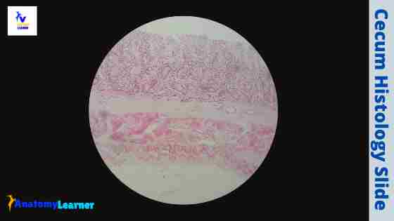
The cecum histology slide follows the general pattern of the small intestine with some exceptional features. You may find a variation in the size of the gross cecum among the different species. But, in all domestic mammals, the cecum histology slide shows a substantial number of lymphatic nodules scattered throughout its length.
Here, I will show you all the histological structures from the cecum with a microscope slide image and labeled diagram. I will also provide the appropriate identification points for the cecum slide under the light microscope. Again, you will get a little information on the specific histological features of the cecum in a different animal.
In addition, at the end of this article, you will learn how you will differentiate the cecum microscope slide from the colon or different parts of the small intestine. So, if you are interested to know the details of the cecum histology slide with labeled images, continue this article till the end.
Cecum histology
In the cecum histology, you will find four layers like a tubular structure. So, the layers of cecum structure are – tunica mucosa, tunica submucosa, tunica muscularis, and tunica serosa. In this part, I will provide a concise description of the histological features of the cecum.
The tunica mucosa of the cecum is lined with the simple columnar epithelium and the goblet cells. You will find both lamina propria and lamina muscularis layers within the tunica mucosa of the cecum. The lamina propria of the cecum consists of numerous intestinal glands.
“There are no villi and plica circularis present in the tunica mucosa of the cecum microscope slide.”
Again, the tunica submucosa of the cecum possesses lymphatic tissue and nodules throughout its length. You will also find numerous adipose tissue in the tunica submucosa of the cecum of some species.
The tunica muscularis of the cecum varies in different species. But, ideally, you will find the inner circular and outer longitudinal smooth muscle layers in the tunica muscularis of the cecum microscope slide.
In addition, the tunica serosa of the cecum consists of a thin layer of loose connective tissue. A single layer of mesothelium lining is present in the outer part of the thin, loose connective tissue layer.
Cecum histology slide identification points
Sometimes, you may be asked to identify the cecum histology slide under the light microscope at your laboratory. Here, I will enlist the essential identification points for the cecum microscope slide under the light microscope.
You may add more identification features for the cecum microscope slide if you want. Okay, let’s identify the cecum slide with the following identifying characteristics.
- The wall of the sample tissue section shows four different layers – tunica mucosa, tunica submucosa, tunica muscular, and tunica serosa.
- There are no villi and the plica circularis on the tunica mucosa of the provided tissue sample.
- The tunica mucosa of the tissue sample lines with the simple columnar epithelium with numerous goblet cells
- Presence of numerous lymphatic tissue and lymphatic nodules in the tunica submucosa of the tissue sample (depends on the species; described in the complete guide section)
- The tunica mucosa of the sample tissue section shows the two distinct smooth muscle layers (inner circular and outer longitudinal; but may vary in different species).
- Again, the outer longitudinal smooth muscle layer forms a large, flat muscle band (taenia ceci).
So, this is a cecum histology slide. The tunica muscular layer and the taenia Ceci band may vary in different species. In the cecum, as mentioned above, microscope image, you will not find any taenia Ceci. Again, the tunica muscle layers are different in the cecum labeled image.
Layers of cecum histology
You know, there are four different layers present in the cecum histology. Most of the histological features of the different layers of a cecum slide are similar to the general pattern of the small intestine. Of course, some salient features are present in the cecum microscope slide (I hope you already got the basic idea).
Let’s discuss the different layers of the cecum microscope slide with the labeled image and diagrams. So, the layers of the cecum are –
- Tunica mucosa layer
- A tunica submucosa layer
- The tunica muscle layer, and
- A tunica serosa layer
I think you have a good piece of knowledge on the general pattern of the tubular organ (essential histology of four different coats of hollow organs). This might help you to understand the cecum structure so quickly.
The tunica mucosa of cecum
In the tunica mucosa of the cecum, you will not find any permanent mucosal folds like the small intestine. There are some temporary mucosal folds present in the tunica mucosa of the cecum. You will not also see the villi and the plica circulars in the tunica mucosa of the cecum.
The tunica mucosa of the cecum histology slide consists of simple columnar epithelium, lamina propria, and lamina muscularis. In the lamina propria of the cecum, you will find longer, straighter, more compact intestinal glands. Sometimes, you may find some diffuse lymphatic tissue in the lamina propria of the cecum microscope slide.
Again, the lamina muscularis layer of the cecum consists of a single but thick layer of smooth muscle. This lamina muscular layer remains continuous in the cecum, but you will find an interrupted lamina muscular layer in the colon.
A tunica submucosa layer of cecum histology slide
Generally, in the cecum slide’s tunica submucosa, you will find the connective tissue cells, fibers, numerous blood vessels, and nerves. The most important feature of the tunica submucosa of a cecum is the presence of lymphatic tissue and nodules.
These lymphatic tissue and nodules remain throughout the length of the cecum. But, the presence of the lymphatic tissue and nodules may vary within the species.
In a dog, pig, and ruminant cecum, you will find numerous lymphatic nodules in the tunica submucosa. These lymphatic nodules are especially multiple around the ileal ostium (opening the ileum into the cecum or colon). Again, in the horse and cat cecum, lymphatic nodules are concentrated near the apex of the cecum.
In addition, you may find numerous adipose tissue in the tunica submucosa of the cecum. But, it also depends on the species. More adipose tissue will discover in the tunica submucosa of the pig cecum.
The tunica muscularis layer of the cecum
Generally, the tunica muscularis layer of the cecum histology slide consists of a thick inner circular and thin outer longitudinal smooth muscle layer. The outer longitudinal smooth muscle layer forms large, flat, smooth muscle bands in pig and horse cecum. This smooth muscle band of the pig and horse cecum is known as the taenia Ceci.
But, this taenia Ceci is not a common feature in other different species. You may also find a significant variation in the pattern of the smooth muscle in the tunica muscular layer of the cecum in different species.
Some species possess the tunica muscular layer’s inner longitudinal and wavy smooth muscle bundles. You may also find some elastic fibers in the tunica muscular layer of the cecum (in some species).
The tunica serosa of a cecum
The tunica serosa of a cecum is a thin layer of loose connective tissue with numerous blood vessels, adipose tissue, and elastic fiber. Again, in the outer part of the thin, loose connective tissue layer, you will find a simple squamous epithelium lining (known as the mesothelium).
Cecum histology slide labeled diagram
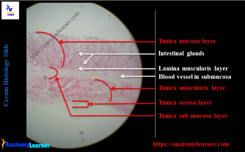
Finally, I will show you all the features of the cecum histology slide with the labeled image. You will also get the cecum microscope slide labeled diagram below.
I tried to show you the lining epithelium with the goblet cells, lamina propria with the intestinal glands, and lamina muscular layer in this cecum slide image. The intestinal glands of the labeled image are numerous, more prolonged, straight, and more compact compared to that of the small intestine glands.
You know, the tunica submucosa contains loose connective tissue, numerous lymphatic nodules, and adipose tissue. But, in this cecum labeled image, there are no lymphatic nodules present in the tunica submucosa. Here, I tried to show you the numerous blood vessels, loose connective tissue from the tunica submucosa of the cecum labeled image.
The tunica muscular layer of the provided cecum labeled image shows two distinct smooth muscle layers – inner longitudinal or oblique bundles and outer wavy bundles. Again, the cecum images show some elastic fibers in this layer.
In addition, the cecum labeled image shows a thin and loose connective tissue layer with numerous blood vessels. Again, the outer surface of this layer shows the mesothelium lining (tunica serosa of cecum).
Normal cecum microscope slide
Now, it is your time to identify all the following structures from the cecum microscope slide image. You may also try to find out these structures from the cecum under the light microscope at your laboratory.
- The lining epithelium (simple columnar) and goblet cells of the tunica mucosa
- A lamina propria (with numerous large, straight intestinal glands)
- The thick lamina muscular layer of the cecum
- Numerous lymphatic nodules and adipose tissue in the tunica submucosa (optional or if present)
- The inner and outer smooth muscle layers of the cecum
- A taenia ceci of cecum (if present), and
- The thin tunica serosa of the cecum
I know you could easily identify these structures from the normal cecum microscope slide image. If you need more labeled pictures and diagrams of a cecum, you may join anatomy learner on social media.
How will you confirm the cecum histology slide under the light microscope?
Sometimes, you may become confused about differentiating the histology slides of different parts of the small and large intestine. This section of the article will show you how to distinguish and confirm the cecum histology slide under the light microscope from the other part of the large intestine.
Fine, so this part of the article will be an excellent note for you to differentiate the cecum microscope slide from different large and small intestine segments. First, you should have a good piece of knowledge on the histology of the various parts of the small and large intestine (or general histological features).
So, let’s differentiate the histology of the large intestine from the small intestine. The most common histological features of the different segments of the small intestine are enlisted below.
- The tunica mucosa of the small intestine possesses permanent and prominent folds (plica circularis) of the mucosa.
- There are numerous villi present on the tunica mucosal surface of the small intestine. So, the tunica mucosal surface of the different segments of the small intestine is not smooth.
- A few small intestine segments contain submucosal glands (duodenum) and lymphatic nodules (ileum).
But, what are the standard histological features that find in the different segments of the large intestine? Let’s know the most common histological features of the various segments of the large intestine that will help you differentiate the cecum from the small intestine.
Standard histological features of the large intestine
Histologically, the segments of the large intestine (except the last part) do not show any prominent and permanent mucosal folds on the tunica mucosa. I mean, you will not find any plica circularis, even the villi in the tunica mucosal surface of the large intestine.
Again, all the segments of the large intestine lack villi, so the tunica mucosal surface of the large intestine is smooth. The lamina propria contains intestinal glands like in the different parts of the small intestine. But, the large intestine’s glands are usually more prominent, straight, and compact compared to that of the intestinal glands of the small intestine.
Now, if you notice the tunica mucosa layer of the provided sample tissue, you will find the typical histological features of the large intestine. The tunica mucosal surface is smooth as no villi are present on its surface. Again, there are no prominent tunica mucosal folds in this tunica mucosal layer.
In addition, the intestinal glands of the lamina propria are larger, straight, and close together in this provided tissue sample. I think these might be an excellent differentiating point for the cecum from the different segments of the small intestine.
But, you may also compare the other tunics (tunica submucosa, tunica muscularis) of the cecum microscope slide with the small intestine.
Final confirmation of cecum microscope slide
Now, you should confirm the cecum microscope slide from the other different parts of the large intestine. You know there are three separate segments in the large intestine of any animal. In the last segment of the large intestine, you will find some tunica mucosal folds without any villi.
In this segment, the longitudinal mucosal folds contain a core of tunica submucosa and covers by the tunica mucosa. The lining epithelium is simple columnar and contains more goblet cells than the cecum mucosa.
Again, the intestinal glands of this segment are numerous and closer together compared to the cecum. Thus, you may differentiate the cecum microscope slide from the last part of the large intestine.
In addition, you will find salient features in the colon histology slide that will help you differentiate the cecum from the colon. The colon’s tunica mucosa is thicker than the small intestine and cecum. This is due to numerous larger, longer, and straight intestinal glands in the lamina propria of the cecum histology slide.
In addition, the intestinal glands of the lamina propria of a colon slide may extend into the tunica submucosa due to the presence of an interrupted lamina muscular layer. But, in the cecum microscope slide, you will not find any such histological features.
However, there is an excellent variation on the tunica muscular layer of the cecum and colon. You will find a thicker smooth muscle layer in the cecum microscope slide compared to the colon microscope slide.
But, there are no outstanding histological features in the tunica serosa layer of the cecum and colon. But, I hope you can differentiate the cecum microscope slide from the colon.
Cecum anatomy in animal
If you have the basic anatomical features of the cecum of a different animal, you may skip this part of the article. The cecum is a blind sac that is continuous with the colon cranially. Again, the caudal blind sac is known as an apex. It extends up to the right side of the pelvic inlet. You may find ventrally curved apex in some animals.
Cecum histology slide image drawing
I hope this simple guide might help you draw the cecum microscope slide so quickly. Here, I will try to show how you will draw the histological features of the cecum from four different layers.
First of all, you might draw the tunica mucosa of the cecum microscope slide. For that, let’s try to draw some long, straight, and compact intestinal glands (as shown on the diagram) in the lamina propria. You should provide the loose connective tissue cells, fibers in the lamina propria of the cecum slide drawing.
Let’s try to draw the single (thick) smooth muscle layer in the lamina muscularis of the cecum. This lamina muscular layer will continue throughout the length of the cecum and divide the tunica submucosa from tunica mucosa.
Now, in the tunica submucosa, you should draw the lymphatic tissue and nodules throughout the length of the cecum. You may draw some adipose tissue, blood vessels, and connective tissue cells, fibers in the tunica submucosa of the cecum slide.
Fine, let’s try to draw the tunica muscular layer of the cecum microscope slide. The arrangement of the smooth muscle of this tunica muscular layer may vary with the species. So, you may draw this layer according to the species it belongs to.
Here, I will draw the inner circular and outer longitudinal smooth muscle layer (different in the microscope slide) in the tunica muscular of the cecum slide. You might provide fibro elastic connective tissue (cells and fibers) in the tunica serosa layer of the cecum. Don’t forget to give the single layer of mesothelium lining at the outer part of the thin tunica serosa of the cecum.
Frequently asked questions on the cecum histology.
In this part, I will try to solve the common inquiries on the cecum histology slide. These questions are commonly asked by the learners who love to know the different interesting fats of a cecum microscope slide. If these questions and answers do not meet your desired expectation, you may also be asked any question on cecum.
What type of epithelium is in the cecum?
Great, in the cecum microscope slide, you will find the simple columnar epithelium lining on the tunica mucosal surface. With this simple columnar lining epithelium, you will also find numerous goblet cells in the tunica mucosal surface of the cecum microscope slide.
I am sure that you have a good knowledge of the structure of a simple columnar epithelium and a good understanding of the goblet cell. You may also memorize these epithelium cells from the article published in anatomy learner previously.
The cell of the simple columnar epithelium is tall or column-like; it possesses an elongated nucleus located at the base of the cell.
What is the cecum made of?
Histologically, the cecum is made of four different tunics or coats – tunica mucosa, tunica submucosa, tunica muscularis, and tunica serosa. If you want to know the detailed description of every single layer of the cecum, you may read the article from the beginning.
In the tunica mucosa, the essential feature is the presence of a large, compact intestinal gland in the lamina propria. Again, there are numerous lymphatic nodules and adipose tissue present in the tunica submucosa of the cecum. The tunica muscularis layer of the cecum may vary with the different species.
What is the cecum?
The cecum is the first segment of the large intestine of any animal where microbial action occurs. This segment of the intestine also helps absorb water, vitamins, electrolytes, and mucus secretion.
What is the histology of the colon?
The colon possesses some salient histological features that make it different from the other segment of the large intestine. Four different layers are also present in the colon histology slide, like the cecum. You will find the simple columnar epithelium lining, longer intestinal glands, and interrupted lamina muscular layer in the tunica mucosa of the colon.
The submucosa and the tunica muscular layer of the colon are less than the cecum microscope slide. You will find a taenia coli structure in the histology of a colon slide. This taenia coli is not commonly found in all species. If you want to know more about the histological features of the colon, you may read the following article.
- Histological features of the colon with the labeled diagram and microscope image
You will learn the details histological features of the colon and compare it with the cecum microscope slide.
Conclusion
I think you got the basics of cecum histology with the slide labeled image. All the cecum microscope slide labeled diagrams might be helpful for you to understand every single feature so easily. You might summarize the cecum histology for your quick identification under the light microscope at your laboratory.
The most characteristic features of the cecum histology slide find in the tunica mucosa and tunica submucosa layer. But, you might practically compare all the cecum layers from the other different parts of the large intestine.