The sacrum of an ox is the single bone or false vertebra located in the axial skeleton. It cranially connects with the last lumbar and caudally with the first coccygeal vertebra.
Here, you will learn the anatomy of the ox sacrum with the labeled diagram. I will show and describe every single osteological feature from it and compare it with other animal’s sacrums.
Quick overview of ox sacrum: sacrum is the fusion of 5 sacral vertebrae in the ox, which form the single triangle bone. You will find 2 surfaces, 2 borders, a base, and an apex with unique osteological features.
The two surfaces of the sacrum bone possess unique features that make it different from other axial skeleton bones. Let’s see the unique anatomical facts of the bovine sacrum with diagrams.
What is the sacrum of an ox?
The sacrum of an ox is the fused bone that is formed by the union of sacral vertebrae. It is roughly triangular in outline and forms the roof of the pelvic cavity.
The articulations of the sacrum bone with other bones are as follows –
- Cranially – articulates with last lumbar vertebra,
- Caudally – articulates with the first caudal vertebra (tail bone), and
- Craniolaterally – articulates with the ilium of the pelvic bone,
Suggested reading for you from anatomy learning regarding the relationship of sacrum bone –
- Cow hip bone – osteological features of pelvic girdle bones of ox with labeled diagrams, and
- Cow tail bone – does cow tail have bones?
These articles might help you to understand – how the sacrum bone articulates with the ilium and form the sacroiliac joint. Again, you will also understand the features of sacrococcygeal articulation between the last part of the sacrum and the first caudal vertebra.
The vertebral rings of sacral vertebrae fuse to form the vertebral canal. This is known as the sacral canal and passes through the sacrum bone.
How many sacral vertebrae does an ox have?
Quick answer: ox have 5 (five) sacral vertebrae, but these fuse to form the single sacrum bone or vertebra. And you know, this is the only false vertebra in the ox skeleton.
“Sometimes two or more vertebrae fuse to form a single vertebra in the animals. This fused single vertebra is the false vertebra.”
From the vertebral formula of an ox, you may also understand the number of sacral bones –
Vertebral formula of ox – C7 T13 L6 S5 C18 – 21
So, you will find –
- The first sacral vertebra – has a distinct cranial articular process and body,
- Second, third, and fourth sacral vertebrae – have body, process, and arches (but are very hard to distinguish), and
- Fifth sacral vertebra – has a distinct body, caudal articular process, and transverse process,
But the number of sacral vertebrae varies in other animals like horses, dogs, sheep, and pigs. You will find 5 sacral vertebrae in the horse’s sacrum bone, but the dog has only 3 sacral vertebrae in their sacrum.
Again, the sacrums of pig, rabbit, and sheep show a similar bone count. Let’s see the number of sacral vertebrae in the sacrum of different animals from Table 1 –
| Animal | Number of Sacral Vertebrae |
| Ox/cow | 5 |
| Horse | 5 |
| Dog | 3 |
| Sheep | 4 |
| Rabbit | 4 |
| Pig | 4 |
The sacrum of ox anatomy
Now, I will discuss the anatomy of an ox sacrum bone with a labeled diagram. Here, I will share the details features of the surfaces (2), borders (2), apex, and base of the bovine sacrum bone.
The bovine sacrum possesses mainly –
- #1. Two surfaces – dorsal and ventral (pelvic),
- #2. Two borders – right and left lateral concave borders,
- #3. A base (is made with the body of the first sacral vertebra), and
- #4. An apex (is made with the body and process of the fifth sacral vertebra),
But, if you think about the individual sacral vertebra of the ox, you will find a body, arch, and 3 types of processes like a typical vertebra. You may get an idea of the body, arch, and processes of the vertebra from the below-mentioned article of anatomy learner –
Ox or cow sacrum bone identification
Here, you will identify the important osteological features of the ox or cow sacrum bone. Let’s see and try to identify these below-mentioned osteological features from the bovine sacrum bone –
- Medial sacral crest on the dorsal aspect (middle),
- Two lateral sacral crests (on both sides),
- Cranial and caudal articular processes,
- Four pairs of dorsal sacral foramina on the dorsal surface of sacrum bone,
- Concave ventral or pelvic surface,
- One longitudinal and four transverse grooves on pelvic surfaces,
- Larger four pairs of ventral sacral foramina,
- Thick concave right and left lateral borders,
- The wide central body of the first sacral vertebrae,
- Wide and almost triangle opening of sacral canal,
- Two lateral wings on the lateral part of the base (possess cranial and caudal oval articular surfaces),
- Elliptical caudal surface of the last sacral body, and
- Caudal triangular opening of the sacral canal,
Again, you will find the sacral tuberosity in the lateral part of the bovine or cattle sacrum bone. The labeled diagram identifies all these osteological features from the ox sacrum bone.
Surfaces of the bovine sacrum bone
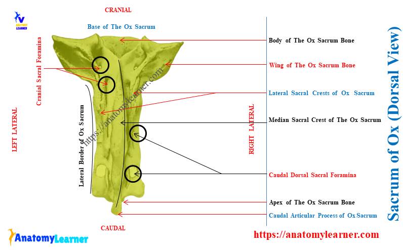
On the dorsal surface of the sacrum bone, you will find the median sacral crest, which is unique in bovines compared to horses. The spinous processes of the ox’s five sacral vertebrae fuse and form the median sacral crest at the midline of this structure.
Again, parallel to the median sacral crest of the sacrum, you will also see the lateral sacral crest. The right and lateral sacral crests of the ox are formed by the fusion of respective articular processes.
The height of these two lateral sacral crests is lower than those of the median sacral crest.
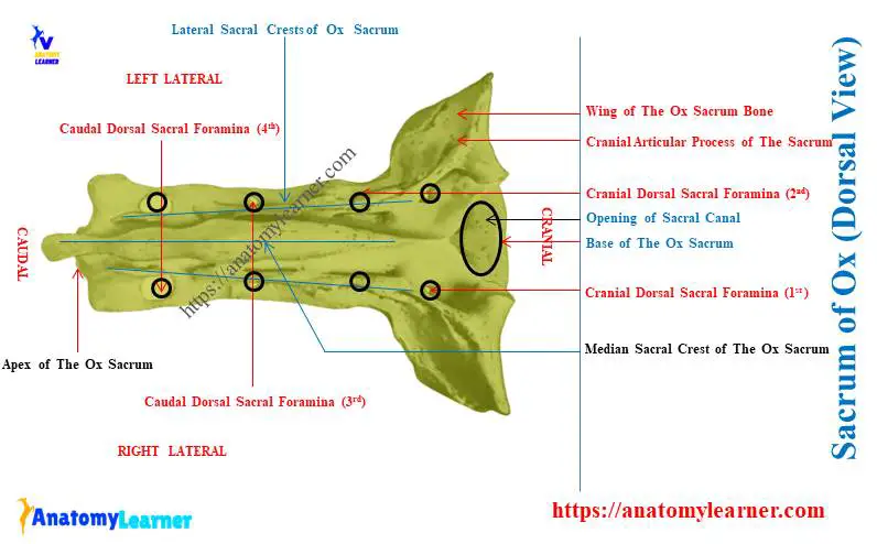
There are four dorsal sacral foramina on the dorsal surface of the sacrum bone. The first two pairs of dorsal sacral foramina are present at the medial aspect of the lateral sacral crest.
Again, the other 2 pairs (caudal) dorsal sacral foramina locate lateral to the corresponding lateral sacral crests. The last two pairs of dorsal sacral foramina are larger than the two cranial pairs of foramina.
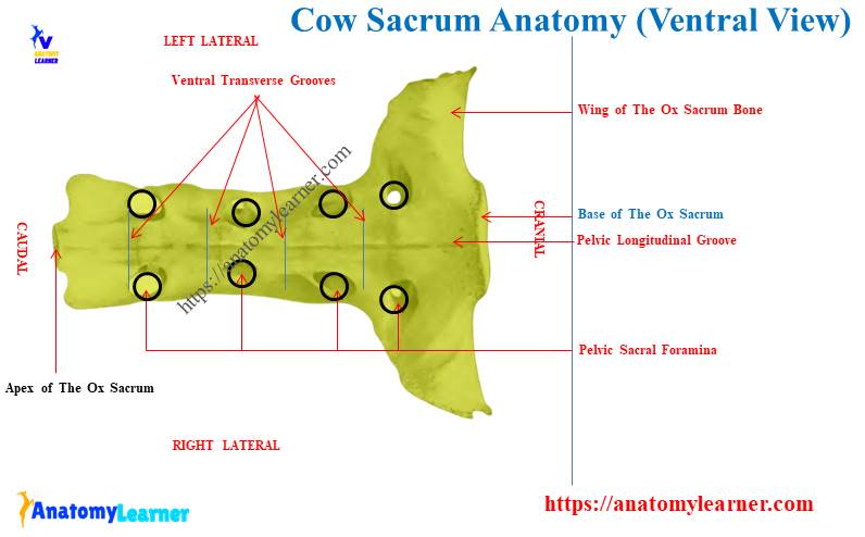
The ventral or pelvic surface of the cow sacrum
The ventral (pelvic) surface of the cow sacrum bone is concave and smooth. This surface is wide cranially and narrow caudally.
There is a longitudinal median groove or line on the pelvic surface of the cow sacrum bone. You will also find four transverse grooves or lines on the ventral surface of the sacrum.
There are four pairs of pelvic (ventral) sacral foramina on the concave surface of the sacrum bone. All these pelvic sacral foramina are larger than the dorsal series.
But, the size of these foramina diminishes from first to last. What structures pass within these ventral sacral foramina of the cow sacrum bone?
The ventral branches of the sacral spinal nerves pass through the pelvic sacral foramina of the sacrum bone.
Do ox sacrum have intervertebral foramen?
Quick answer: yes, the dorsal and pelvic sacral foramina are equivalent to the usual intervertebral foramen in the ox sacrum. Here, the dorsal branches of sacral spinal nerves pass through the dorsal sacral foramina, whereas the ventral branches pass through the pelvic sacral foramina.
The dorsal and pelvic sacral foramina of the bovine sacrum bone communicate with the sacral canal.
The base of the ox sacrum bone
The sacrum of an ox possesses a very wide and cranially directed base. You will see the central body of the first sacral vertebra in the structure of the cow sacrum bone.
The body of the cranial part of the sacrum bone possesses the following –
- It is wide transversely,
- The body of the sacrum is flattened dorsoventrally,
- Have rounded articular surface which articulates with the body of the last lumbar vertebra through intervertebral disc or cartilage,
You will see one pair of cranial articular processes dorsal to the body. Again, this cranial pair of articular processes articulate with the caudal articular process of the last lumbar vertebra.
Dorsal to the body of the sacrum bone, you will also see the opening of the sacral canal. It is also transversely wide and looks like a triangular structure.
The ventral margin of the central body of the cow sacrum bone projects slightly. This slight projection of the sacrum is the promontory used to measure the pelvic conjugated diameter.
The lateral part of the sacrum base represents the alae or wings (two lateral). A cranial aspect of both wings presents smooth surfaces.
But, in the horse sacrum, you will find the large, oval, and convex articular surface on the cranial aspect of the wing. It articulates with the caudal concave articular surface of the transverse process (TP) of the last lumbar vertebra.
The caudal part of the wing possesses an elongated oval area that articulates with the ilium bone. This caudal part of the wing faces dorsolateral in the bovine sacrum bone.
The apex of the cow sacrum bone
Apex is the caudal aspect of the last sacral vertebral segment. It is small and possesses an elliptical flattened caudal surface in the body.
Dorsal to this caudal body of the sacrum, you will find 2 distinct features –
- Dorsally triangle caudal opening of the sacral canal through which the cauda equina passes, and
- Two distinct caudal articular processes of the last sacral segment,
You will also see the pairs of narrow notches between the body and arches of the last sacral segment. The transverse process of the last sacral segment is visible and longer than the previous part.
Borders of the ox sacral vertebrae
The right and left borders of the ox sacral vertebrae are rough and concave. But how are these borders formed in the ox sacrum bone?
The transverse processes of the sacral vertebrae modify and fuse from both the right and left lateral borders. Both the right and left lateral borders of the sacrum bone are thick cranially and thin caudally.
You will find several sacral tuberosities in the lateral borders of the bovine sacrum bone.
How to differentiate the sacrum of a horse from a cow?
Quick answer: you may easily differentiate the sacrum of a horse from a cow with the appearance of their wings and median sacral crest. When compared with the cows, you will not find any median sacral crest in the horse sacrum bone.
Five sacral bones fuse in the horse sacrum, but the spinous processes don’t fuse. They remain separate and form individual spinous processes. Again, the lateral sacral crests in the horse sacrum are ill-developed.
The wing of the horse sacrum is pointed and prismatic. Again, the pelvic surface of the horse sacrum bone is less concave than the cow sacrum bone.
Let’s see the main osteological differences between the horse and ox sacrums from Table 2 –
| Features | Horse sacrum | Ox sacrum |
| Number of bones | 5 | 5 |
| Spinous process | Separated sacral spine Direction – caudo-dorsal Bifid | United Form median sacral crest |
| Wings | Pointed Prismatic shape Convex dorsal surface | Curved ventro-caudally Quadriangular shape Compressed cranio-caudally |
| Transverse process | Prominent | Rudiment |
| Sacral tuberosity | Prominent | Less developed |
How to differentiate the sacrum of a dog from an ox?
Quick answer: you may easily differentiate the sacrum bone of a dog from a cow on the base of the number of sacral segment fusion. There are only 3 sacral vertebrae in the structure of the dog sacrum bone.
Thus, the sacrum of a dog is small than a cow’s. The median sacral crest in the dog sacrum is notched.
You will find two pairs of dorsal and pelvic sacral foramina in the structure of a dog sacrum bone. The articular surfaces on the wings are wide than the ox. Again, the pelvic surface of the dog sacrum is more concave than the oxes.
Let’s see the main osteological differences between the dog and ox sacrums from Table 3 –
| Features | Dog Sacrum | Ox Sacrum Bone |
| Number of Bones | 3 | 5 |
| Shape | Triangular | Quadrangular |
| Wings | Quadrangular Curved bone | Very high Prismatic |
| Median sacral crest | Longer | Notched |
| Transverse process | Less developed | Project caudally |
The sacrum of ox labeled diagram
Now, let’s see the all-important osteological features of the ox sacrum bone from the labeled diagrams. Here, I tried to show the median and lateral sacral crests from the dorsal surface of the bovine sacrum bone.
I also identified the longitudinal and transverse grooves in the ox sacrum labeled diagram from the ventral surface. The base and apex and their osteological structures are also identified in the labeled diagram.
You will find more ox sacrum-labeled diagrams on social media of anatomy learners.
Conclusion
The sacrum of an ox possesses exceptional median and two lateral sacral crests on its dorsal surface. Again, the dorsal surface of the sacrum bone represents four pairs of sacral foramina.
The ventral or pelvic surface of the bovine sacrum is concave and represents various grooves and foramina. Here, the base and apex of the cow sacrum bone are more or less exceptional than other animals’ sacrums.