The cow tail bone consists of incomplete caudal vertebrae. These incomplete vertebrae form the skeleton of the cow tail.
Here, I will show you the details anatomical features of cow tail skeleton along with their number. You will find the features of the body, arch, and processes from the ox tail bones with the labeled diagrams.
Quick overview of cow’s tail: cow tail bones are the atypical caudal vertebrae which range from 18 to 21. You will see complete arches and spinous processes in the first five or six tail bones of a cow.
Again, I will show you the differentiating features of ox tail bones from other species like horses, dogs, and pigs.
Let’s continue this article to learn the anatomical facts of the bovine tail bones (caudal vertebrae).
Cow tail bone
These are the caudal vertebrae, also known as the coccygeal vertebrae. The number of caudal or coccygeal vertebrae varies in animals, including cows.
Typically, you will find 18 – 21 caudal vertebrae in the structure of a cow tail. But, this number may vary in different breeds of cows.
The tail bone of a cow (caudal vertebrae) is relatively larger and better developed than these of horses. You will not typically find the complete arch and processes in each caudal vertebrae of the cow’s tail.
The first few coccygeal vertebrae of a cow show arches and processes. But the rest of these vertebrae only show the cylindrical body.
Again, the transverse process of the first few caudal or coccygeal vertebrae is comparatively larger. You will also find the larger cranial articular process in the anterior series of cow’s coccygeal vertebrae.
The first few coccygeal vertebrae possess a pair of ventral spines that form the ventral median groove.
Does a cow tail have bones?
Yes, the cow tail has bones that are known as coccygeal vertebrae. These are the caudal segment of the cow vertebral column.
But, most of these bones do not possess the osteological features of a typical vertebrae. You may know the details of the osteological facts of the animal typical vertebrae from a below-mentioned article of anatomy learner –
As mentioned earlier, the article might help you understand how a cow’s caudal or coccygeal vertebrae differ from its normal structure.
Except for the bones in a cow tail, you will also find the muscles, vessels, and nerves. Among the vessels of a cow tail, the most important is the coccygeal artery.
Cow tail muscles
The cow tail muscles are almost similar to the horses and enclosed by strong coccygeal fascia. This strong fascia continues with the gluteal fascia and blends with a lateral sacroiliac ligament.
You will find the following muscles in the cow tail bone –
- #1. Coccygeus muscle of the cow tail,
- #2. Sacro-coccygeus dorsalis muscle of the cow tail,
- #3. Sacro-coccygeus lateralis muscle of the tail,
- #4. Intertransversales caudae muscle of the tail, and
- #5. Sacro-coccygeus ventralis muscle of the ox tail,
Here, the coccygeus muscle of the cow tail is more well-developed than the horse. It is a flat and triangular muscle that lies between the sacro-sciatic ligament and the caudal part of the hip bone.
This coccygeus muscle of the cow tail arises from the pelvic surface of the sacro-sciatic ligament near the ischiatic spine. You may know the details of anatomical facts of the cow hip bone (including the ischiatic spine) from the below-mentioned article of anatomy learner –
The action of the coccygeus muscle is to flex the cow tail and compress it over the perineum. Laterally, you will find a relationship between the coccygeus muscle of a cow with the semimembranous muscle.
Other muscles of the ox tail
The sacrococcygeal dorsalis (sometimes known as the medialis) muscle lies along the dorso-median aspect of the ox tail. This muscle arises from the sacral spine of the last three coccygeal vertebrae of the cows.
Again, they insert on the dorsal surface of the coccygeal or caudal vertebrae of the cow. Laterally, this muscle is related to the sacrococcygeus lateralis muscle.
Thus, the sacrococcygeus lateralis muscle of the cow tail immediately lies lateral to the preceding muscle. This muscle also arises from the sacral spine and the transverse process of the sacral and coccygeal vertebrae.
The sacrococcygeus lateral and dorsalis muscles together elevate the cow tail. Ventrally, this sacrococcygeus lateralis muscle is related to the intertransversalae caudae.
The intertransversale caudae muscle arises from the lateral edge of the sacrum bone. It (intertransversale caudae) assists in lateral flexion of the tail.
The sacrococcygeaus ventralis muscle arises from the ventral surface of the sacrum bone. Again, this muscle inserts into the ventral surface of the coccygeal vertebrae.
Cow tail nerves
You will find 3 – 5 caudal or coccygeal spine nerves that attach to the spinal cord of the cow. The last part of the spinal cord of a cow looks like the tail of a horse (also known as the cauda equina).
The dorsal and ventral branches of these 3 – 5 pairs of caudal spinal nerves anastomoses from the respective trunks on either side. They extend at the tip of the cow’s tail and supply its muscles.
Here, the dorsal trunk of the caudal spinal nerve runs with the dorsolateral artery between the sacrococcygeus dorsalis and intertransversalae caudae muscles. The ventral branch accompanies the ventrolateral artery below the intertransversalae.
How many tail bones does an ox have?
Quick answer: the number is variable, but you may find 18 – 21 bones in the tail of an ox. Most local breeds of cows have 15 – 18 bones in their tail.
First, five or six caudal vertebrae or bones of the cow tail show the typical features. But the rest of the caudal vertebrae or bones don’t have any arches or processes.
These five or six caudal bones of the cow tail show the typical neural rings. But, the rest bones of the tail gradually reduce in size and only show the typical rod shape feature.
You will find variations in the number of the tail bone in various animals. Let’s see the variation in the number of tail bones in different animals from Table 1 –
| Species | Number of Tail bones |
| Ox | 18 – 21 |
| Horse | 15 – 22 |
| Dog | 18 – 25 |
| Pig | 18 – 24 |
| Rabbit | 15 – 19 |
I hope you got an idea of the tail bone number from different species.
Are cow tails bone or cartilage?
Quick answer: the cow tails possess bones, not cartilage or cartilaginous structure. All these hardest structures of the cow tail possess the typical features of the bones.
These are the type of irregular bones of the cow skeleton. These irregular bones locate in the midline of the body and are considered part of the axial skeleton.
Some of these bones show bony projections that help attach various muscles. In the structure of the tail bone, you will find the spongy substance with a thin covering of compact substance.
Suggested article from anatomy learner (regarding this ox tail bones) –
Cow tail bone anatomy
You already got a little idea of the cow tail bone anatomy from the above discussion. The tail possesses incomplete vertebral bone that varies in number.
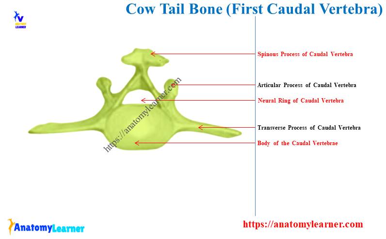
Let’s see the anatomical facts of the first three to six caudal or coccygeal vertebrae from the cow tail. You know, the vertebrae possess three (3) important parts – a cylindrical body, an arch (consisting of pedicle and lamina), and three processes.
Here, these first five or six caudal vertebrae of cow tail have the developed body, neural ring, and processes. The body of these tail bones is flattened dorsoventrally.
They show the typical constriction at the ventral medial part of the body. Like the other typical vertebrae of the cow vertebral column, they (caudal vertebrae) show less typical osteological features.
Both cranial and caudal extremities of the body show convex articular surfaces. Their body looks like the elliptical shape (first five caudal vertebrae).
The ventral surface of these caudal vertebrae presents an important osteological feature. Two median crests form the median or longitudinal groove ventrally.
And you know, this longitudinal ventral groove on the ventral surface of the caudal vertebrae is for accommodating the coccygeal artery.
The arches of these coccygeal vertebrae of cow tails don’t show the typical features. You will find the small and triangular arches in the coccygeal vertebrae of the cow tail.
The two flat plates form the arch of the coccygeal vertebrae. This plate articulates dorsally and forms the small spinous process in the caudal vertebrae.
These five or six caudal bones of the cow tail show the little caudal notch on their arches. But, you will not find any cranial notch on their arches.
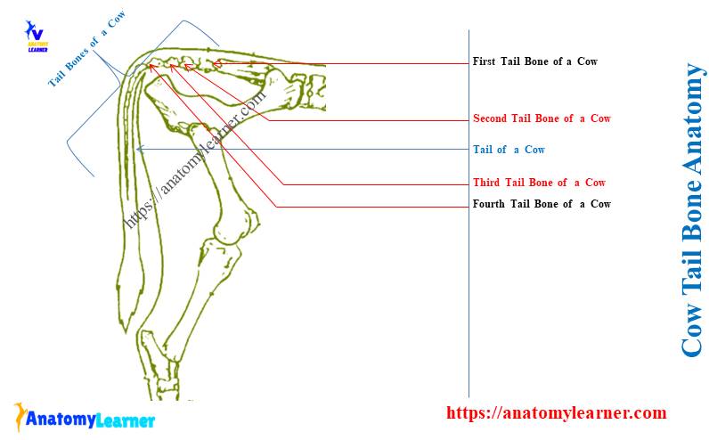
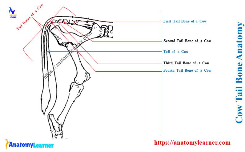
Cow tail bone labeled diagram
Here, you will see the ox tail bones with the labeled diagram. You will see the chronological changes of these tail bones from the cow.
I tried to identify the arches and processes from the few caudal vertebrae of the cow’s tail. Again, the labeled diagram also identifies the neural rings from the first few vertebrae.
You may see more labeled diagrams on the cow tail bones (caudal or coccygeal vertebrae) on the social media of anatomy learners.
Conclusion
The cow tail bone is made of 18 – 21 incomplete coccygeal vertebrae. The first five or six tail bones show the typical long bodies and arches in the cow.
The provided labeled diagrams help you to identify these 18 or 21 caudal vertebral bones from the cow tail.