The atlas of an ox is the first cervical vertebra which is highly modified in conformity. And this modification of the ox atlas is for the unique function of support and movement of the head.
In this article, I will describe the osteological features of cow or ox atlas bone with a labeled diagram. You will find the unique features of the atlas vertebra that might help you to differentiate it from other cervical vertebrae.
A quick guide on ox atlas: this is the first cervical vertebra with a strong ring made of two wings. There are two cranial deep oval-shaped articular surfaces and caudal saddle-shaped fovea dentis in the ox atlas.
Again, I will also show the basic structural differences of the bovine atlas bones of various species like horses and dogs. Okay, let’s see the anatomical facts of the bovine atlas bone with the labeled diagrams.
Atlas bone of an ox
The atlas bone of an ox differs from the osteological features of a typical vertebra. First, let’s see the unique osteological features of the ox atlas bone –
- #1. Strong rings of the atlas bone,
- #2. Two lateral curved plates or wings of the atlas,
- #3. Two deep and oval cranial articular surfaces,
- #4. Caudal single saddle-shaped articular surface (fovea dentis),
- #5. Median dorsal tubercle of the dorsal arch,
- #6. Thick and narrow ventral arch of the cow atlas bone,
- #7. Ventral tubercle of the ventral arch,
- #8. Depression and two foramina (intervertebral and alar) on the dorsal aspect of wings, and
- #9. Atlantal fossa (fossa Atlantis) on the ventral surface of the wings,
The labeled diagram identifies all these above-mentioned osteological features from the ox atlas bone. I hope you will quickly identify these features from the ox atlas.
But, you will learn the details of the anatomical facts of bovine atlas bone in the next section of this article.
What is the atlas of the ox?
Quick answer: It is the first cervical vertebra of an ox vertebral column. Among the 7 cervical vertebrae, the atlas (first vertebra) shows exceptionally modified osteological features.
This vertebra carries the round head in humans; therefore, the name atlas has been given to it. The ox atlas bone comprises a large neural ring and two lateral wings.
Again, you will see the thick plate-like body in the ox atlas bone. This body of the atlas bone forms the ventral wall of the neural ring.
Here, you might have an idea on –
- How is the vertebral column formed in an ox? And
- How many bones does the cow vertebral column have?
Well, you will find your inquiries on the formation of vertebral columns with their number (vertebral formula) in the article’s next section.
Vertebral formula of ox
You know the ox body has two skeleton types – axial and appendicular. When a series of vertebrae bones articulate together to form a long column along the axial skeleton, it is known as the vertebral column.
Let’s see the axial skeleton of a goat or ox; you will find a series of vertebral bones. They form the vertebral column, also known as the spine of the ox.
These vertebrae are grouped into five regions based on their position and shape (external appearance). Each of these regions shows a variable number of bones in various species.
In ox, 48 – 52 vertebral bones are found in the vertebral column. The number of the various vertebral bones is expressed in the vertebral formula.
- Let’s see the vertebral formula of ox – C7 T13 L6 S5 C18 – 21
Here, C = Cervical vertebrae, T = Thoracic vertebrae, L = Lumbar vertebrae, S = Sacral vertebrae, and C = coccygeal or caudal vertebrae.
Number of Vertebrae in Animals
Table 1 shows the number of vertebrae in different regions of the ox spine of some common domestic animals –
| Regions of spine | Ox/ Cow | Horse | Dog |
| Cervical | 7 | 7 | 7 |
| Thoracic | 13 | 18 | 13 |
| Lumbar | 6 | 6 | 7 |
| Sacral | 5 | 5 | 3 |
| Caudal | 18 – 21 | 16 – 22 | 18 – 25 |
Suggesting reading from anatomy learner (based on axial skeleton including vertebrae) –
The bodies of the vertebral bones and intervertebral discs form a solid rod. On this solid structure, vertebral arches are placed to form the hollow canal. And you know, within this hollow canal, the spinal cord of the ox passes.
Two intervertebral foramina are formed on two sides when the adjacent vertebrae are articulated. They give passes to the spinal nerves and vessels.
Unique characteristics of ox atlas
Here, I will enlist the unique osteological features of the ox atlas bone. But, you might have an idea of the typical vertebra of the ox that will help you to understand the atlas’ unique features easily.
You may read the below-mentioned article to get an overview of the typical vertebra –
Now, let’s see the unique features or characteristics of atlas bone –
- #1. The ox atlas bone possesses a thick plate-like body (some authors stated that there is no body) but doesn’t have any processes,
- #2. There is a strong and larger neural ring and two lateral (right and left) wings or modified transverse processes in the ox atlas bone,
- #3. You will find two arches (dorsal and ventral) on the structure of the atlas bone,
- #4. There are 2 main articular surfaces (deep cranial oval and caudal fovea dentis) in the ox atlas bone,
- #5. The dorsal surface of the lateral wings presents two important foramina (intervertebral and alar), where the ventral surface represents only the fossa atlantal,
So, you got the unique characteristics of the bovine atlas bone with the diagram.
Bovine (cows or oxes) atlas bone anatomy description
Let’s describe the anatomy of the cow or ox atlas bone with the labeled diagram. Here, I will describe the following structures from the atlas bone –
- Larger vertebral rings, with their formation,
- Dorsal and ventral arches of the atlas bone, and
- Anatomical features of the atlas bone’s wings,
Typically, you will not find any body in the ox atlas vertebral bone. Sometimes the thick ventral plate is considered the body. But, there are no typical vertebral processes in the ox atlas bone.
The strong vertebral ring of the atlas bone encloses very large foramen. Again, two curved plate arises from the lateral aspect of the vertebral ring.
These curved plates of the atlas vertebra are known as the wings. It is believed that these wings of the atlas vertebra are the modified transverse processes.
Here, the dorsal and ventral arches connect two wings (also known as the lateral masses). The cranial aspect of the atlas bone represents two deep oval articular surfaces for the occipital condyle of the ox skull.
You may learn the details of the anatomical features of the ox skull bones along with the structure of the occipital condyle from the below-mentioned article –
These two deep oval articular surfaces are separated by a wide notch dorsally and a narrow notch ventrally.
Dorsal and ventral arches of the atlas bone
The dorsal arch of the bovine atlas bone presents the median dorsal tubercle on its dorsal surface. Again, the ventral surface of the dorsal arch is concave and has no tubercle.
The ventral arch of the cow atlas bone is thick, narrow, and less curved. The ventral aspect of the ventral arch represents a ventral tubercle. On this ventral tubercle, you will find the insertion of the longus colli muscle.
The dorsal surface of the ventral arch presents the caudal transverse concave articular surface. This concave caudal transverse articular surface is known as the fovea dentis (or dens). On this fovea dentis of the atlas bone, the odontoid process of axis bone rest.
Again, you will find the ridge and transverse excavation on the cranial surface of the ventral arch. On this ridge, you will find the longitudinal ligament of the atlas bone in its fresh condition.
Wings of the ox atlas bone
The modification of the transverse processes forms these wings. Anatomically, you will find 2 distinct surfaces of the atlas bone’s wings – dorsal and ventral.
The dorsal surface of the wings is concave compared to the other species. You will see a depression in the cranial aspect of the dorsal surface.
There are two foramina present within this small depression. The foramen that locates medially and connects with the vertebral ring is the intervertebral foramen.
What structure passes from the intervertebral foramen of the oxes atlas? The first pair of spinal nerves and branches of the vertebral artery passes through the intervertebral foramen of the atlas bone.
The lateral foramen that passes to the ventral aspect of the wing is the alar foramen. Within this alar foramen, the dorsal branch of the first spinal nerve passes.
But, there is no transverse foramen in the wing of the bovine atlas bone. This transverse foramen is at the caudal aspect of the horse’s atlas vertebra.
Under the ventral surface of the atlas wing, you will find the deep oval depression or cavity. This deep oval depression is known as the atlantal fossa.
A larger foramen on the atlantal fossa opens into the vertebral canal.
How to differentiate the horse atlas from the ox?
Quick answer: you may easily differentiate the horse atlas vertebra from the ox with the help of their external appearance of wings and the length of the bodies. The wing of the cow atlas bone is less curved than the horse atlas.
The most differentiating osteological feature between the horse and ox atlas vertebrae is the presence of transverse foramen. The caudal part of the wings (of horse atlas bone) represents the distinct larger transverse foramen.
Again, some other osteological features might help you to differentiate the bovine atlas bone from the horse. Let’s see the main differentiating points for the ox and horse atlas bones from Table 2 –
| Features | Horse atlas | Ox atlas |
| Wings | Modified More curved | Less curved |
| Foramen on wings | 3 in number | 2 in number |
| Body | Longer | Smaller |
| Ventral arch | Thicker Larger ventral tubercle | Less thick Small ventral tubercle |
| Dorsal arch | Small | Larger |
| Transverse foramen | Caudally | Absent |
| Fovea dentis | More extended | Well-developed |
I hope you will quickly differentiate the horse atlas vertebra bone from the ox atlas.
How to differentiate the dog atlas vertebra from the ox?
Quick answer: the body and wings of the dog atlas vertebra show some of the typical features compared to the oxes. The foramen transversarium (transverse foramen) is present in the dog atlas bone at its caudal aspect.
The body is comparatively less developed in the dog atlas compared to the ox. Again, you will find the exceptional alar notch on the cranial aspect of the flattened wings (on both sides).
Let’s find the other important differentiating osteological features between ox and dogs atlas vertebrae from Table 3 –
| Features | Dog atlas | Ox atlas |
| Wings | Flattened Wide horizontally | Curved Concave in middle (less) |
| Body | Less developed | Well-developed |
| Dorsal arch | Small Less developed | Larger Distinct |
| Ventral arch | Not developed | Developed |
| Foramen on wings | 2 | 2 |
| Alar notch | Cranial aspect of wings | Not present |
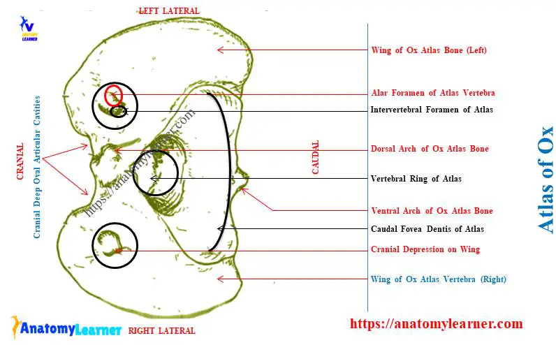
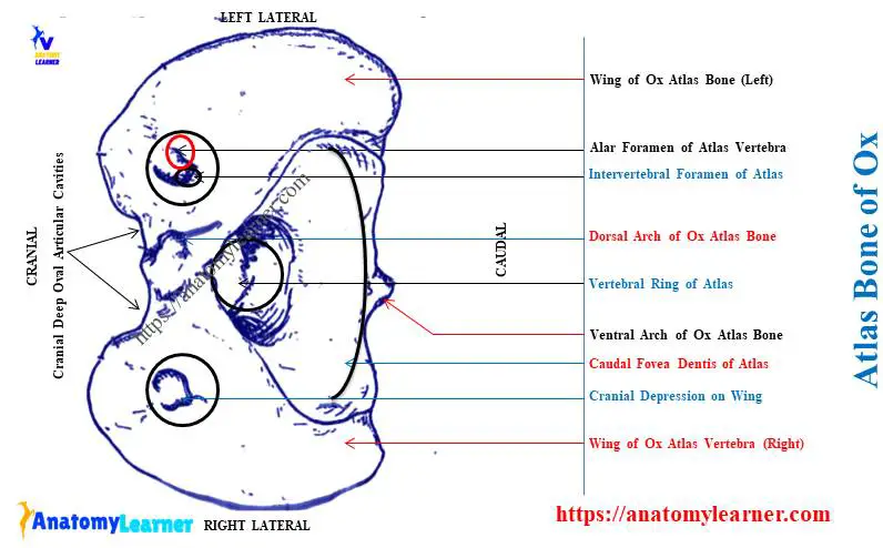
Ox atlas vertebra labeled diagram
In this section, I tried to show you the dorsal and ventral views of the ox atlas vertebra bone with the labeled diagrams. Two lateral wings, dorsal arch, and ventral arches are identified from the ox atlas vertebra labeled diagram.
Here, two foramina from the cranial depression of the bovine atlas bone are also identified in the diagram. I tried to show you the atlantal fossa with the foramen from the ventral view of the atlas bone.
You may find more labeled diagrams on the ox atlas vertebra on social media of anatomy learners. Again, you will find more diagrams where I tried to compare the ox atlas with other animals’ atlas bones.
Conclusion
The atlas of the ox or first cervical vertebra possesses modified wings and two arches. Two lateral wings of the atlas vertebra present two important foramina on its cranial aspect.
The cranial deep oval articular fossae on the ox atlas are exceptional features compared to other vertebral bones. Other features of ox atlas bone provided in this article might help you differentiate it from other species atlas vertebra.