The spermatic cord contents in animals like bulls, tomcats, dogs, and stallions are almost similar. It is a structure found in male animals and is considered an essential part of their reproductive system.
Here, I will show you the location of the spermatic cord in animals with their contents. You will clearly understand the number and extension of the spermatic cord from the animals.
Quick overview of spermatic cords: they are 2 in number in male animals, and each of them consists of 7 more structures. It extends from the base of the animal’s testis and passes through the inguinal canal.
Here, I will describe all the structures of the spermatic cord from a bull with a labeled diagram. But, you will get the comparative anatomy of the spermatic cords from various animals in the last.
Let’s see the spermatic cord anatomy of the bull with a diagram.
What is the spermatic cord of the ox?
The spermatic cord of an ox consists of ductus deferens, nerves, vessels, lymphatics, and serous covering. It begins at the internal deep abdominal inguinal ring, where its constituent parts come together.
Then the spermatic cords migrate through the oblique inguinal canal from the abdominal cavity. It passes over the side of the copulatory organ and ends at the attached border of each testis.
So, there are 2 spermatic cords in male animals like bulls, tomcat, dog, and stallion. These 2 cords are from the single spermatic bundle in an animal.
Thus, you will see only one single spermatic bundle externally in any animal. Here, the diagram shows the number of spermatic cords and bundles from the bull.
Now, let’s see the contents of each of these spermatic cords.
Spermatic cord contents in bull and dog
Table 1 shows the spermatic cord contents in a bull, dog, and other animals –
| Serial | What does the spermatic cord contain in a bull? |
| 1 | Spermatic artery – single in each cord |
| 2 | Spermatic veins – form pampiniform plexus |
| 3 | Lymphatics – accompany the veins |
| 4 | Spermatic nerves – runs with artery |
| 5 | Ductus deferens – single and runs caudally |
| 6 | Internal cremaster muscle – consists of unstriped muscles |
| 7 | Visceral layer of tunics – part of the parietal peritoneum |
So, there are 7 contents in the structure of each spermatic cord of the animals. These 7 structures of the cord comprise two bundles –
- Cranial bundle of the spermatic cord, and
- Caudal bundle of the spermatic cord,
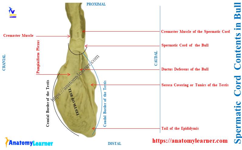
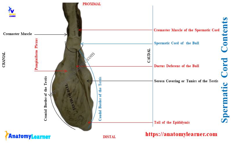
Both the cranial and caudal bundles of the spermatic cord are enclosed by the serous covering (tunics).
The cranial bundle of the cord consists of arteries, veins, lymphatics, and nerves. Again, the cremaster muscle is also included in this cranial bundle of the spermatic cord. However, some anatomists stated the cremaster muscle is not a part of the spermatic cord.
The caudal bundle of the bull spermatic cord contains only the ductus deferens. And you know, this ductus deferens is the caudal continuation of the epididymis.
Here, the bull spermatic cord labeled diagram shows the various structures from it. Let’s find a more labeled diagram of the animal’s spermatic cords here.
Spermatic cord anatomy in animals
The spermatic cord anatomy in all mammals is almost similar. You might describe the following under the spermatic cord anatomy in animals –
- Location of the animal’s spermatic cord and
- Structures of the spermatic cord,
So, let’s see the location of the spermatic cord in different animals first. Then, I will describe every single structure of the animal spermatic cord with a diagram.
Where is the spermatic cord located in a bull?
The spermatic cord of a bull is located at the inguinal region. This structure passes through the inguinal canal during the descent of the testis.
The inguinal region is the lower part of the lateral abdominal wall of a bull. Again, the term inguinal canal is an oblique passage through the lower abdominal wall between 2 inguinal rings.
The 2 inguinal rings of the inguinal canal are –
- Deep inguinal ring (internal abdominal part), and
- Superficial inguinal ring (external abdominal part),
Let’s get the detailed guide on animal inguinal canals from the below-mentioned article –
Here, the diagram shows the exact location of the spermatic cord in a bull. This diagram also indicates the oblique inguinal canal with deep and superficial rings.
What is the structure of the spermatic cord in animals?
The structures of the animal’s spermatic cord divide into cranial and caudal bundles. 4 out of 7 structures of the spermatic cord remain in the cranial bundle.
Again, only the ductus deferens remain in the caudal bundle of the animal spermatic cord. Let’s discuss these structures of the animal spermatic cord with the diagram.
Bull spermatic artery
The spermatic artery is the convoluted structure of the spermatic cord that supplies the bull’s testis. This spermatic artery arises directly from the abdominal aorta of the ox.
It originates from the ventral surface of the abdominal aorta. Then, the spermatic artery arises cranial to the origin of the caudal mesenteric artery.
It runs laterally and caudally, crossing the ventral surface of the ureter. In this area, the spermatic artery joins with the spermatic vein and nerve.
Spermatic veins of the bull
You will find 3 – 4 spermatic veins in the bull spermatic cord. They arise from the head of the epididymis and the upper end of the testis.
Here, the left spermatic vein empties into the left renal vein. Whereas the right spermatic vein empties into the caudal vena cava.
You will find a unique structure here, known as the pampiniform plexus. Let’s know how is the pampiniform plexus formed.
What is the pampiniform plexus in a bull or dog?
Quick answer: the pampiniform plexus is a structure of the spermatic cord that is formed by the anastomoses of spermatic veins. A longitudinal section of the spermatic cord shows the torturous bends of veins that wrap around the artery.
The unique features of the pampiniform plexus in a bull and a dog –
- They are formed by the colis of spermatic veins that surround the spermatic artery,
- It makes the bulk of the spermatic cord and
- It functions to draw heat from the spermatic artery and cool the circulation before it reaches the testis,
Lymphatics of the spermatic cord
The spermatic veins accompany the lymphatics of the spermatic cord. There are several lymphatic capillaries in the structure of the spermatic cord in animals.
The lymphatic spermatic cord drains lymph into different lymph nodes. Finally, the filtrated lymph through the spermatic lymphatic will empty into the lumbar and para-aortic nodes.
You may know the details of how is the lymph forms and transported in animals. The below-mentioned article will help you to get an idea of lymph formation and transformation –
Spermatic nerves in the ox spermatic cord
The plexus of the animal spermatic nerves arises from the area of the sympathetic trunk. It is located between the third and sixth lumbar sympathetic trunk ganglia.
So, the spermatic plexus proceeds from the following –
- Aortic plexus, and
- Branches from the posterior mesenteric ganglion,
The corresponding internal spermatic artery accompanies each of the spermatic nerves. The genitofemoral nerve also innervates the cremaster muscle of the animal spermatic cord.
This spinal nerve of the animal can be found coursing through the superficial inguinal ring. Let’s know the ideal formation of animal spinal nerves from the below-mentioned article –
Animal spermatic cord and ductus deferens
The ductus deferens is one of the essential parts of the spermatic cord contents in a bull. It is a thin tube that starts from the tail of the epididymis and passes upward along the caudal border of the testis.
You will see the close association of the ductus deferens with the medial aspect of the epididymal body. Within this course, the ductus deferens show a tortuous appearance.
Finally, the ductus deferens get incorporated into the caudal bundle of the animal spermatic cord. The tube now passes through the inguinal canal and separates itself from other structures of the cord.
It turns backwards and crosses the external iliac artery and corresponding ureter. Now, the ductus deferens of the ox reaches the caudo-dorsal aspect of the urinary bladder.
Finally, both the right and left ductus deferens disappear under the body of the prostate gland. The terminal part of both tubes is a little dilated and is known as the ampulla.
The terminal part or ampullae are placed side by side under the bodies of the seminal vesicle. They open at the root of the beginning of the urethra on either side of the seminal colliculus.
The seminal colliculus is the slit-like opening where the ducts of the seminal vesicle open.
Branches of urogenital artery that arise from the internal iliac supply the ampulla of ductus deferens. Again, the rest part of the ductus deferens is supplied by the differential branches of the umbilical artery.
The innervation to the animal ductus deferens comes from the pelvic plexus.
Internal cremaster muscle of the spermatic cord
The cremaster muscle of the bull or dog spermatic cord is a ribbon-like strip of skeletal muscle. It originates from the caudal boundary of the internal oblique abdominal muscles.
You may know the details of the abdominal muscles from the dog and cow. The below-mentioned articles will provide the full guide on abdominal muscles –
- Dog abdomen anatomy – abdominal muscles and the organs and
- Cow muscle anatomy – bovine myology identification with the diagram,
The cremaster muscle of the spermatic cord of an ox course on the superficial surface of parietal tunics. Again, this cremaster muscle is slightly covered by the spermatic fascia.
Some authors do not consider this muscle to be part of the animal spermatic cord. Like the tunica dartos muscle, the cremaster muscle is also involved in thermoregulation. It voluntarily draws the testis close to the animal body when it contracts.
The external cremaster muscle of the spermatic cord lies on the lateral and posterior parts of the tunics.
Visceral layer of the tunics of animal spermatic cord
The tunics are the flask-like serous sacs that extend through the inguinal canal to the bottom of the testis. It includes all the peritoneal structures that have descended from the abdominal cavity.
So, the tunics of the dog or ox spermatic cord (serous covering) divide into – the parietal and visceral layers. These two layers are frequently called the common tunic of the animal spermatic cord.
The parietal layer is reflected from the posterior wall of the inguinal canal. It surrounds the structure of the spermatic cord of animals and forms the mesorchium.
Let’s summarize the tunics of the animal spermatic cord –
It is the double-layered serous membrane around the spermatic cord, testis, and round ligament. The visceral, parietal, and mesorchium are the parts or layers of this tunic.
- Visceral tunics – is the continuation of the abdominal visceral peritoneum. It tightly invests the structures of the spermatic cord and testis.
- Parietal tunics – it is the continuation of the abdominal parietal peritoneum through the inguinal canal. It surrounds the visceral tunic-covered spermatic cord and testis.
- Mesorchium or connecting tunic – is the serous fold connecting the visceral tunics with the parietal tunics. The structure of the mesorchium tunics is similar to the serous covering.
Thus, the mesorchium is similar to –
- The parietal peritoneum lining of the abdominal wall and
- The visceral peritoneum covering the gastrointestinal tracts
Mesoductus and mesofuniculus of spermatic cord
Again, you will also find another 2 terms related to the tunics – mesoductus and mesofuniculus. Let’s see what are the mesoductus and mesofuniculus.
- Mesoductus – is the fold connecting the tunics between the mesochium and ductus deferens and
- Mesofuniculus – it is the part of the mesochium. It lies between the parietal part of the tunics and where the mesoductus arises.
You will also find a cavity between the two layers of the tunics. It is filled with fluid and continues with the peritoneal cavity at the inguinal rings.
Who supplies and innervates the spermatic cord of a bull?
Quick answer: The spermatic artery from the aorta and the artery of the cord from the external iliac supply the bull spermatic cord. Again, the artery of the vas from the umbilical artery supply to a spermatic cord of the bull.
Again, the spermatic plexus innervates the bull’s spermatic cord.
What is the function of the spermatic cord in a bull?
Quick answer: the primary function of the spermatic cord in a bull is to connect the testis with the abdominal wall. Again, the bull’s spermatic cord houses the ductus deferens, cremaster muscle, and pampiniform plexus.
Here, the ductus deferens of the bull carry the sperm from the tail of the epididymis and empties into the urethra. Again, the cremaster muscle is the primary muscle that supports the testis.
It is also responsible for facilitating the circulation flow in the testis. Again, the pampiniform plexus of the spermatic cord is necessary to cool the circulation.
How will you differentiate between bull and horse’s spermatic cords?
Quick answer: the spermatic cord and tunics are longer in the bull compared to the horses. The extra-inguinal part of the spermatic cord in a bull is about eight to ten inches in length.
Again, the cremaster muscle of the bull’s spermatic cord is more well-developed than the horses. It almost completely encloses the tunics of the spermatic end part.
The internal and external inguinal rings of the bull are smaller than the horse. Again, the internal cremaster muscle of the bull’s spermatic cord is feeble than the horses.
Frequently asked questions about spermatic cord contents
I enlist some of the commonly asked questions on the spermatic cord contents in various animals. But please go through the full guide to know the details of the animal spermatic cords.
Okay, let’s see the frequently asked questions on the spermatic cord structures –
What is in the spermatic cord of a dog?
Quick answer: the dog spermatic cord is composed of two bundles – cranial and caudal, which are enclosed with tunics. Here, the cranial bundle of the dog spermatic cord contains an artery, vein, lymphatic, and nerve.
Again, the caudal bundle of the dog spermatic cord contain the ductus deferens or vas deferens.
What are the unique features of a pig’s spermatic cord?
Quick answer: the pig’s spermatic cord is very long compared to the horse’s. You will find the flexous ductus deferens in the pig’s testicular part.
There are no distinct ampullae at the end part of the pig’s ductus deferens. The cremaster muscle is well-developed in the spermatic cord of a pig.
What is the uterus musculinus?
Quick answer: uterus musculinus is the remnant of the paramesonephric duct. Typically, you will find this structure in a horse between the layers of genital folds.
Again, you may also find this uterus musculinus between the ampulla of the ductus deferens of a horse. This structure may also be found in the bull.
What is the cremaster muscle in a dog?
Quick answer: the cremaster muscle is the bundle of unstriped muscular tissue in a dog. It locates. The internal cremaster muscle remains in the cranial part of the dog’s spermatic cord.
The connective tissue unites the structures of the cranial bundle of a dog’s spermatic cord. Again, these structures are interspersed with the bundle of internal cremaster muscle.
What is special in a dog’s spermatic cord?
Quick answer: both the spermatic cord and tunics are longer in the dog. They cross the side of the copulatory organ very obliquely.
The upper extremity of the dog’s tunic is closed. You will find the narrow ampullae at the end part of the dog’s ductus deferens.
Conclusion
So, the spermatic cord contents of bull and dog are similar, with few exceptions. In each animal, this spermatic cord is located in the inguinal region and passes obliquely through the inguinal canal.
The animal spermatic cord is the primary structure that contains ductus deferens, nerves, and vessels. A serous covering and internal cremaster muscles are other important contents of the spermatic cords.