While reading about the distribution of the spinal nerves from various animals, you might know the basic structure of it. Here, I will show you the dog spinal nerve formation and its branches with labeled diagram.
Quick answer: each spinal nerve from the dog is formed by the union of dorsal and ventral roots just outside the intervertebral foramen. After forming the spinal nerve, it immediately divides again into a dorsal and ventral ramus.
But how these roots of the spinal nerves are formed? What are these branches of spinal nerves? If you want to know these features of the dog’s spinal nerve, continue this article till the end.
Here, I will explain every feature of a typical spinal nerve from a dog with the labeled diagram. Again, you will get an idea of the different spinal nerves from different animals, including dogs, cats, horses, and cows.
So, let’s learn the structure of a spinal nerve.
Spinal nerve formation
You will find a great variation in the number of spinal nerves in different animals (like dogs, cats, goats, horses, and cows), but the formation pattern is similar. Again, the innervation of these spinal nerves is almost similar except for the followings –
- Thoracic thirteen spinal nerve (cost abdominal nerve),
- Lumbar one (iliohypogastric), lumbar 2 (genitofemoral), and lumbar 3 (ilioinguinal), and
- Spinal nerves that contribute to forming the brachial and lumbosacral plexus,
“If you read the formation of brachial and lumbosacral plexus, you will understand why the spinal nerve structure is important for you.”
Suggested reading for you from anatomy learner –
- Formation of brachial plexus and distribution of its nerves with diagram, and
- Lumbosacral plexus formation with labeled diagram,
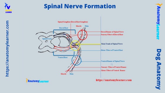
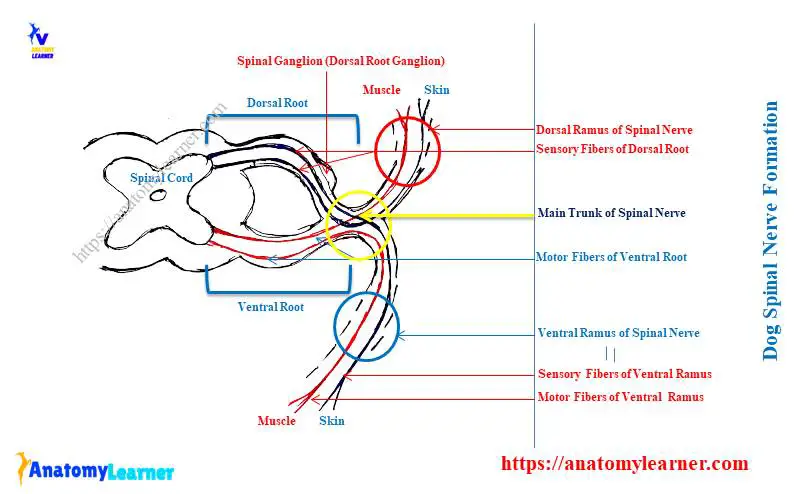
Okay, let’s see the labeled diagram on spinal nerve formation and try to identify the following features –
- Dorsal and ventral roots that originated from the corresponding parts of the spinal cord,
- Dorsal root ganglia,
- Spinal nerve (outside of intervertebral foramen),
- Major dorsal and ventral branches of a spinal nerve, and
- Meningeal and communicating branches of a spinal nerve,
The dorsal and ventral roots (trunk) of the spinal nerve are formed by the union of numerous rootlets that arise from the corresponding aspect of the spinal cord. Here, the dorsal root of the spinal nerve structure is sensory, whereas the ventral root is motor.
Again, the dorsal root of the spinal nerve possesses a dorsal root ganglion. This dorsal root ganglion contains unipolar neurons and is encapsulated by the connective tissue.
But, you will not see any such type of spinal ganglion on the ventral root of the spinal nerve.
How is a spinal nerve formed in a dog?
The dorsal and ventral roots that come from the corresponding aspect of the spinal cord join together just outside of the intervertebral foramen. Here, the dorsal sensory fibre and ventral motor fibres exchange their fibres and form a small spinal nerve.
Now, the spinal nerve immediately divides into 2 main trunks –
- Dorsal trunk or ramus – that will contain both sensory and motor fibres (as the ventral and dorsal roots exchange their fibres), and
- The ventral trunk or ramus – also contains both sensory and motor fibres,
Now, let’s try to understand the following from the spinal nerve structure –
- Dorsal and ventral roots of the spinal nerve – remain within the vertebral canal,
- Spinal nerve – formed just outside of the intervertebral foramen, and
- The dorsal and ventral ramus of the spinal nerve – remain outside of the intervertebral foramen and go for further divisions,
But where will you find the communication and meningeal branches of the spinal nerve structure? Well, the meningeal branch of the spinal nerve remains within the intervertebral foramen.
On the other hand, the communicating branch is formed from the spinal nerve just outside of the intervertebral foramen. Then this communicating branch of the spinal nerve continues with the larger ventral ramus.
The dorsal ramus or trunk divides into medial and lateral branches. Here, the medial one is motor and supplies to the epaxial muscle. Again, the lateral one is sensory and innervated to the skin near the dorsal midline of a dog.
The ventral ramus divides into medial and lateral branches and innervates to the hypaxial muscle of the body wall. Again, these medial and lateral branches further divide into lateral and ventral branches that innervate the skin and ventral aspect of the dog’s body wall.
Typical spinal nerve description – branches and ganglion
So, you already got an idea of the structure of a typical spinal nerve from the dog. Now, you may learn the details of these structures that are associated with forming the spinal nerve in a dog.
From the previous discussion, a typical spinal nerve has the followings four/ five segments from proximal to distal –
- Roots of the spinal nerves (both dorsal and ventral),
- Main trunk or spinal nerve,
- Four primary branches – dorsal, ventral, meningeal, and communicating, and
- Peripheral branches of the dorsal and ventral ramus (numerous branches, mainly lateral and medial)
Now, let’s describe all these structures from the spinal nerve with the labeled diagram. First, let’s start with the dorsal and ventral roots of the spinal nerve.
Dorsal and ventral roots of spinal nerve
The roots of the spinal nerve run within the vertebral canal and possess 3 major features –
- A dorsal root,
- The ganglion on the end part of the dorsal root, and
- A ventral root,
You will see several rootlets forming both the spinal nerve’s dorsal and ventral roots. Here, the dorsal root attaché to the dorsal horn of the spinal cord. In comparison, the ventral roots come from the ventral horn of the spinal cord.
The spinal ganglion (dorsal root ganglion) locate just near the junction of dorsal and ventral roots (near the intervertebral foramen). Actually, the dorsal root of the spinal nerve sends axons into the spinal cord on its dorsolateral aspect.
Again, the ventral root emerges from the wide, distinct ventrolateral aspect of the spinal cord. The number of rootlets of both dorsal and ventral roots varies in different species.
In a dog’s first 5 cervical spinal nerves, each dorsal and ventral roots possess average 6 rootlets. These rootless of dorsal and ventral roots may increase in size and also in number from the six cervical spinal nerves.
But, from the third thoracic to thirteen thoracic spinal nerves, both the dorsal and ventral roots possess only 3 rootlets. Each of these dorsal and ventral roots is surrounded by the pia and arachnoid maters and possesses cerebrospinal fluid.
In almost all species, these spinal nerves form in different regions (cervical, thoracic, lumbar, sacral, and caudal). You will find the details of these spinal nerves (especially their number and formation in different animals) in the next section of this article.
Spinal or dorsal root ganglion
Before spinal nerve formation, you will always find each side’s spinal ganglion on the dorsal root. They are the oval structure and the aggregation of unipolar nerve cell bodies.
The spinal ganglion (dorsal root ganglion) contains neurons that are arranged in a group. They are separated by bundles of nerve fibres and intraganglionic connective tissue.
Each neuron possesses a single dendro-axonal process. This process divides into 2 parts – peripheral afferent and central parts.
Here, the peripheral part of the dendro-axonal process is very long and conducts sensation forward to the cell body. Thus this is functionally an elongated Dendron.
Main trunk or spinal nerve of a dog
Now, the dorsal and ventral branches join to form the main trunk of the spinal nerve just outside of the intervertebral foramen. Sometimes, you may find the starting part of the spinal nerve within the intervertebral foramen.
But how long is this main trunk of the spinal nerve? Well, the length of the main trunk of a spinal nerve may vary. Averagely, you may find the spinal nerve in the cervical, and thoracic region of a dog is about 1 millimetre.
Here, within the spinal nerve, the sensory dorsal and moto ventral roots exchange their fibres and immediately divide into 2 primary branches – dorsal and ventral ramus.
The dorsal and ventral ramus of the spinal nerve (primary branches)
So, you will find the sensory and motor fibres in both the dorsal and ventral ramus. Here, the dorsal ramus of the spinal nerve passes upward between the transverse process.
Now, the dorsal ramus reaches the dorsal muscles of the back and further divides into peripheral branches. Typically, you will see 2 major branches of the dorsal ramus of the spinal nerve – lateral and medial branches.
The medial peripheral branch of the dorsal ramus innervates the muscle of the dorsal aspect of the dog’s body. Again, the lateral peripheral branch of the dorsal ramus innervates the skin of the dorsal aspect of the dog’s body.
These lateral and medial peripheral branches of the dorsal ramus are smaller than the branches of the ventral ramus. Let’s see the anatomical facts of the ventral ramus of the dog’s spinal nerve.
The ventral ramus of the dog spinal nerve is comparatively longer than the dorsal ramus. Typically, the ventral ramus of the spinal nerves divides into different peripheral branches.
But, for your easy understanding, I will tell you only the 2 main branches of the ventral ramus. Like the dorsal ramus, here, you will also find the lateral and medial branches of the ventral ramus.
Again, the lateral branch of the ventral ramus innervates the skin of the lateral abdomen. At the same time, the medial ramus innervates the lateral and internal muscles of the abdomen.
But, the ventral branches of the spinal nerves form different plexus (typically in the fore and hind limbs). In this case, these ventral branches of the spinal nerve innervate different specific body parts.
I have already described all these innervations from the brachial (forelimb) and lumbosacral (hindlimb) plexus of the animal.
Spinal nerve list and number
As the vertebrae (intervertebral foramen) are different in animals, so they also have variations in the number of spinal nerves. Here, I will provide the number of spinal nerves from different animals like dogs, cows, horses, and pigs in table 1 –
| Animals Spinal Nerves | Cervical | Thoracic | Lumbar | Sacral | Caudal |
| Dog | 8 | 13 | 7 | 3 | 5 |
| Cow | 8 | 13 | 6 | 5 | 5 |
| Horse | 8 | 18 | 6 | 5 | 5 |
| Pig | 8 | 15 | 7 | 4 | 5 |
So, you will find 36 pairs of spinal nerves in a dog. The cow possesses 37 pairs of spinal nerves, whereas the horse has 42 pairs of spinal nerves. Again, pigs possess 39 pairs of spinal nerves.
Now, let’s enlist the spinal nerves from the different regions of the different animals. Let’s say – how many spinal nerves do dogs have? Do you know the number? They have 36 pairs (72) of spinal nerves in their body.
So, dog spinal nerve list –
- Cervical spinal nerves (8 pairs),
- Thoracic spinal nerves (13 pairs),
- Lumbar spinal nerves (8 pairs),
- Sacral spinal nerves (3 pairs), and
- Caudal spinal nerves (5 pairs),
36 pairs of spinal nerves in a dog
I have already enlisted the 36 pairs of spinal nerves in the dog. Now, let’s learn a little about some of the important spinal nerves in dogs.
You will find only 7 cervical vertebrae in the axial skeleton of a dog. But, there are 8 pairs of spinal nerves in a dog? How this occurs in a dog?
Well, the first cervical nerve arises from the first segment of the spinal cord. This nerve is located just caudal to the foramen magnum and surrounded by the cranial part of the atlas.
But, other cervical nerves typically arise in between the 2 corresponding vertebrae (intervertebral foramen).
The second cervical spinal nerve is larger and atypical than the other cervical spinal nerve. Here, the dorsal and ventral roots of the second cervical spinal nerve join peripheral to the second intervertebral foramen.
Again, the spinal ganglion of the second cervical spinal nerve locates completely outside of the vertebral canal. Thus, this second cervical spinal nerve is an exception and atypical.
Spinal nerves and brachial plexus in a dog
The ventral branches of the last three cervical and first thoracic spinal nerves give rise to the brachial plexus in a dog. Here, you will find 15+ major nerves that innervate the different muscles of the forelimb of a dog.
The radial, median, and ulnar are the important nerves of the brachial plexus that innervate up to the digits of the dog.
Thoracic spinal nerves of the dog
You will find 13 pairs of a thoracic spinal nerves in a dog. Each pair of thoracic spinal nerves exits from the intervertebral foramina.
All these thoracic spinal nerves of the dog show a similar structure that you have already found in a typical spinal nerve formation. So, each of these thoracic spinal nerves of the dog gives off dorsal and ventral ramus, which will further divide into lateral and medial peripheral branches.
In the thoracic spinal nerve section, you will find the important nerve termed cost abdominal. It is the ventral branch of the last or thirteenth thoracic spinal nerve.
This cost abdominal branch of the spinal nerve innervates to the abdominal wall band adjacent to the caudal border of the last rib. Again, this nerve also innervates to the last ribs and caudal arch in the abdominal wall.
Lumbar and sacral spinal nerves
The lumbar spinal nerves are seven on each side of the dog. In contrast, the number of sacral spinal nerves in a dog is 3 pairs.
Here, the distribution and branches of the lumbar and sacral spinal nerves are very complicated in a dog. It would help if you had a separate guide to understanding the anatomical facts of the lumbar and sacral spinal nerves from the dog.
For that, follow the articles that I previously suggested here.
Here, the ventral branches of the fifth, sixth, seventh, and 3 sacral spinal nerves from the lumbosacral plexus in the dog. The major nerves from the lumbosacral plexus are the sciatic, femoral, tibial, and fibular, along with their small branches.
These nerves from the lumbosacral plexus of a dog innervate to the specific area (muscles and skin) of the hind leg.
Let’s learn a little about the ilioinguinal, genitofemoral, and lateral cutaneous femoral nerves from the dog. These nerves originate from the ventral branches of the lumbar (3 – 5) spinal nerves.
The ilioinguinal spinal nerve – is the direct ventrolateral continuation of the ventral branch of a third lumbar spinal nerve. It gives off a medial branch that innervates to the psoas major, psoas minor, and ilicus muscles. Again, the ilioinguinal spinal nerve communicates with the fourth lumbar spinal nerve and divides into lateral and medial branches.
The genitofemoral spinal nerve – arises from the ventral branches of the third and fourth spinal nerve of the dog. This nerve innervates the medial part of the psoas major muscles, cremaster muscle, and inguinal rings.
You may know the details of these ilioinguinal, genitofemoral, and iliohypogastric spinal nerves from other different articles on anatomy learners.
The number of the caudal spinal nerve varies from 4 – 7 in a dog.
Spinal nerves or cord forming cauda equina
So, the different spinal cord segments give off the spinal nerve roots. The spinal cord of a dog is a cylindrical structure that runs within the vertebral column.
This spinal cord is covered by meninges and extends from the foramen magnum to the middle of the dog’s sacrum bone. You will find the idea on the spinal cord of a dog along with the vertebral column in the below-mentioned article –
- Dog spine anatomy – anatomical features of canine vertebrae, intervertebral discs, and spinal cord,
The caudal end of the dog’s spinal cord (at the level of the second sacral vertebrae) terminates in a tapering and pointed structure. You know this pointed and tapered structure of the dog’s spinal cord is the conus medullaries.
Again, there is a very thin cord that extends from the conus medullaries to the first caudal vertebrae. This structure of the dog’s spinal cord is the filum terminale.
Some other different roots arise from the conus medullaries (as shown in the diagram). These roots increase in length and travel caudally within the vertebral column.
Thus, you will see a long lash of spinal nerve roots in the terminal part of the vertebral column. This structure of the spinal cord looks like the tail of a horse and is termed the cauda equina.
Dog spinal nerve formation labeled diagram
Now, I will show you the labeled diagrams of the dog spinal nerve formation. First, let’s see the labeled diagram that shows the different segments of the spinal cord, roots of the spinal nerve, and different segments of the typical vertebrae.
You may also know the different segments of the typical vertebrae of an animal from the below-mentioned article –
- Typical vertebrae of ox – the body, arch, and process anatomy
Here, the dorsal and ventral horns from each side of the dog’s spinal cord are identified in the labeled diagram. The diagram also shows the dorsal and ventral roots of the spinal nerve (from both lateral aspects of spinal cord).
The spinal ganglion from the dorsal root of the spinal nerve is identified in the labeled diagram. A small main trunk of the spinal nerve (formed by the uniting of dorsal and ventral roots) is shown in the labeled diagram.
The diagram also shows the 4 branches of the spinal nerve – meningeal, communicating, and 2 rami. Each of the dorsal and ventral rami of the spinal nerve shows the lateral and medial branches (shown in the diagram).
Now, let’s see the cervical spinal nerves and formation of the brachial plexus in a dog. Here, the diagram shows the dorsal and ventral branches of the cervical nerves from the dog spine.
Again, the formation of the dog brachial plexus is shown in the labeled diagram. Other spinal nerves from the thoracic, lumbar, sacral, and coccygeal regions of a dog are also identified in the labeled diagram.
Again, the diagram also shows the cauda equina from the end part of the dog’s spinal cord. You will find more labeled diagrams on the dog spinal nerves on the social media of anatomy learners.
Frequently asked questions on spinal nerve structure
Now, let’s see the commonly asked questions on the spinal nerve structure of any animal. You will find the frequently asked questions on the spinal nerves of animals with their concise answer.
But, it is recommended to read this whole article to get a basic idea of every single feature of a typical spinal nerve. Okay, let’s see the commonly asked question on the spinal nerves by anatomy learners –
Where is the spinal nerve formed?
The spinal nerve is formed by the union of dorsal and ventral roots (which come from the spinal cord’s horn) just outside of the intervertebral foramen. This spinal nerve is a few millimetres long (typically 0.5 – 1 millimetre) and immediately divides into 2 rami.
Both the dorsal (1) and ventral rami of the spinal nerve possess lateral and medial branches. The ventral branch of the spinal nerve forms different plexus in the various regions of the dog’s body.
What are the 4 divisions of a spinal nerve?
When the spinal nerve is formed just outside of the intervertebral foramen, it divides into 4 branches –
- Meningeal branch – remains within the intervertebral foramen,
- Communicating branch – remain outside of the intervertebral foramen and join with the ventral branch,
- The dorsal branch of the spinal nerve – is small than the ventral branch and possesses lateral and medial peripheral branches, and
- The ventral branch of the spinal nerve – is longer and also possesses lateral and medial branches,
I have already provided the labeled diagram, which clearly shows these 4 divisions of the spinal nerve of the dogs.
Where do spinal nerves originate and end?
The roots of the spinal nerves originate from the dorsal and ventral horns (2a) of the spinal cord. This nerve divides and reaches to the different muscles and skin of the body.
So, the branches of the spinal nerves end on muscles and skin. You may find the details of the origin and distribution of a typical spinal nerve in the previous section of this article.
What 3 structures does the spinal nerve form?
Sensory fibre, motor fibres, and sensory neurons in the dorsal root ganglion are the 3 structures that form the spinal nerve. You know, both the dorsal sensory and ventral motor fibres exchange their fibres and form the spinal nerve.
Thus, the dorsal and ventral rami of the spinal nerve contain both sensory and motor fibres. The diagram of a dog’s typical spinal nerve might help you understand the structures that form the nerves.
What are the 5 types of spinal nerves?
You know the spinal nerves of the dog originate from the specific regions of the spinal cord. Again, the spinal cord of a dog shows 5 different segments cervical, thoracic, lumbar, sacral, and caudal.
So, you will also find 5 types of spinal nerves in a dog. According to the spinal cord segments, you will also find – cervical, thoracic, lumbar, sacral, and caudal spinal nerves in a dog.
Conclusion
The spinal nerve formation in a dog or other animal is simple, but the branches or distribution is somewhat complex. Here, I tried to show the simple form of the spinal nerve structure with the labeled diagram.
Each spinal nerve of the dog has a similar structure and possesses dorsal and ventral branches. Again, all spinal nerves of the dog body have a longer ventral branch than the dorsal branch.