The dog mammary gland anatomy consists of glandular tissue and papilla. You will find a significant variation in the number and distribution of dog mammae compared to cows and goats.
Here, I will show you the basic anatomical features of the dog’s mammary glands with a labeled diagram. You will also know the exact location of the dog mammae from this article with the actual numbers.
Quick summary of canine mammary gland anatomy: the dog has 10 – 12 mammae that distribute from thoracic to inguinal regions. Anatomically, you will find the glandular tissue, connective tissue stroma, and the papillary system.
Let’s know the anatomical facts of canine mammary glands with a diagram with their actual numbers and distribution.
Dog mammary gland anatomy
Like other mammals, a dog also possesses mammary glands that secrete milk for nursing their young. These mammary glands of the dogs are also known as the mammae.
Each mamma consists of an epithelial complex (glandular and connective tissues) and a single papillary system. the skin covers the whole structure of the canine mammary gland.
First, let’s see the different features of the dog mammary gland anatomy from the labeled diagram. You will find the below-mentioned structures in each mamma of the dogs –
- Internal connective tissue stroma and glandular tissue from the dog mamma,
- Papillary ducts and the ostium of the canine mammary gland, and
- Mammary lymphatics and vessels,
The basic structure of the dog mamma is similar to the cows, but the associated structures are somewhat different. Again, the number and location of the cow’s mammary glands are different compared to the dogs.
You will find the details guide on the cows’ mammary glands in the below-mentioned article –
Now, let’s see the summary of the anatomical facts of canine mammary glands (mammae) from table 1 –
| Dog mammary glands | Information (short) |
| Number of dog mammary glands | 10 – 12 |
| Location of dog mammary glands | Thoracic to inguinal regions |
| Segments or structure of dog mamma | Glandular and papillary parts |
| Papillary ducts in each mamma of a dog | 8 – 16 |
| Supernumamarry glands of a dog | Locates on thoracic and abdominal regions |
Now, let’s know the details features of the canine mammary glands. But, first, let’s know the location and exact numbers of the dog mammae.
Where is the mammary gland in a dog
The dog mammary glands are typically located in 2 bilateral symmetric rows that extend from the thoracic to the inguinal regions. You may quickly identify these mammary glandular and papillary parts in the lactating female dog.
But, in male and nonlactating females, you can only identify the location of the mammary glands with their papillary parts. Generally, these mammary glands remain rudimentary throughout life.
In female dogs, these mammary glands go a conspicuous change during pregnancy and during and after lactation.
Now, let’s identify the mammary glands from the ventral surface of the thoracic to the inguinal region of your female dogs.
Dog mammary gland numbers – how many mammary glands are in a dog?
The number of dog mammary glands varies in different species. Averagely, you may find 8 – 12 mammary glands in a female dog.
A dog typically possesses 10 mammary glands (5 pairs arranged into 2 rows). But, you may rarely see 8 mammary glands in the dogs (4 pairs). Sometimes, you may also find 12 mammary glands in some species of dogs.
So, the typical number of dog mammary glands: 10 (5 pairs),
Dog mammary gland anatomy names
Do these mammary glands of the dogs have any special names? If you find 10 mammary glands in your dog, then you may name these glands as follows (table 2) –
| Regions | Numbers of mammae | Name of dog mammary gland |
| Thoracic | 4 (2 pairs); small | Cranial thoracic mammary glands (2) Caudal thoracic mammary glands (2) |
| Abdominal | 4 (2 pairs); medium | Cranial abdominal mammary glands (2) Caudal abdominal mammary glands (2) |
| Inguinal | 2 (1 pairs); larger | Inguinal mammary glands (2) |
So, 4 cranial thoracic mammary glands of the female dog are relatively smaller. The abdominal mammary glands of the dog are medium in size. Again, the caudal 2 inguinal mammary glands are larger in the female dog.
But, in pregnancy and during lactation, these mammary glands may increase in size. Sometimes the caudal abdominal glands may be larger than these of the inguinal mammae.
The female dog shows some supramammary glands in their thoracic and abdominal regions. You may also find a great variation in the supramammary glands in female, male, and young dogs.
With the increase of the age of the female dog, these supernumerary glands become disappear. Only you may see the supernumerary thoracic glands in older female dogs.
Development of dog mammae
You know the mammary glands of any mammal are the modified accessory gland of the skin. They (mammae) resemble sweat glands in their mode of development.
The development process of the dog mammary gland anatomy is complex. But, in this section, I will provide a simple guide on mammae development in the female dog.
In the embryonic development period, a mammary ridge is present in the fetus’s ventral aspect. This parental development of the mammary ridge is responsible for the location of regular and supernumerary mammae in female dogs.
The parental development of the mammary ridge extends from the axillary to the inguinal region of female dogs. This mammary ridge is well developed in the dog compared to the other mammals.
In embryonic development, paired individual papillae mammae can be seen along the parental mammary ridge. Typically, the formation of these mammae is symmetric in the dog that arranges into 2 rows.
But, uneven numbers of mammae can occur in female dogs. The most cranial thoracic mammary gland of the female dog may be absent in some cases.
Again, in the male fetus of the dog, you will not find the caudal inguinal mammary glands.
Canine mammary gland anatomy
The mammary gland of the canine is considered an accessory organ or gland. This is because it develops with the influence of hormones during puberty and becomes functioning at the later stage of pregnancy.
These mammary glands become more developed during late pregnancy and after the parturition. Now, these mammary glands enlarge through the action of estrogen and progesterone.
They cause (estrogen and progesterone) fat deposits, stromal development, and growth of the lobule, ducts, and alveoli. Overall the anatomical fact of the dog mammary glands divide into –
- Epithelial glandular tissue, along with the connective tissue stroma, and
- The papillary duct system of the dog’s mammary glands,
A glandular tissue of dog mammary glands
It is very hard to see the details structures from the glandular part of the dog mammary gland grossly. You may easily see the details structural features of the glandular part of the dog mammae with a histology study.
For that, you may find the details guide on the dog mammae (internal features) in the below-mentioned article –
Like other glands of the dog, you will find both the epithelial and connective tissue components. And you know, the epithelial component of the dog mamma is the parenchyma, whereas the connective tissue component is the stroma.
If you examine these structures from the functional dog mammary glands, you will find more epithelial components than the stroma. In contrast, the inactive mamma of the dog shows less epithelial component (parenchyma).
- Internally, you will see the following from the dog mammae –
- Different lobules and alveoli,
- Alveolar ducts and excretory lobular ducts in the mammae,
- Larger lobular ducts and milk sinus,
- Teat sinus and
- Lactiferous ducts,
But, you will find more developed glandular parts in the cow or goat mammary glands than in dogs. I have already mentioned that article where you will find the details guide on the cow’s mammary gland anatomy.
External to the glandular tissue, you will see the facia followed by the external skin.
Papillary part of dog mammary gland anatomy
Under the papillary part of the dog mammary gland anatomy, I will show you the features of the papillary duct and lactiferous sinus. You will find the variation in the number of opening ducts on the papilla of each mamma.
Some of the mammae of the female dog show 8 – 16 ducts on their papillae. In comparison, some female dogs’ mammary glands show 15 – 22 ducts on their papilla.
The length of the papillary ducts is also variable in the dog’s mammae. Primarily the duct occupies one-third of the length of the papilla of the mammae.
Histologically, you will find the stratified squamous epithelium lining on the papillary ducts of the mammary glands. Usually, this epithelium lies in the folds near the margin of the papillary sinuses of the mammae.
The lactiferous duct of the dog mammae consists of both the glandular and papillary parts. This lactiferous sinus of the dog mamma extends from the papillary duct to the parenchyma of the gland.
If you examine the larger mammary gland from the older dogs, you will find the lactiferous sinus grossly from the sectioned mammae. Again, you will find gradual changes in the lining epithelium from the papillary duct to the lactiferous sinus histologically.
The histological study of the dog mammae shows the simple columnar epithelium lining in the lactiferous sinuses.
Sphincters of the lactiferous ducts
In the middle part of the papilla of each mamma, you will find the smooth muscle fibres and connective tissue elements. These elements run circularly and radiate from the axis of the central zone of the ducts.
These smooth muscles of the duct encircle and condense from the sphincters. Histologically, you will also find the elastic fibre that radiates from the papillary ducts of the dog’s mammae.
These elastic fibres of the dog’s mammae (papillary ducts) form an extensive network. Again, under the light microscope, you will find an identifiable layer on the upper part of the papilla (tunica propria).
When the milk comes into the lactiferous sinus, they unfold and not shows any constriction or circular folds between 2 divisions of the sinuses.
You will find the connective tissue septa in each lactiferous sinus structure from dog mamma. With the help of the connective tissue septa, each gland sinus is seperated from surrounding sinuses.
Externally, you will find the skin surrounding the papillary part of the dog’s mammae. The epidermis of the skin of the papillary part of the dog’s mammae may be pigmented.
Vessels and nerves of dog mammary gland anatomy
The dog’s mammary glands are highly vascular. You will find more extensive veins compared to the arteries.
Followings are the main arteries that supply the dog’s mammae –
- Cranial branches of the internal thoracic arteries,
- Intercostal and lateral thoracic arteries,
- Mammary branches of superficial epigastric arteries,
- Cranial superficial epigastric arteries, and
- Branches from the external pudendal artery,
As the mammary glands distribute from the thoracic to inguinal regions of the female dog, you might know the exact artery that supplies to the specific mamma. Here, I will discuss the specific arterial supply of the dog’s mammary glands.
Arteries of dog thoracic mammary glands
Here, the thoracic mammary glands of female dogs receive the arterial supply from the cranial branches of internal thoracic arteries. These arteries penetrate the intercostal spaces of the dog’s thorax and supply the thoracic mammary glands.
Again, the intercostal and lateral thoracic arteries supply the cranial and caudal pairs of dog mammary glands. So, the summary of the arterial supply for the dog’s thoracic mammary glands are –
- Internal thoracic arteries (branches), and
- Branches from intercostal and lateral thoracic arteries,
Now, let’s see the arterial supply to the abdominal mammary glands of female dogs.
Arterial supply to the dog’s abdominal mammary glands
If you see the dog abdominal mammary gland anatomy, you will find the mammary branches of the superficial epigastric arteries. Here, the cranial superficial epigastric artery arises from the cranial epigastric artery.
This artery penetrates the straight abdominal muscle of the female dog and sends mammary branches to the cranial abdominal mammae. This artery also anastomoses the caudal superficial epigastric artery of the dogs.
So, branches from the epigastric artery are the main arterial supply to the abdominal mammary glands of female dogs. Now, let’s see the arterial supply to the paired inguinal mammae of the dogs.
Arteries to dog inguinal mammae
The mammary branches of the superficial epigastric arteries also supply blood to the paired inguinal mammae of the dogs. Again, the external pudendal artery branch also supplies blood to the dog’s inguinal mammary glands.
Here, the branch of the external pudendal artery of the dog runs cranially on the surface of the straight abdominal muscle. Then it (external pudendal artery) passes deep into the inguinal mammary glands.
This artery also continues cranially and supplies to the caudal abdominal mammary glands of the dog. It gives off different branches and anastomoses with the branches of the cranial superficial epigastric artery of the dogs.
Veins of the dog’s mammary glands
The veins of the dog’s mammary glands run parallel to the arteries (almost 90%). The branches of the cranial and caudal superficial epigastric veins are the prominent ones that drain blood from the dog’s mammary glands.
Here, the branches of caudal superficial epigastric veins drain blood from the dogs’ abdominal and inguinal mammary glands. Again, the cranial superficial epigastric veins drain blood from the thoracic mammary glands of the female dogs.
The branches of the internal thoracic veins also drain blood from the thoracic mammary glands of the dogs at the level of the fifth intercostal space. The labelled diagrams show all these arterial and venous supplies to the dog’s mammary glands.
Nerves innervation to dog mammary gland
You will find the below-mentioned nerves that innervate the dog mammary glands –
- Lateral cutaneous branches of fourth, fifth, and sixth thoracic spinal nerves,
- Branches of genitofemoral nerves,
- Ventral branches of first three lumbar spinal nerves – cranial hypogastric, caudal iliohypogastric, and ilioinguinal nerves.’
- Some sympathetic fibres also innervate to the dog’s mammae,
Now, let’s see which nerves specifically supply the specific mammary glands of female dogs.
Cranial thoracic mammary gland of the dogs – the lateral cutaneous branches of the fourth, fifth, and sixth thoracic spinal nerves innervate these mammae of the dogs.
Caudal thoracic mammary glands of the dog – the lateral cutaneous branches of the sixth and seventh thoracic spinal nerves innervate the caudal thoracic mammary glands of the dog.
Abdominal and inguinal mammary glands of the dog – these mammary glands of the dogs are innervated by the genitofemoral, cranial and caudal iliohypogastric, and ilioinguinal nerves.
You will find different fibres from the sympathetic nerve that are distributed in the different mammary glands of female dogs. If you see the internal and external structure of the dog mamma, you will find these nerves distributed to the different areas of these mammae.
They mainly distribute to the parenchyma of the glandular tissue and to the blood vessels. Again, they extend to the smooth muscle fibres of the mammary glands and the skin.
Different hormones and nervous control also influence the female dog mammary glands. They are secreted mainly from the hypophysis and other organs of the dog’s body and carried out to the mammae through blood circulation.
Other species of mammary glands compare to dog
You know the basic structure of the mammal’s mammary glands are almost similar. But, the location and number of mammary glands significantly vary in different species.
Here, I will show some of the essential features of the mammary glands of different species, like horses, cows, goats, rabbits, and pigs. Let’s see the different facts of mammae from various animals –
Goat and cow mammary gland
You will find some signification variation by comparing the dog mammary gland anatomy with the goat and cow. You will find only 2 mammary glands in sheep and goats, whereas the dog contains 8 – 12 mammae.
In goats or sheep, these 2 mammary glands are located in the inguinal region. But, you have already seen the distribution of the dog mammary glands from thoracic to inguinal regions.
These 2 glands are well-developed and larger in goats and sheep compared to these dogs. Again, you will find 4 well-developed mammary glands in cows arranged in one udder.
Different arteries supply the various mammary glands of the dog. But, pudendoepigastric is one of the main arteries that supply mammary glands in cows or goats.
Again, the nerve innervation in the cow and goat mammary glands is somewhat different than these of the dogs. A venous circle is formed around the goat mammary gland, whereas the venous circulation in dog mammary glands is complicated.
Mammary glands of horse or mare
The mammary glands of the horse or mare are also somewhat different in case of their location and number. You will find only 2 mammary glands in a mare, and their size is comparatively smaller.
The papilla of each mammary gland of the mare shows 2 lactiferous ducts, which is less than a dog. Here, the lactiferous ducts are very small in the papilla of the mare’s mammary gland.
You will see a significant variation in the location of the mare’s mammary glands compared to the dogs. The horse or mare only contains its mammary glands in the pre-pubic area.
The length of the papilla part of the mare mammary gland is comparatively shorter than these of cows and dogs. These papillae of the mare’s mammary gland almost flattened transversely.
Pig mammary gland anatomy
The number of mammary glands in the pig also varies. But, on average, you will find 10 – 12 (5 to 6 pairs) of mammary glands in the ventral aspect of the pig.
The location of these mammary glands in the pig is also similar to these of the dogs. Here, the mammary glands of the pigs also extend from the thoracic to inguinal regions.
But, the internal structure of the pig mammary glands shows some differences compare to the dogs. You will find only 2 lactiferous ducts in each papilla of the pig mammary glands.
Again, the lactiferous sinus in the pig mammary gland is not as developed as you find in dogs. But, the papilla length is more in pig mammary glands than in dogs.
Rabbit mammary glands compared to dogs
The rabbit possesses 3 – 4 pairs (6 – 8) of mammary glands that are distributed through the ventral aspect of the thoracic to the inguinal region. If there are 3 pairs of mammary glands in a rabbit, you will probably find them in the thoracic, abdomen, and inguinal regions.
The size of the rabbit mammary glands is comparatively smaller. Again, the papillar part of a rabbit’s mammary gland is also smaller than a dog’s.
The lactiferous ducts and sinus of the rabbit mammary glands are not so well-developed.
I hope this information might help you to differentiate the features of mammary glands from different species, including dogs, cows, goats, horses, pigs, and rabbits.
Dog mammary gland anatomy labeled diagram
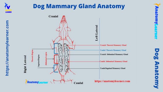
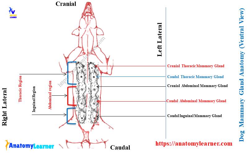
Now, I will provide more labeled diagrams on dog mammary gland anatomy. First, let’s see the location of the dog’s mammary glands from the ventral aspect of the dog.
Here, the diagram shows the 4 thoracics (cranial and caudal), 4 abdominal (cranial and caudal), and 2 large inguinal mammary glands from the ventral aspect of the dog.
The diagram also shows the smaller mammary glands in the thoracic region of the dog. Again, the larger mammary glands (inguinal mammae) are identified from the inguinal region of the dogs.
Now, let’s see the internal and external anatomical facts from the individual mammary gland of dogs. Here, the diagram shows the internal features (glandular parts) of the dog mammary gland.
Again, the lobules and lobular ducts from the dog mammary glands are identified in the labeled diagram. The papillary part of the dog mamma is identified in the labeled diagram.
Here, the lactiferous ducts and sinus are also identified in the dog mammary gland labeled diagram.
The arterial and venous supply to every single mammary gland of the dog is also shown in the labeled diagram. Let’s find more labeled diagrams on dog mammary glands and also from other species like horses, pigs, and rabbits on the social media of anatomylearner.
Canine mammary gland lymphatic drainage
Lymphatic drainage on the canine (dog) mammary gland varies on each side. Each mammary gland of the dog has its own plexus of lymphatic channels.
These lymphatic channels anastomose and encircle the base of the papillary part of the dog mammary glands. You will also see the distribution of the lymphatic channels in the parenchyma and subcutaneous tissue of the dog mammae.
One to three main channels from each gland leave and pass superficially to the nearest lymph nodes. Let’s see the lymphatic drainage of different mamame from the female dog.
The thoracic mammary glands of the dog drain lymph directly to the axillary lymph nodes. Again, the caudal abdominal mammary glands drain lymph in 2 ways.
It directly drains the lymph into the superficial inguinal lymph node. Again, the caudal abdominal mammae drain lymph into the lymphatic meshwork of the inguinal mammary gland.
Finally, the inguinal mammary glands of the dog have an extensive interlocking lymphatic plexus. These plexus drain the lymph into the adjacent superficial inguinal lymph nodes.
The axillary lymph nodes of the dog drain lymph by the sternal nodes into the thoracic cavity. Again, the superficial inguinal lymph nodes of the dog drain lymph into the iliac lymph node through the lymphatic of the inguinal canal.
The different authors describe the pathways of lymphatic drainage in different ways. If you want to know the basic information on lymph, its formation, and drainage, the below-mentioned article will help you to achieve your goal –
Frequently asked questions on dog mammary gland anatomy
Dog anatomy learners ask various questions regarding their mammary glands. Here, I tried to enlist the questions only on the dog mammary gland anatomy with their concise answer.
But, you should know the details of dog mammae’s shape, size, numbers, location, and internal and external features. I have already described these features of the dog mammary glands here in this article.
Okay, let’s see the common questions on dog mammary glands that are asked by anatomy learners –
Where are mammary glands located in dogs?
The mammary glands are located at the ventral aspect of the thoracic to inguinal regions in dogs. So, you may tell the mammary glands of the female dog extends from the pectoral to inguinal regions.
Mostly you will find the paired mammary glands in the female dog. So, these paired glands typically arrange into 2 rows (bilaterally symmetric) on the ventral aspect.
How many mammary glands does a cat have?
Typically, you will find 4 pairs (8) of mammary glands in the female cat. But, this number of mammary glands also varies from 4 – 5 pairs (8 – 10).
Like the female dog, the mammary glands are also arranged into 2 rows on the ventral aspect of the cat. You will find these mammary glands on the ventral aspect of the pectoral to the inguinal region of the cat.
How many mammary glands does a dog have?
Typically, dogs have 5 pairs of mammary glands on the ventral aspect. But you may also find 4 or 6 pairs of mammary glands in the dog.
Four pairs of mammary glands rarely occur in the dog. All these mammary glands of the dog arrange into 2 rows on the ventral aspect.
In the pectoral or thoracic region, you will find 2 pairs (4) of mammary glands. Again, the abdominal region of the dog contains 2 pairs (4) mammary glands. Finally, you will find only one pair of mammary glands on the ventral aspect of the dog’s inguinal region.
What is the functions of mammary glands in dogs?
The main function of the mammary glands in dogs is to produce milk for the newborn’s feeding. These mammary glands accumulate fat reserves over time and provide a reliable source of sustained nutrients for the young.
The glandular tissue of the dog’s mammary glands significantly develops only during pregnancy, pseudopregnancy, and during lactation. They also remain well developed in the nursing time of the young and for 5o days following weaning.
But, after the postpartum period, the alveoli and lobules of the dog mammary glands become shrunken.
Do mammary glands feel like lumps in dogs?
Yes, sometimes you may feel like the lumps along the mammary glands in the dogs. But, this situation does not frequently occur in the dog mammary glands.
After the oestrous cycle, some of the dogs show the lumps structure along with the chain of their mammary glands.
Is mammary gland painful for dogs?
Typically, a healthy mammary gland is not painful for dogs. But, if improper drainage or other disease condition is present in the dog’s mammary glands, it will be very painful for the dogs.
In improper drainage of milk, the dog mammary gland shows sollowen features.
Conclusion
I hope you got the basic idea of the dog mammary gland anatomy. Here, the location, number, and structure are important for you from the dog mammary glands.
The 5 pairs of dog mammary glands are arranged in 2 rows on the ventral aspect and show different sizes. Typically, the caudal inguinal pair of the mammary gland is larger than the dog’s other mamamae.