The sternum of an ox is a long osteo-cartilagenous plate. You will find this sternum bone at the midline of the floor of the cow’s thoracic cavity.
Here, I will share the osteological features of the cow sternum bone with the labeled diagram.
Summary of the ox sternum bone: it consists of seven (7) sternebrae, most developed from two lateral centers. The first segment of the sternum is wedge-shaped manubrium, whereas the last part is thin xiphoid cartilage.
You will know how different sternebrae joins together and form diarthrodial joints. Again, I will also show how the various ribs (sternal part of sternal and asternal ribs) join with the costal facets of the ox sternum bone.
Sternum of ox – what is the sternum bone?
Sternum is the flat long plate-like osteo-cartilagenous bone in the bovine (cow or ox). This plate-like bone is located in the ventral part of the thoracic cavity.
First, let’s identify the essential osteological features from the sternum bone of ox –
- #1. Body with various sternebrae (identified in the labeled diagram with their number),
- #2. Manubrium of the cow sternum (first segment),
- #3. Xiphoid cartilage (Last cartilaginous structure of sternum),
- #4. Facets for the first ribs on the manubrium of the sternum,
- #5. Various facets on the lateral aspect of the sternal body or sternebrae, and
- #6. Wide dorsal and flattened ventral surfaces of the bovine sternum,
The provided labeled diagram identifies all these osteological features of the cow sternum bone.
How many sternum bones (sternebrae) does an ox have?
The ox sternum has seven (7) bones, also known as the sternebrae. These sternebrae of the sternum fuse to form diarthrodial articulation.
Together they (sternebrae) form the wider, flatter, and relatively longer structure in an ox than the horse. But the question is – how are these sternebrae formed in an ox?
During development, the costal cartilages proceed toward the midline of the thoracic floor. The ventral extremities reach towards their fellows of the opposite site on this thoracic floor.
Now, the mesenchyme between the distal end of the costal cartilages appears. Then they get ossified and form the sternebrae and subsequently form the sternum bone in the cow.
Ox sternum bone anatomy
The anatomy of an ox sternum bone possesses the following –
- #1. A long body that is formed by the sternebrae, and
- #2. Two extremities that are also formed by the first and last sternebrae,
Now, let’s know the anatomical facts of the sternum body with a labeled diagram.
Body of cow sternum bone
The cranial part of the sternal body is wider, flatter, and longer. But, it becomes much narrower behind the last pair of costal facets.
The body of the cow sternum bone typically possesses –
- #1. Two surfaces – dorsal and ventral,
- #2. Two borders – right and left dorsolateral borders,
Here, the dorsal surface of the sternal body is wide and remains covered with the transverse thoracic muscles. The dorsal surface is concave longitudinally and flattened transversely.
You will also find the superior longitudinal ligaments on the dorsal surface of the sternal body along with thoracic muscles.
The ventral surface of the sternal body is flat but concave. This surface gives the origin of the pectoral muscles of the ox.
You will not find any ventral crest or keel on the ventral surface of the ox sternal body.
How does costal cartilage attach to the ox sternum?
The dorsolateral surface of the cow sternum body possesses seven pairs of sternal facets on either side of intersternal articulation. Second to eight pairs of costal cartilage of sternal ribs articulate to these facets.
The cartilage of the first pair of the sternal rib articulates with the extensive facets of the manubrium. The rest costal cartilage of asternal ribs articulates to the sixth to seventh sternebrae and the seventh pairs of costal cartilage.
You may learn more about the osteological features of the cow ribs and costal cartilage from the below-mentioned article of anatomy learner –
Manubrium of ox sternum
The first sternebra forms the cranial end (known as manubrium). This manubrium of cow sternum bone is somewhat wedge-shaped and laterally compressed.
The base of the ox manubrium forms a diarthrodial articulation with the body of the second sternebrae.
The dorsal surface of the ox manubrium is concave. You will find extensive lateral facets on the manubrium of the cow sternum bone.
The first pairs of costal cartilage articulate to the extensive lateral facets of the manubrium. Again, the thoracic and neck muscles also attach to the manubrium of the cow sternum bone.
Xiphoid process of the cow sternum bone
The xiphoid cartilage forms the caudal extremity of the cow sternum bone. This is a thin plate-like structure that attaches to the last sternebrae.
The xiphoid cartilage is much smaller in ox compared to the horses. You will find two other important attachments of this xiphoid cartilage –
- Dorsally – the xiphoid cartilage gives attachment to the diaphragm, and
- Ventrally – the xiphoid cartilage attaches to the ventral abdominal muscles,
Here, the dorsal surface of the xiphoid cartilage is concave and attached to the diaphragm. Again, the ventral surface of the xiphoid process is convex.
This convex ventral surface not only attaches to the ventral abdominal muscle but also attaches to the linea alba.
What are the 3 parts of the ox sternum?
From the above discussion, you may easily understand that there are 3 main parts in the ox sternum bone. These three parts of the ox sternum bone are –
- #1. A long body that possesses five sternebrae,
- #2. Proximal or cranial extremity that consists of manubrium, and
- #3. Distal or caudal extremity that consists of xiphoid process,
How to differentiate the sternum of a horse from a cow’s sternum?
The external appearance of a horse’s sternum bone is exceptional compared to the cow’s. Thus, you may easily identify or differentiate the horse sternum bone from the cow or ruminant’s sternum.
Though the horse sternum bone also possesses seven (7) sternebrae, the shape is different. You will see the boat-shaped sternum on the floor of the horse’s thoracic cavity.
This sternum shows the laterally compressed cranial end. In contrast, the caudal extremity of the horse sternum is dorsoventrally flattened.
Again, the ventral surface of the horse sternum shows a typical crest (known as keel). The lateral (both right and left lateral) is more concave than the cow’s sternum bone.
Again, the shape and formation of the xiphoid and manubrium of the horse sternum are also different than the cows. Here, the xiphoid cartilage in the horse sternum bone is flat and rounded.
The manubrium is oval and possesses a cranial cartilaginous extension. Now, let’s see these differences between horse and cow sternums from Table 1 –
| Features | Ox sternum | Horse sternum |
| Shape | Wider Flatter Relatively longer | Boat shape Compressed (cranially) Flattened (caudally) |
| Sternebrae | Seven | Seven |
| Manubrium | Concave Have extensive facets | Posses extension |
| Xiphoid process | Thin plate Concave | Flat Rounded |
| Body | Flat No ventral crest | Concave Posses ventral crest |
Again, the provided labeled diagram of the horse and cow’s sternum might help you understand and differentiate them. You may find more such labeled diagrams of various animal’s sternums here on social media of anatomy learners.
How to differentiate cow sternum from dog sternum bone?
The dog sternum bone is also unique in its shape and formation. Thus, you will easily identify the dog sternum bone from the oxes.
You will find eight (8) sternebrae in the structure of a dog sternum bone. But, these eight sternebrae of the dog sternum do not usually fuse.
Sometimes, these sternebrae in a dog may fuse at a very older age.
You will see the exceptionally very longest first sternebrae (manubrium) in the dog sternum bone. The cranial end of the first sternebrae of the dog is blunt. Again, the xiphoid process of the dog sternum is narrow compared to the cow.
The sternum of the dog plays an important role in forming the rib cage or thoracic cage. You will find the details features of the rib cage along with the features of the dog sternum bone from the below-mentioned article –
Cow sternum function
The cow sternum has various functions. Here, I will enlist some of the (essential) important functions of the cow sternum –
- The cow sternum forms the ventral boundary of the thoracic cavity,
- It furnishes the attachment of transverse thoracic muscle and superior ligament on its dorsal surface,
- The dorsolateral border of the cow sternum possesses seven pairs of facets where the costal cartilage of sternal and asternal ribs attach,
- It’s the first sternebra–manubrium furnishes attachment to the thoracic and neck muscles,
- The convex xiphoid process of cow sternum furnishes attachment to the transverse abdominis muscle and then linea alba,
I hope you got the basic functions of the cow sternum bone. It actually plays an important role in informing the thoracic cavity of the cow, where you will find different vital organs.
How strong is the ox sternum bone?
The sternum of the ox is considered the strongest bone in the thoracic cavity. Seven segments of sternum bones fuse to form the diarthrodial articulation, strengthening the sternum.
This bone of the cow’s thoracic cavity protects the visceral organs of the thorax. It also forms the strongest articulation with the costal cartilages from the asternal and sternal ribs.
What is on the top of the cow sternum?
You will see the costal cartilages that attach to the top of the cow sternum. This articulation extends from the manubrium to the last facest behind the xiphoid process of the cow sternum bone.
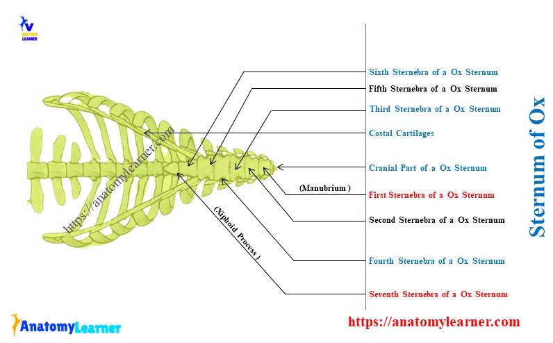
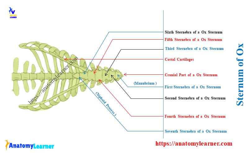
Ox sternum labeled diagram
Let’s see every sternebra and other features from the cow sternum bone labeled diagram. Here, I will show you the seven segments of the sternum bone along with the manubrium and xiphoid cartilage in the labeled diagram.
The labeled diagram of the cow sternum identifies the extensive facets from the first sternebra. Again, the diagram also identifies a thin plate-like xiphoid process from the caudal end of the sternum.
The labeled diagram also identifies the articulation of costal cartilages with seven sets of sternal facets.
Conclusion
So, the sternum of an ox possesses a long body and two identifiable distinct extremities. The appearance of the manubrium and xiphoid cartilage varies in a cow compared to horses.
The flattened and broader body of the cow sternum bone possesses seven pairs of facets for costal cartilage. The attachment of muscles and ligaments on the sternum is also essential when describing the anatomy of an ox’s sternum bone.