Cow Metatarsal Bone – How Many Metatarsals Does a Cow Have
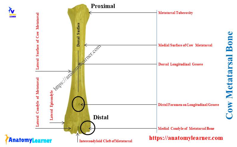
The cow metatarsal bone is the modified long bone on its hind limb. You will find two types of metatarsal bones in the pes segment of the cow’s hind limb. Here, I will show you the number of cow metatarsal bones and their details anatomical facts. Again, you can make the difference between metatarsal and … Read more