The terms ‘condyle’ and ‘epicondyle’ mean the type of articular surface in the bone. Here, I will answer the question – what is the difference between a condyle and an epicondyle?
Quick answer and explanation: the knuckled-shaped paired articular surface of the bone is condyle. In contrast, a small projection near the condyle is the epicondyle.
I will explain the terms ‘condyle’ and epicondyle’ from the animal bones. Thus, you will quickly identify the condyles and epicondyles from the various bones of the animal skeleton.
You will also differentiate the condyles from trochlea based on their appearance. So, let’s learn the features of the condyle and epicondyle from the animal bones.
What is the difference between a condyle and an epicondyle?
Explanation of the answer: The bones of the animal skeleton have different types of articular surfaces. According to their external appearance, these articular surfaces are condyle, epicondyle, trochlea, facet, and fovea.
Here, the condyle and epicondyle are the articular surfaces of the particular bone. However, the appearance of their articular surfaces is different in the same bone.
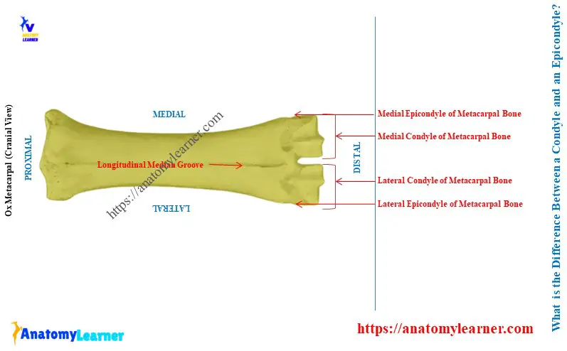
With examples, let’s clarify the terms ‘condyle’ and ‘epicondyle.’ Here, the diagram shows the distal extremity of the ox metacarpal bone.
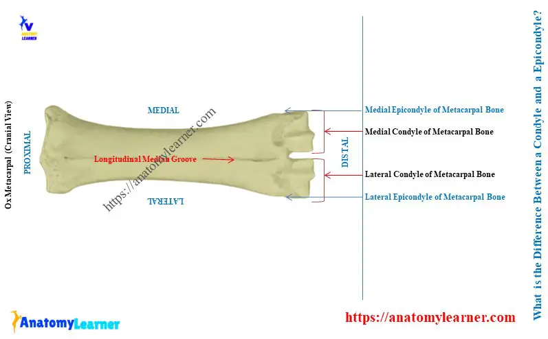
This diagram identified two condyles from its distal extremity – lateral condyle and medial condyle. And you know, these condyles of the ox metacarpal bone attach with the corresponding first phalanx.
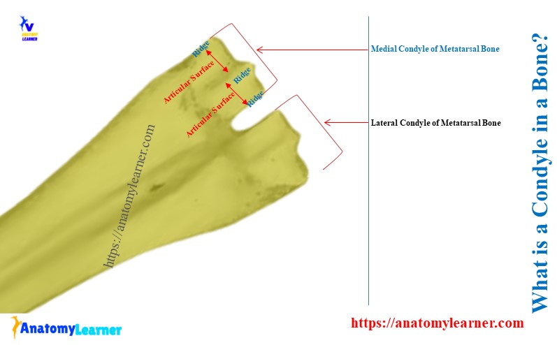
Let’s notice the articular surface of each articular surface of the condyles. Each condyle shows two articular surfaces.
Thus, two articular surfaces are on each condyle divided by the middle ridge. This type of paired articular surface of any bone is the condyle.
Again, these articular surfaces look knuckled. The knuckles are the type of joint found in your fingers.
Now, let’s see, there is a small projection near each condyle of the ox metatarsal bone. These small projections are also identified in the ox metatarsal bone labeled diagram. They are termed as the corresponding epicondyles of the metatarsal bone.
So, the summary of the terms ‘condyle’ and ‘epicondyle’ are –
- Condyle: knuckled-shaped (figure joint-like) paired articular surface of the bone.
- Epicondyle: small bony projection near the condyle of a similar bone.
Examples of condyles and epicondyles in animal bones
Condyles in animal bones:
- Condyles of the metacarpal and metatarsal bones of ox,
- Lateral and medial condyles of the ox humerus bone,
- Distal condyles of the animal femur bones,
Epicondyles in animal bones:
- Lateral and medial epicondyles near the corresponding condyles of the metatarsal and metacarpal bones,
- Epicondyles near the lateral and median condyles of the animal humerus bones,
- Lateral and medial epicondyles near the corresponding condyles of animal femur bones,
What are the condyles and epicondyles of the animal humerus?
Answer: the distal extremity of the animal humerus bone has lateral and medial condyles. These condyles articulate with the humeral articular circumference of the radius bone.
Here, the medial condyle of the humerus bone is known as the trochlea. Meanwhile, the lateral condyle of the humerus is known as the capitulum.
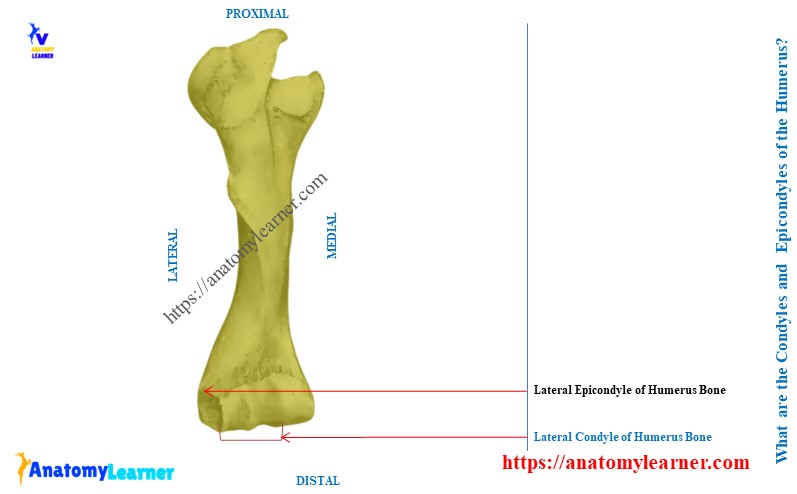
Again, the animal humerus bone also has lateral and medial epicondyles. A small bony projection is seen at the dorsolateral aspect of the lateral condyle of the animal humerus bone. This is the lateral epicondyle of the animal humerus bone.
In the same way, a small bony projection arises at the dorsomedial aspect of the medial condyle. This is the medial epicondyle of the humerus bone.
What is the condyle of the femur and epicondyle?
Answer: the caudal aspect of the distal extremity of the ox femur has the lateral and medial condyles. These condyles of the femur attach with the proximal articular surfaces of the tibia.
Here, the diagram identifies the lateral and medial condyles from the distal extremity of the ox femur. Between these two condyles, a deep depression is present, which is known as the intercondylar fossa.

Again, epicondyles present on the corresponding aspects of the lateral and medial condyles of the femur. The diagram also identifies the lateral and medial epicondyles from the caudo-distal part of the animal femur.
Here, the medial epicondyle is prominent in the femur bone. Meanwhile, the lateral epicondyle is less prominent in the ox femur bone.
Let’s differentiate the condyles and epicondyles of the ox femur bone in Table 1 –
| Condyle of Ox Femur | Epicondyle of Ox femur |
| Have paired articular surfaces | No such paired articular surfaces |
| Knuckled shaped structure | Just an outgrowth of the condyle |
| Articulates with tibia bone | Articulates with tendon and ligament |
Conclusion
So, the main differences between a condyle and an epicondyle are in their articular surfaces and appearance. The condyle must have paired articular surfaces, but the epicondyle has rough surfaces. Again, epicondyle always arises near the condyle of any particular bone.