I think you are looking for the best guide to learn seminal vesicle histology. In this article I am going to discuss on the important characteristics of seminal vesicle histology slide with labeled diagram. After reading this article you will able to know the seminal vesicle epithelium histology.
Hey, welcome again to anatomylearn and thanks for getting into this article. Probably this will be the easiest guide to learn seminal vesicle histology of animal online. I will also share my seminal vesicles histology drawing along with real slide.
You will also get other male genital organs histology like – vas deferens histology, epididymis histology and urethra histology from this anatomylearn blog. Let’s find other accessory glands histology like prostate histology at here.
Okay, fine; let’s get into today’s discussion – seminal vesicle histology real slide labeled diagram.
Seminal vesicle histology
You know, the eja-culates from male genital system of animal consists spermatozoa and seminal fluids. This seminal fluid contains the secretion of epididymis and male accessory glands.
Do you know about accessory glands of male genital system of animal? Fine, there are three major accessory glands in male genital system of animal.
#1. Seminal vesicle or it is also known as vesicular gland in animal
#2. Prostate gland of animal and
#3. The bulbourethral gland of animal; this gland also known as cowpers gland
In this article I will enlist the common characteristics of seminal vesicle histology. I will also show you the main difference in the histological features of seminal vesicle from different animals. If you interest to learn the histology of seminal vesicles then continue this article.
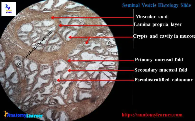
Seminal vesicle histology labeled diagram
I would like to provide you the list of structures that you should identify under the light microscope at laboratory from the vesicular gland histology slide
#1. Lining epithelium of glandular part of folded mucosa of seminal vesicle and lining epithelium of secretory ducts
#2. Interlobular septa in seminal vesicle in different animal
#3. Outer capsule of seminal vesicle in different animal
#4. Thin or thick tunica muscularis layer in different animal
So, let’s find these important structures from the seminal vesicle histology labeled diagram.
Characteristics of seminal vesicle histology slide of animal
You will find the four main layers (mucosa, propria mucosa, tunica muscualris and tunica adventitia layer) in the vesicular gland histology slide of any animal. I am going to enlist the common characteristics of seminal vesicle from different animal.
#1. The mucosa of seminal vesicle is markedly folded in most of the animals. Primary fold of mucosa branched again into secondary and tertiary folds. These branched folds of mucosa are the most exceptional histological features in seminal vesicle.
#2. The glandular epithelium (fold of mucosa) is lined by the pseudostratified columnar epithelium.
#3. There are many interlobular and main secretory ducts which are lined by the simple cuboidal epithelium in seminal vesicle of animal. Sometime you may find stratified columnar epithelium in seminal vesicle histology of few animals
#4. There is no distinct lamina muscularis layer in seminal vesicle; so you will find the propria submucosa with highly vascularized loose connective tissue.
#5. There are dense connective tissue trabeculae found in the seminal vesicle of animal. These trabeculae subdivided the vesicular gland into different lobes and lobules.
#6. Tunica muscularis is varies in seminal vesicle histology of different animals. You will find inner circular and outer longitudinal layers of smooth muscle layers in the vesicular gland histology.
#7. There is and external tunica adventitia layer which is consists of dense connective tissue along with elastic fibers.
Histological difference of seminal vesicles from different animals
Do you want to learn the histological difference of seminal vesicle from different animals? Well, let’s find the main difference of seminal vesicle histology from the slide of different animals.
Let’s find the histological features of seminal vesicle form bull.
#1. The vesicular gland of bull is compact and lobulated. You will find primary, secondary and tertiary folds of mucosa in the seminal vesicle histology of bull. The lining epithelium of these glandular parts of seminal vesicle is pseudostartified columnar epithelium.
#2. Few of these columnar cells possess the lipid droplet or light bleblike epical projections in bull seminal epithelium
#3. There is distinct well-developed interlobular septa which is derived from the thick tunica muscularis layer
#4. The tunica muscularis layer of bull seminal vesicle is thik than other animals. This thick muscularis layer is surrounded by a thick capsule of dense connective tissue with few smooth muscle cells.
In boar seminal vesicle histology, the secretory epithelium is also folded and lined by pseudostartified columnar epithelium. The tunica muscularis layer in boar seminal vesicle is thin than bull. Interlobular septa of boar vesicular gland are also thin and contain predominant smooth muscle cells.
In horse seminal vesicle, you will find the short branched mucosal folds. There is also thin connective tissue septa present in the horse seminal vesicle. The smooth muscle cells in horse seminal vesicle are arranged irregularly.
The histology of seminal vesicle of buck and ram are similar with the histology of bull vesicular gland. But lipid droplet in the basal cells of lining epithelium is absent in buck seminal vesicle.
Seminal vesicle histology drawing
I am so happy to share the seminal vesicle histology drawing with you. You may follow this drawing and try to perform better than this drawing.
Just focus on the every single structure of seminal vesicle. You may draw one specific part from animal seminal vesicle. You don’t need to be perfect; practice of drawing will make you perfect.
Do you want to know the function of seminal vesicle?
You know seminal vesicle secrete yellowish viscous fluid that contains numerous substances. Seminal fluid contains fructose, globulin and prostaglandins. Fructose is so important to maintain the nutrition of the spermatozoa in animal.
But make sure, the seminal vesicle is not the site of storage of spermatozoa.
If you want then you may also learn the histology of other accessory glands of male genital system of animal from anatomylearner. You might like other article related to prostate gland histology, epididymis histology, ductus deferens histology and testis histology.
Again you should find more labeled diagram of different histological slide from male genital system of animal at here in social media of anatomylearner.
Conclusion
I know this guide will help you a lot to learn seminal vesicle histology. If you need more information about vesicular gland histology then let me inform. I will try to provide more information on seminal vesicle from different animals.
If you like this article and think that was helpful for you; I would like to request to share this article with your friends who want to know about the histology of vesicular gland of animal.
I will highly appreciate your valuable suggestion for this seminal vesicle histology article.