
Do you want to learn testis histology slide with labeled diagram? Testis histology labeled diagram is so important to understand the all structures of testis.
In this article I am going to discuss on testis histology of animal. You will get an ideal concept of seminiferous tubule histology with a labeled diagram. I am going to share the real testis histology labeled slide with you.
I will also provide histology of testis ppt for your practices. You will also get testis histology drawing at the end of this article.
Do you know the testis anatomy? Let’s get a complete guide to learn testis anatomy at here in anatomylearner blog
Let’s get into testis histology description.
Testis histology
You know testis is the male reproductive organ that is surrounded by the serous sac of tunica va(g)zi-nealis and testicular envelopes.
You will find the parietal layer of tunica va(g)zi-nealis that attached with the scrotum after removing testis from scrotum. Again the visceral layer of tunica va(g)zinealis attached with the testis. This tunica vagzi(nea)lis is derived from the visceral layer of peritoneum.
Deep to this tunica vag-zi(nea)lis you will find a thick white fibrous connective tissue capsule called tunica albuginea, Tunica albuginea primarily consists of collegen fibers, few elastic fibers and myofibronlast. You will also find branches of testicular artery and a network of anastomosing vein on the vascular layer of tunica albuginea.
The tunica albuginea continuous with the connective tissue trabeculae that is known as septula testis or septae
The septual testis divides the testicular parenchyma into a variying number of testicular lobules. Each of these lobules contains various numbers of highly convoluted seminiferous tubules
The septula testis communicates with mediastinum testis. But it is difficult to understand the septa in ruminant testis. You may get complete septa in dog and boar testis.
Mediastinum testis contains a network of irregular tubules which is known as rete testis. You will find mediastinum testis in central position of testis in ruminant and dog. In other animals it is located at the marginal part of testis.
These rete testes actually connected with the seminiferous tubules through the straight tubule. You will learn about seminiferous tubule histology in details in this article.
In testis histology, deep to the tunica albuginea there is a layer of vascular loose connective tissue. This vascular loose connective tissue layer is known as tunica vasculosa. It extends into the interior inferential connective tissue that is surrounded by seminiferous tubules.
Testis histology slide
I would like to enlist the structures that you should identify under the light microscope from testis histology slide at laboratory.
#1. Tunica vagzi(nea)lis layer of testis of animal
#2. Tunica albuginea layer of testis of animal
#3. Mediastinum and rete testis of animal
#4. Seminiferous tubule of animal testis
#5. Different types of spermatogenic cells of seminiferous tubule (Spermatogonia, primary spermatocytes, secondary spermatocytes, spermatid cells and spermatozoa)
#6. Sustenttacular cells of animal testis or sertoli cell
#7. Interstitial cell or leydig cell
#8. Myoid cells of testis of animal
#9. Basal lamina or lamina propria at seminiferous tubule of testis
Let’s find these structures and cells from the testis histology slide labeled diagram. Hope this testis labeled slide will help you to understand the complete testis histological structures.
Convoluted seminiferous tubule histology
The common characteristics of seminiferous tubule histology from different animals are –
#1. Seminiferous tubules are highly coiled packed tubules
#2. The tubules are lined by the stratified germinal epithelium (like spermatogenic cells, sustantacular or sertoli cells)
#3. Presence of three important components – lamina propria, sustantacular cells and spermatogenic cells.
Do you want to know the seminiferous tubule histology in details?
There is a lamina propria that surrounds the seminiferous tubule of testis. You will find a basal lamina at the innermost area of lamina propria. There are some flatten myoid cells adhering to basal lamina of seminiferous tubule. These myoid cells have the properties of smooth muscles cell.
Sustantacular cells of seminiferous tubule of testis histology
Sustantacular cells include somatic and supportive cell or sertoli cells. Most of the sustantacular cells of seminiferous tubule of testis are irregular in outline and elongated. The broad base of sustantacular cells rest on the basal lamina of seminiferous tubule. I would like to describe about the most important supportive cells of seminiferous tubule – sertoli cell.
Sertoli cells are tall columnar cells in seminiferous tubule of testis histology. The supportive cells extend from the basal lamina to the lumen of the seminiferous tubules. Sertoli cell have pear shaped nucleus and they are located generally aat the basal part of the cells.
Adjacent supporting cells are joining with each other by tight junction twith their basal cytoplasmic process. These tight junctions help to from blood testis barrier in testis.
The sertoli cell has the following important functions –
#1. They have nutritive, protective and supportive function for the spermatogenic cells
#2. Sertoli cells are also responsible for phagocytosis and degenerating spermatogenic cells
#3. They help to release spermatozoa into the lumen of seminiferous tubule
#5. Sertoli cells mediate the action of follicular stimulating hormone and testosterone
#7. They produce androgen binding protein and secrete constitutents of intertubular fluid like inhibin
#8. Responsible for structural and metabolic support to the spermatogenic cells
#9. Form the blood testis barrier on animal body
Spermatogenic cell of seminiferous tubule histology
Spermatogenic celss are arranged developmentally higher order from the basement membrane to the lumen. Sequence of events of development of spermatozoa from spermatogenic cells known as spermatogenesis
The spermatogenic cells of seminiferous tubules are –
#1. Spermatogonia cells – small, diploid primitive germ cells
#2. Primary spermatocytes – larger cell with larger nucleus
#3. Secondary spermatocytes – smaller cell with dense nucleus
#4. Spermatid cells – smaller and stay in group
Again, spermatogenesis is subdivides into three phages –
#1. Spermatocytogenesis – the process during spermatogonia develop into spermatocytes
#2. Meiosis – the maturation division of spermatocytes that results in spermatid
#3. Spermiogenesis – the process of transformation of spermatids into spermatozoa
You know there are various spermatogenic cells that represent different phases of development and differentiation of spermatozoon. There are two main type of spermatogonia and they are –
#1. The type A spermatogonia – have darker nucleus
#2. Type B spermatogonia (light stained nucleus)
Again type B spermatogonia undergo the maturation and produce the primary spermatocytes.
How you may identify these spermatogenic cells under light microscope at laboratory?
Identification of spermatogenic cells under light microscope
In testis histology, Type A spermatogonia are the largest spermatogonia. These spermatogonia share an extensive contact area with the tubular basal lamina. They (spermatogonia A) possess prominent nucleoli with pale nuclei.
In the other hand, B spermatogonia have spherical nuclei with more chromatic particle and less prominent nucleoli.
The primary spermatocytes are the largest spermatogenic cells in the tubular epithelium of testis in animal. They located in intermediate position between spermatogonia and spermatid in animal’s testis.
Secondary spermatocyte varies in size and intermediate between primary spermatocytes and spherical spermatids in animal seminiferous tubule.
You know, spermiogenesis in animal mainly divided into golgi phase, cap phase, acrosomal phase and maturation phase.
Spematozoa in different animal’s testis vary in length. You will find two main parts of animal spermatozoa – head and tail. Again the tail of animal spermatozoa divides into neck, middle piece, principal piece and end piece.
If you want to more about spermatogenic cell histology and the complete process of spermatogenesis then find the complete guide here in this blog.
Interstitial cell or leydig cell from testis
The intertubular space in testis histology contains loose connective tissue, blood vessels, fibrocytes and interstitial endocrine leydig cells.
Leydig cells are responsible to produce testicular androgen in boar and large amount of estrogen as well. These cells occurs in cords or cluster in testis histology. Leydig cells are larger in size, polyhydral in shape and have acidophilic cytoplasm.
The functions of leydig cell of testis are –
#1. Promotion of normal sexual behavior of animal
#2. Enhance the growth and maintenance of the functions of pen-(is), male accessory sex organs and secondary sex characteristics
#3. Control of spermatogenesis in animal testis
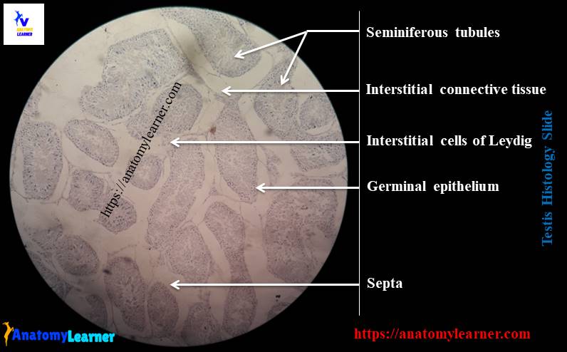
Testis histology labeled diagram
I would like to provide the summary of testis histology slide labeled diagram. You should identify testis slide with following characteristics –
#1. Presence of seminiferous tubules in testis section
#2. Spermatogenic cells of various stage of maturation
#3. Presence of sertoli and leydig cells in testis histology section
Conclusion
I am sure you got the best guide to learn testis histology with real testis histology slide pictures. Now you will able to identify the important histological structures from testis histology labeled diagram.
If you need histology of testis ppt or pdf for practice let me inform. You may get more testis histology drawing diagram here in anatomylearner social media.
Do you want to learn more about spermatogenic cells from seminiferous tubule histology? Don’t forget to share this article with your friends who want to learn histological features of animal testis.
Get ready to learn epididymis histology and ductus deferens histology from anatomylearner.