
It is so important to know testis histology and epididymis histology before to start learning ductus deferens histology. After a sharp bend at the end of ductus epididymis, it become straight and acquires the histological features of ductus deferens.
Hey, do you want to learn ductus deferens histology with labeled diagram? You are now in the right place where you are going to learn the complete ductus deferens histology slide with labeled drawing.
In this article I am going to discuss on ductus deferens histology layers along with their features. You will also learn about ejaculatory duct histology in this article.
If you know vas deferens anatomy then it will be easy to understand the vas deferens histology diagram.
So, interest to learn vas or ductus deferens histology? Let’s get into the details histology of vas deferens slide.
Ductus deferens histology
You should identify the ductus deferens under light microscope with the following histological features. Hope, these will give an idea what actually you should learn in details from ductus deferens.
#1. Lining epithelium of mucosa from different part of animal’s ductus deferens
#2. Propria submucosa with tubuloalveolar glands in few animals’ ductus deferens
#3. Different layers of smooth muscle layers at tunica muscularis layer of vas deferens
#4. Identify the tunica serosa or tunica adventitia of vas deferens of animal
#5. Find out spermatozoa at the ejaculatory duct of different domestic animals
Let’s try to find out these structures from vas deferens histology labeled diagram. Do you want to learn details histological features from different layers of ductus deferens?
Ductus deferens histology slide
I would like to explain ductus deference histological layers from the real slide. It is so easy to learn vas deferens histology if you already known the histological features of tubular organ. You should only point out the specific features in different layers of vas deference.
You will find the following four layers in the histology of vas deferens from different animals.
#1. Mucosa layer of vas deferens of animal
#2. Propria submucosa of vas deferens of animal
#3. Tunica muscularis layer of vas deferens of animal
#4. Tunica adventitia of vas deferens of animal
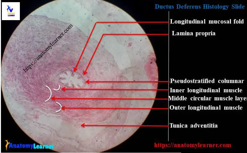
The mucosa of vas deferens is folded and lined by the pseudostratified columnar epithelium. But this lining epithelium is varies with the different part of ductus deferens in different animal. You will find simple columnar epithelium towards the end of the duct.
Sometime short and branched microvilli may be found in the columnar epithelium lining of ductus deferens. In bull, there is some lipid droplet present in the basal cells of lining epithelium.
The lamina muscularis layer is absent in the mucosa layer of ductus deferens of animal. You will find the propria submucosa layer which is highly vascularized. This submucosa layer of ductus deferens is also rich in fibroblast and elastic tissue in animal.
The smooth muscle layer of tunica muscularis of ductus deferens histology is varies. You may get two layers or three layers of smooth muscles layer in tunica muscularis layer of ductus deferens.
Generally found circular, longitudinal and obliquely smooth muscle layers in most of the species. I will discuss the differences of ductus deferens in different animals below.
Tunica adventitia contains the highly vascularized loose connective tissue in ductus deferens of animal.
Ductus deferens histology labeled diagram
I have the real ductus deferens histology slide labeled diagram for you. Hope these histology slides pictures will help you a lots to understand the histological features of vas deferens.
Ductus deferens histology drawing
I am so happy to share this ductus deferens histology drawing with you. I tried to focus the every single structure from the histology of vas deferens. In this ductus deferens diagram I only used the simple line drawing with pencil. You might try to draw the better histological picture of ductus deferens than this drawing.
Ductus deferens histological features in different animal
The terminal part of the ductus deferens connects with the vesicular gland in different animal like stallion, dog and ruminant. There is dilated portion at the terminal part of the each ductus deferens of animal called the ampulla. But in boar and cat there is no ampulla present at the terminal end of the ductus deferens.
In the ductus deferens histology, you may find some simple branched tubuloalveolar glands in the propria submucosa in boar and cat. These tubuloalveolar glands occupy entire propria submucosa in stallion, bull and ram which is rich in the smooth muscle cell. But in dog and buck these gland are surrounded by the dense connective tissue layer which is devoid of smooth muscle cells.
Tunica muscularis consists of circular, longitudinal and oblique layers of smooth muscle in the ductus deferens histology of stallion, bulls and boar.
But in small ruminant and dog, the tunica muscularis consists of inner circular and outer longitudinal smooth muscle layers.
You will find considerable numbers of spermatozoa in the lumen of ductus deferens at the terminal part.
Ejaculatory duct histology
In some animal like stallion and ruminant, the ductus deferens connects with the execratory duct of seminal vesicle. Thus it forms the ejaculatory ducts in stallion and ruminant. Do you want to know the ejaculatory duct histology?
Ejaculatory duct is the straight continuation of the ductus deferens beyond where it receives the duct if the vesicular gland. The mucosa of ejaculatory duct is lined by the pseudostratified epithelium. Ejaculatory duct lacks of muscular layer.
This ejaculatory duct enters into the prostate gland and end at the seminalis colliculus. In boar, the ductus deferens and execratory duct opens separately into the urethra. But there is no vesicular gland in carnivore; so the ductus deferens opens alone into the urethra.
I am sure that you are also interest to learn histology of the following organs from different animal –
#1. Complete histological features of lung parenchyma of animals
#2. Best guide to know the histology of gallbladder in details
#3. General organizational pattern of the hollow organ (most important to learn histology)
You may find all of these articles at here in this blog; don’t forget to learn vas deferens anatomy from anatomylearn.
Conclusion
I hope you learn the ductus deferens histology in details with the real histology slide. Now you are able to identify the important structures from ductus deferens histology labeled slide. Let’s practice ductus deferens histology drawing.
If you need more information about ductus deferens histological layers let me inform. I will discuss more on ejaculatory duct histology in coming article. Get more ductus deferens histology slide picture at here in the social media of anatomylearn.
Share this article with your friends if you really got any value from vas deferens histology article. Thanks for staying with anatomylearner and learn ductus deferens histology with me.