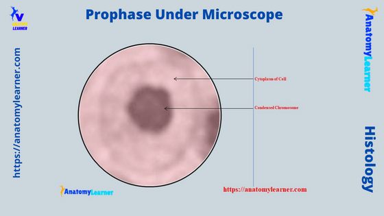
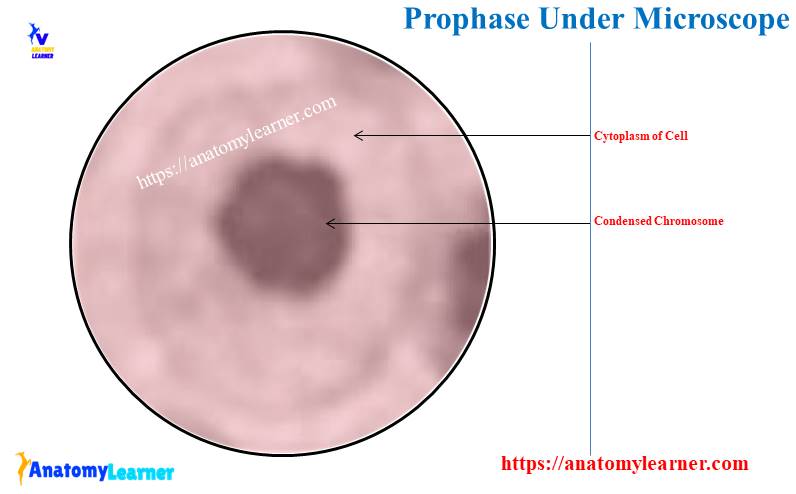
The prophase under a microscope shows the gradually becoming condensed chromatin, resulting in the formation of the individual chromosome. You know this prophase is the first stage of mitosis cell division which may quickly identify with the help of a light microscope.
Again, you will also see the prophase 1 and prophase 2 stages in the meiosis cell division in animals. The prophase of meiosis I last for a long time and divides into 5 distinct stages.
This article might help you to understand the basic structural changes in the prophase stage of mitosis and meiosis cell divisions. It might be helpful for you to identify the prophase of mitosis and meiosis cell division under the light microscope perfectly.
Again, I will provide the basic concept of the different stages of mitosis and meiosis cell division with the labeled diagram. So, if you want to learn and identify the prophase stage from the microscope slide, let’s continue this article till the end.
Prophase under microscope
First, I would like to show you the main features of the prophase under a light microscope with the labeled diagram. You will find the following characteristics in the prophase stage of mitosis cell division –
- You will see the gradual coiling of the nuclear chromatin and giving rise to several individual bodies (coiled chromosomes),
- The nuclear membrane and the nucleolus disappear,
- Now, the chromatin granules resolve into chromosomes,
- Centrosome divides into 2 (paired) chromatids that lie side by side and united at a point,
- Centrosomes with their centrioles separate, and a centrosome migrates to each pole of the cell, and
- The centrioles are connected by the microtubules, which form the mitotic spindle within the cell,
But, you may also find a little difference between the early and later prophase of the mitosis cell division.
In meiosis cell division, you will also find the prophase stage – prophase 1 and prophase 2. Meiosis I is the first event of meiosis cell division, also known as the reductional division.
In this event, the homologous pairs of a chromosome line up, and each pair separates and go to each daughter cell. Thus, each daughter cell receives half of the number of chromosomes (haploid number).
In constant, meiosis II is the second event of meiosis cell division, also known as the equatorial division. This event shows that 2 chromatids of each chromosome are separated and migrated to each daughter cell.
In the prophase of meiosis 1, you will see the following 5 stages –
- Leptotene stage,
- Zygotene stage,
- Pachytene stage,
- Diplotene stage, and
- Diakinesis stage,
I will describe all these stages of prophase from meiosis 1 and 2 in the next section of this article with the labeled diagram.
Stages of mitosis under microscope labeled
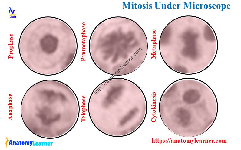
In the mitosis cell division, the cytoplasm and nucleus of a cell divide equally into 2 identical daughter cells. The process of mitosis divides into a series of 4 or 5 stages under the microscope –
- Prophase (stage 1) – condensing and replicating chromosomes,
- Prometaphase (stage 2) – nuclear membrane breaks down, and mitotic spindle forms,
- Metaphase (stage 3) – chromosome become oriented in the equatorial plane perpendicular to the centrosome,
- Anaphase (stage 4) – two sister chromatids (2) separate from each other, and
- Telophase (stage 5) – nuclear envelope reappears, and chromosomes decondense,
In addition, cytokinesis occurs concomitantly with the telophase stage of the mitosis cell division. I have already enlisted the basic microscopic features of the prophase of mitosis cell division under a light microscope. So, let’s start to learn the other different stages of mitosis cell division with the diagram.
Prometaphase under a light microscope
In the prometaphase stage of the meiosis cell division, you will find the following essential features under the light microscope. Sometimes, it is very difficult to differentiate the microscopic figures between the prophase and prometaphase.
Let’s see the characteristics features of the prometaphase of the mitosis cell division under the microscope –
- The disappearance of the nuclear membrane,
- The mitotic spindle is clearly formed in this prometaphase stage of the mitosis cell division,
- You will see 3 types of microtubules in this stage – astral, kinetochore, and spindle microtubules,
The astral microtubules grow from the poles to the plasma membrane. They anchor the mitotic spindle in the center of the cell.
Again, the kinetochore microtubules grow from the poles and attach to the chromosome. Finally, the spindle can attach to the arms of the chromosome.
In this prometaphase stage of mitosis cell division, the movement of the chromosome is highly dynamic.
Metaphase under a light microscope
During the metaphase of the mitosis cell division, you will find the below-mentioned essential features –
- The chromosome becomes oriented in the equatorial plane perpendicular to the centrosome,
- Each chromosome gives closely apposed, identical halves (chromatid or sister chromatid),
- Again, the chromatid connects at the centromere,
The centromere is a constricted, pale staining structure containing kinetochore microtubules (protein complex). This kinetochore is an attachment point for the microtubules emanating from opposite centrosomes.
Anaphase under microscope
Anaphase is one of the important stages of mitotic cell division, where you will find the following essential changes –
- The centromere divides into 2 sister chromatids completely and separates from each other in this anaphase stage,
- The centrosome also moves away from the center, pulling along the remaining of chromosomes,
- Now, the mitotic spindle tubules pull the separated group of chromatids to opposite poles,
- Here, the 2 groups of chromatids (now chromosomes) are identical.
The pair of chromosome aline in the spindle checkpoint and kinetochore-microtubule rapidly shorten. And together with the molecular motor (kinesin and dynein), the pairs of a chromosome are separated.
Each of the daughter chromosomes moves apart to the poles that vary rapidly. This phase of mitosis is very rapid and takes few minutes only.
Telophase and cytokinesis under a light microscope
Telophase is the last stage of mitotic cell division, where the nuclear envelope reappears. The main microscopic features of the telophase of the mitosis cell division are –
- Nuclei reappear in the daughter cells,
- The chromosome become less distinct, and a new nuclear membrane appears around each of these daughter cells,
- Now, the chromosome decondense, and nucleoli appear,
You will also find the cytokinesis stage within this telophase stage of the mitosis cell division. What happens in the cytokinesis stage under the light microscope?
A constriction develops at the equatorial plane of the parent cell and progresses. That means a cleavage furrow find in the cytokinesis stage around the equatorial plane of the cell.
This furrow divides the cytoplasm into two halves surrounding each newly formed nucleus.
Prophase vs. telophase under a microscope
You may easily differentiate the microscopic figures of the prophase from the telophase. Let’s see the main differentiating features of the prophase and telophase under the light microscope.
Here, in the prophase, the nuclear chromatin gradually coils and gives rise to several individual bodies. The nuclear membrane breaks, and the nucleolus disintegrates.
Again, the centrosome with their centrioles separate, and a centrosome migrates to each pole of the cell. Finally, the microtubules of the mitotic spindle appear between the 2 centrosomes.
On the other hand, the nuclei reappear, and chromosomes recondense. The nuclear membrane also appears around the daughter chromosome.
Finally, a cleavage furrow appears at the equatorial plane of the cells and progresses until the cytoplasm and organelles divide into two.
Prophase vs interphase
The interphase is a long period of total mitosis cell division. During this long time, the cell doubles (2) in size and DNA content.
The interphase and mitosis are two major events in the cell cycle. You know the alternation between mitosis and interphase is the cell cycle.
In the interphase event of the cell cycle, you will find the followings 3 phases –
- Presynthesis (G 1phase),
- DNA synthesis (S phase), and
- Post DNA duplication (G 2 phase),
In the pre-synthesis phase, you will find the following important features –
- You know the daughter cell is formed during mitosis and now enters into the pre-synthesis phase of interphase,
- In this phase of interphase, cell synthesis RNA, a regulatory protein essential to DNA replication and enzyme for these synthetic activities,
- In the S 2 phase of interphase, genom beome double. Now, the cell contains twice the normal component of its DNA.
Finally, in the G 2 phase, RNA and essential proteins for cell division are synthesized.
Meiosis stages under a microscope
Meiosis is a special type of cell division that produces haploid daughter cells from the haploid parent cell. This meiosis cell division occurs only in the germ cells during the maturation of gamate.
You will find 2 events in the meiosis cell division –
- Meiosis I – first division, reductional division, and
- Meiosis II – second division, also known as the equatorial division,
Now, let’s see the different meiosis I and II stages.
Here, meiosis I, or reductional division, shows the following stages –
- Prophase I – includes leptotene, zygotene, pachytene, diplotene, and diakinesis,
- Metaphase I,
- Anaphase I, and
- Telophase I – similar to the telophase of mitosis cell division,
Again, you will also find 4/5 stages in the meiosis II or equatorial division – prophase II (important), metaphase II, anaphase II, telophase II, and cytokinesis.
Here, I will describe only some of the stages of meiosis I and II. I prefer to describe prophase I and II from the meiosis cell division.
Prophase 1 under a microscope
As I told you before, prophase 1 under the microscope, shows 4/5 main stages like – leptotene, zygotene, pachytene, diplotene, and diakinesis. Now, I will show you these different stages of prophase 1 microscopic features with the labeled diagram.
Let’s see what happens in the leptotene of prophase 1 –
- The 2 sister chromatids of an individual chromosome join at the centromere,
- These sister chromatids can easily be visible as a long strand in the nucleus,
- These chromatids of the nucleus take deep stains with the basic dyes,
So, under a microscope, you will see deeply stained strands (chromatids) in the leptotene stage of prophase 1. Now, let’s see the special features of the zygotene stage under the light microscope.
In the zygotene stage of prophase 1, homologous chromosomes become attracted to each other and lie side by side. Thus they form the conjugation or synapse (known as synapsis).
Here, one member of the pair is maternal, and the other membrane is parental in origin.
In the pachytene stage, 2 chromosomes together constitute a bivalent. Each bivalent posses 4 chromatids and thus forms the tetrad (chromosome splits into 2 chromatids except at centromere).
2 central chromatids become coiled over each other and form chiasmata. You may also call this chiasma as the crossing over.
By this crossing over, a random exchange of genetic material occurs between the homologous chromosome.
The chromosome continues to condense and begin to separate in the diplotene stage of prophase 1. Now, the homologous centromeres pull apart, and the chromosomes are separated.
At the same time, the nucleolus disappears, and the nuclear membrane.
Prophase 2 under a microscope
At telophase 1 of meiosis 1 will produce two daughter cells. Each of these two new daughter cells enters meiosis II.
Meiosis II is a second meiotic division separating the sister chromatids resulting in haploid cells. The chromosomes of 2 daughter cells condense again in preparation for the second meiotic division.
So, in the prophase 2 of meiosis cell division, you will see –
Gradual condensation of chromatin results in individual, discernible chromosome formation. Again, the nucleoli disappear, and the centrosome divides into 2 centrosomes.
These centrosomes migrate to the opposite poles of the cell. Now they begin to organize microtubules into the meiotic spindle.
Prophase under microscope labeled diagram
I will show you the prophase under a microscope-labeled diagram in this article section. From both mitosis and meiosis (I and II), I will show the microscopic figures of the prophase stage.
First, let’s see the labeled diagram of the prophase stage from the mitosis cell division. In this diagram, you will find the gradually coiled nuclear chromatin, broken nuclear envelope, and centrosome with their centrioles.
The diagram also shows the migrated centrosome to each pole of the cell. Again, you will see the microtubules of the mitotic spindle between the 2 centrosomes in the labeled diagram.
Again, the other diagram shows the different stages of mitosis cell division (except prophase). Now, I will show you the labeled diagrams on the different stages of prophase 1 of meiosis I.
The sister chromatids from leptotene of prophase 1 (long strand) are identified in the labeled diagram. Again, the synapse between the homologous chromosomes is identified from the zygotene of prophase 1 in the labeled diagram.
The most important events in the pachytene stage of prophase 1 are bivalent formation and crossing over. The labeled diagram shows the bivalent formation in the pachytene stage of prophase 1.
The diplotene stage of prophase 1 labeled diagram shows the recondensed chromosomes. Now, in the labeled diagram, let’s see the furrow between homologous chromosomes from the diakinesis stage of prophase.
At the same time, the diakinesis of the prophase 1 labeled diagram shows the 2 separated chromosomes. Let’s find more labeled diagrams on the prophase of mitosis and meiosis on social media of anatomy learners.
Common inquiries on prophase under a microscope
In this section, I will provide concise information on the inquiries related to the prophase under the light microscope. But, reading the whole article (phase of mitosis – especially the changes in prophase and prophase 1 in meiosis) is recommended to get the basic idea on prophase.
Let’s see the common inquiries on the prophase stage asked by the histology learners.
What does prophase look like under the microscope?
You know the prophase is the first stage of mitosis cell division which may easily identify from the microscopic slide. Under the microscope, prophase shows the gradually condensed chromatin.
With hematoxylin or other basic dyes, the prophase stage (gradually condensed chromatin) takes deep or intensified stained. Sometimes you may see the individual chromosome in this stage under a light microscope.
In addition, the nucleoli may disappear in this stage of cell division under the light microscope. You may read the information I provided earlier to learn more about microscopic features.
How can prophase be distinguished from interphase under a microscope?
To distinguish the microscopic features of prophase from interphase, you might have a good piece of knowledge. Under the microscope, you will not find any changes in the interphase stage of a cell (as in this phase, the cell does not undergo division).
But, it shows (interphase) the increased size and contents under the microscope. You will find the distinct nucleolus and normal stages of chromosomes in the interphase stage of the cell under a light microscope.
But, you will see the condensed nuclear chromatin in the early prophase stage. Again, the later stage of prophase shows the broken nuclear envelope. The microscopic figure of prophase also shows the centrosomes with centrioles and the mitotic spindle.
Thus, you may easily distinguish prophase from interphase under a microscope.
Can you see chromosomes in prophase under a microscope?
Yes, you can see the individual chromosome in prophase under the microscope. In normal hematoxylin and basic dye staining, you will see the deeply stained chromosomes under the microscope.
In the early stage of prophase, you can also see the condensed chromosome in the microscopic view. On the other hand, you will also see the clear centrioles (2 pairs) in this stage under the light microscope.
How do you determine the stages of meiosis under a microscope?
To determine the stages of meiosis under a light microscope, ensure you have a good piece of knowledge (info) on the full events of this cell division. The meiosis posses 2 important events – meiosis I and meiosis II.
The stages of meiosis I cell division (prophase 1, metaphase 1, anaphase 1, and telophase 1) can easily identify with the help of a microscope. Again, prophase 1 shows different stages with some unique microscopic features.
Long-strand chromatids are the main microscopic features of the leptotene stage of prophase 1. Again, you can easily identify synapses, crossing over, and homologous chromosomes from the different stages of prophase 1 with the help of a microscope.
Conclusion
So, the prophase under a microscope shows different unique features like a condensation of nuclear chromatin and disappears of the nuclear envelope. Each chromosome duplicates and forms paired sister chromatids that lie side by side.
Centrosomes in the prophase stage with their centrioles separate, and the centrosome migrates to each pole of the cell. The microscope view of prophase also shows the mitotic spindle between two chromosomes.